section name header

Disease Prologue

Summary

Cancers Staged Using This Staging System

The staging system applies to all primary carcinomas that arise in the stomach. Adenocarcinoma is the most common histologic type, whereas other histologic types are observed less frequently.

Cancers Not Staged Using This Staging System

These histopathologic types of cancer…Are staged according to the classification for…and can be found in chapter…
Gastrointestinal stromal tumorsGastrointestinal stromal tumors43
Other sarcomasSoft tissue sarcoma of the abdomen and thoracic visceral organs42
LymphomasHodgkin and non-Hodgkin lymphomas79
Well-differentiated neuroendocrine tumors (G1 and G2)Neuroendocrine tumors of the stomach29

Summary of Changes

ChangeDetails of ChangeLevel of Evidence
Anatomy—Primary Site(s)Anatomic boundary between esophagus and stomach: tumors involving the esophagogastric junction (EGJ) with the tumor epicenter no more than 2 cm into the proximal stomach are staged as esophageal cancers; EGJ tumors with their epicenter located greater than 2 cm into the proximal stomach are staged as stomach cancers. Cardia cancer not involving the EGJ is staged as stomach cancer.III
Definition of Regional Lymph Node (N)N3 has been subdivided into N3a and N3b.II
AJCC Prognostic Stage GroupscTNM: stage groupings for cTNM differ from those of pTNM. New cTNM groupings and their cooresponding prognostic information are presented in this edition.III
AJCC Prognostic Stage GroupsypTNM: stage groupings are the same as those for pTNM; however, prognostic information is presented using only the four broad stage categrories (Stages I-IV).III
AJCC Prognostic Stage GroupsIn pathological classification (pTNM), T4aN2 and T4bN0 are now classified as Stage IIIA.II

ICD-O-3 Topography Codes

CodeDescription
C16.0Cardia, esophagogastric junction
C16.1Fundus of stomach
C16.2Body of stomach
C16.3Gastric antrum
C16.4Pylorus
C16.5Lesser curvature of stomach, NOS
C16.6Greater curvature of stomach, NOS
C16.8Overlapping lesion of stomach
C16.9Stomach, NOS

Tumors that involve the esophagastric junction with their tumor epicenter greater than 2 cm into the proximal stomach are staged as stomach cancers.

WHO Classification of Tumors

This list includes histology codes and preferred terms from the WHO Classification of Tumors and the International Classification of Diseases for Oncology (ICD-O). Most of the terms in this list represent malignant behavior. For cancer reporting purposes, behavior codes /3 (denoting malignant neoplasms), /2 (denoting in situ neoplasms), and in some cases /1 (denoting neoplasms with uncertain and unknown behavior) may be appended to the 4-digit histology codes to create a complete morphology code.

CodeDescription
8000Neoplasm, malignant
8010Carcinoma, NOS
8013Large cell neuroendocrine carcinoma (NEC)
8020Undifferentiated carcinoma
8041Small cell neuroendocrine carcinoma (NEC)
8070Squamous cell carcinoma
8082Lymphoepithelial carcinoma
8140Adenocarcinoma
8142Linitis plastica
8144Adenocarcinoma, intestinal type
8145Carcinoma, diffuse type
8148Intraepithelial neoplasia (dysplasia), high grade
8211Tubular adenocarcinoma
8214Parietal cell carcinoma
8244Mixed adenoneuroendocrine carcinoma
8246Neuroendocrine carcinoma (NEC)
8255Mixed adenocarcinoma
8260Papillary adenocarcinoma
8480Mucinous adenocarcinoma
8490Signet ring cell carcinoma
8510Medullary carcinoma, NOS
8512Carcinoma with lymphoid stroma (medullary carcinoma)
8560Adenosquamous carcinoma
8576Hepatoid adenocarcinoma

Histology is not ideal for clinical use in patient care, as it describes an unspecified or outdated diagnosis. Data collectors may use this code only if there is not enough information in the medical record to document a more specific diagnosis.

Bosman FT, Carneiro F, Hruban RH, Theise ND, eds. World Health Organization Classification of Tumours of the Digestive System. Lyon: IARC; 2010. Used with permission.

International Agency for Research on Cancer, World Health Organization. International Classification of Diseases for Oncology. ICD-O-3-Online.http://codes.iarc.fr/home. Accessed September 29, 2017. Used with permission.

Introduction

Gastric adenocarcinoma is rampant around the world1 and often is diagnosed in advanced stages.1 Earlier stages (Stage 1 or less) are treated either endoscopically or surgically, but the intermediate stages (Stages II and III) are treated with multimodality therapies.2 Stage IV gastric adenocarcinoma is uniformly incurable, and therapies are palliative. Some progress has been made in the treatment of gastric adenocarcinoma, including laparoscopic surgery, better techniques for endoscopic resection of early tumors, the establishment of postoperative adjuvant therapy, a better understand ing of molecular subtypes, and further understand ing of carcinogenesis and prevention.

This edition of gastric adenocarcinoma provides additional resources that were not available in the AJCC Cancer Staging Manual, 7th Edition. When patients are diagnosed with gastric adenocarcinoma, they invariably undergo various diagnostic/staging tests to establish a “clinical stage.” This clinical information guides the treating physician(s) in making initial therapy decisions. Because of the lack of an official clinical stage classification in the past, treating physicians have used the pathological stage (pStage) classification proposed in previous AJCC classification versions to clinically stage patients. However, application of pStage to designate a clinical stage may not be appropriate. Additionally, the use of pStage to establish the clinical stage has not been validated. Moreover, one is almost always uncertain about the type of treatment that should be given to a patient (e.g., he or she may not receive surgery, may receive preoperative therapy, or may develop metastatic tumor within weeks of initial evaluation); therefore, applying pStage to each of these patients is problematic. The assumption that pTNM can be used for clinical stage grouping may lead to inappropriate therapies. Therefore, to avoid the continued use of pTNM groups for clinical staging, we added clinical stage groups based on two datasets: the National Cancer Data Base (NCDB), representing patients diagnosed in the United States (surgical and nonsurgical), and the Shizuoka Cancer Center dataset, representing surgically treated patients in Japan, for a total of 4,091 patients. Clinical stage groupings are different from stage groupings used for pathological or postneoadjuvant therapy (ypTNM). In particular, prognosis for cT4bNXM0 was found to be extremely poor, likely reflecting understaging in the clinical setting, and is designated as clinical Stage IV.

The stage groupings and prognostic information for pathological stage are now based on greater than 25,000 gastric adenocarcinoma patients in the International Gastric Cancer Association (IGCA) database, which includes both Asian and Western patients who had gastric cancer surgery with adequate lymph node removal and pathological assessments and were followed up for a minimum of 5 years.3 Patients treated neoadjuvantly with chemotherapy or radiation before surgery were not included in this analysis.3

During the past several years, there has been increasing awareness of the documented benefits from preoperative therapy in patients with localized gastric cancer.4 As a result, more patients are receiving preoperative (neoadjuvant) therapy. Because there has been no postneoadjuvant therapy classification for stage grouping, pTNM groupings have been applied in such patients; however, this practice is not validated and may not be appropriate. Therefore, in the AJCC Cancer Staging Manual, 8th Edition staging classification, we provide meaningful prognostic information that may be used for patients who have received therapy before surgery (see Survival Data in this chapter). Because of the limited number of patients available for this analysis (n = ~700), broad stage groupings were created to provide prognostic information. This ypTNM stage grouping system fulfills an unmet need in the clinics.

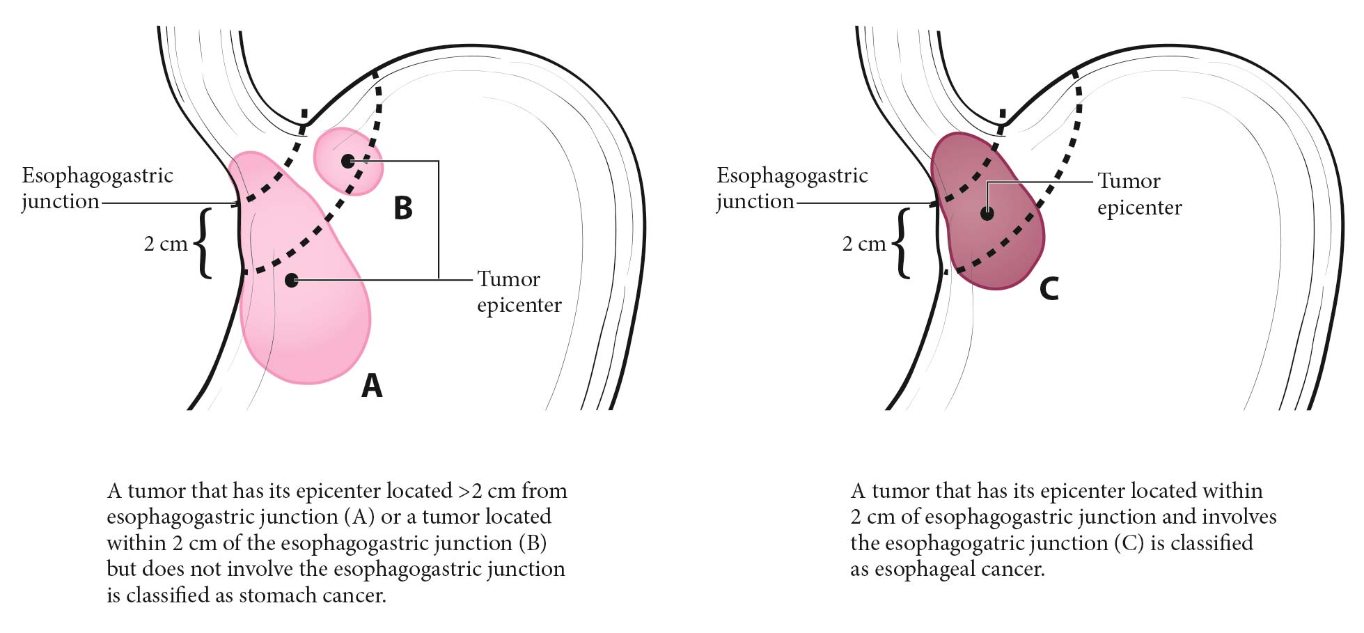
In this edition, there is a modification regarding tumors located at the esophagogastric junction (EGJ) and in the cardia of the stomach. If a tumor involves the EGJ and its epicenter is less than or equal to 2 cm into the proximal stomach (i.e, less than or equal to 2 cm distal to the EGJ), we recommend using the esophageal cancer schema for stage groupings. Tumors involving the EGJ with their epicenter greater than 2 cm into the proximal stomach (i.e., greater than 2 cm distal to the EGJ) are now classified using the stomach schema. Cardia cancers that do not invade the EGJ should be classified based on the stomach cancer schema for stage groupings. Thus, determining the exact location of the EGJ and whether it is involved by the tumor is critically important for assessing tumors in this region.

In summary, the current classifications provide more comprehensive tools (cTNM, ypTNM, and pTNM) for stage grouping of gastric cancer patients under different circumstances. The three classifications should provide the resources needed for the stage grouping required in the clinic and may prove more useful than the one staging system applied to different situations that was provided previously. Some deficiencies are acknowledged, such as a lack of uniformity in initial clinical stage assessments (including nonstand ardized radiology reports and endoscopic assessments/descriptions), the lack of a uniform surgical approach (particularly in the United States), and pathology assessments of yp categories. However, we will make a concerted effort to improve these assessments and evaluate their value based on the emerging data for future revisions of the staging system for gastric adenocarcinoma.

Anatomy

Primary Site(s)

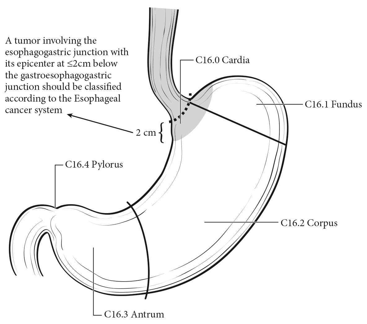
The stomach is the first division of the abdominal portion of the alimentary tract, beginning at the EGJ and extending to the pylorus. The proximal stomach is located immediately below the diaphragm and is termed the cardia. The remaining portions are the fundus and body of the stomach, and the distal portion of the stomach is known as the antrum. The pylorus is a muscular ring that controls the flow of ingested contents from the stomach into the first portion of the duodenum. The medial and lateral curvatures of the stomach are known as the lesser and greater curvatures, respectively. Histologically, the wall of the stomach has five layers: mucosal, submucosal, muscular, subserosal, and serosal.

In the 8th Edition, cancers crossing the EGJ with their epicenter in the proximal 2 cm of the stomach are incorporated into the esophagus chapter, whereas cancers crossing the EGJ with their epicenter in the proximal 2 to 5 cm of the stomach are addressed in the stomach chapter. All tumors in the stomach that do not cross the EGJ are classified in the stomach chapter (Figures 17.1 and 17.2).

17.1 Anatomic subsites of the stomach.

17.2 (A) EGJ tumors with their epicenter located greater than 2 cm into the proximal stomach are staged as stomach cancers. (B) Cardia cancers not involving the EGJ are staged as stomach cancers. (C) Tumors involving the EGJ with thier epicenter less than 2 cm into the proximal stomach are staged as esophageal cancers.

Regional Lymph Nodes

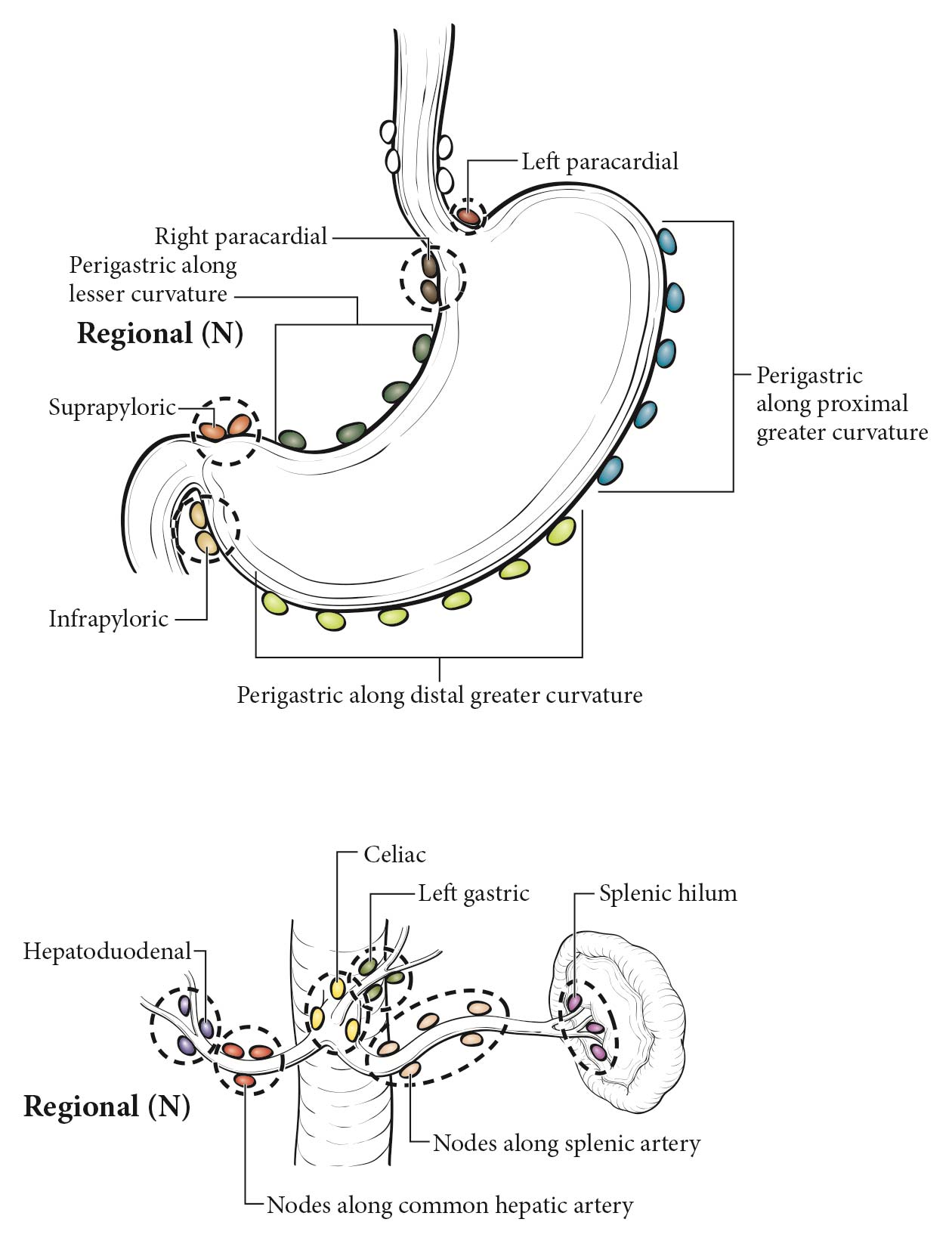
Several groups of regional lymph nodes drain the wall of the stomach. Perigastric nodes are found along the lesser and greater curvatures (Figure 17.3). Other major nodal groups follow the main arterial and venous vessels from the celiac artery and its branches and the portal circulation. Adequate nodal dissection of these regional nodal areas is important to ensure appropriate designation of the N (ypN or pN) category.5 Although it is suggested that at least 16 regional nodes be removed/assessed pathologically, removal/evaluation of more nodes (>=30) is desirable.2

The specific regional nodal areas are as follows:

  • Perigastric along the greater curvature (including greater curvature, greater omental)
  • Perigastric along the lesser curvature (including lesser curvature, lesser omental)
  • Right and left paracardial (cardioesophageal)
  • Suprapyloric (including gastroduodenal)
  • Infrapyloric (including gastroepiploic)
  • Left gastric artery
  • Celiac artery
  • Common hepatic artery
  • Hepatoduodenal (along the proper hepatic artery, including portal)
  • Splenic artery
  • Splenic hilum

17.3 Regional lymph nodes of the stomach.

Metastatic Sites

The most common metastatic distribution is to the liver, peritoneal surfaces, and nonregional/distant lymph nodes. Central nervous system and pulmonary metastases occur but are less frequent. Tumors found in these locations are considered metastatic disease (M1). In contrast, direct extension of bulky tumors to the liver, transverse colon, pancreas, and /or undersurface of the diaphragm is considered as tumor invading adjacent structures/organs (T4b) not M1. Positive peritoneal cytology is classified as metastatic disease (M1).

Distant Nodal Groups.Involvement of other (nonregional) intra-abdominal lymph nodes, such as the retropancreatic, pancreaticoduodenal, peripancreatic, superior mesenteric, middle colic, para-aortic, or retroperitoneal nodes, translates into a patient having metastatic disease (M1).

Classification Rules

Clinical Classification

Designated as cTNM, clinical stage is based on evidence of extent of disease present before therapy is instituted. It includes physical examination, laboratory testing, radiologic imaging, endoscopy (possibly including endoscopic ultrasonography [EUS] with fine-needle aspiration [FNA] for cytologic assessment), and biopsy (for histologic confirmation), and may include diagnostic laparoscopy with peritoneal washings for cytologic/histologic assessment.

Clinical T Category

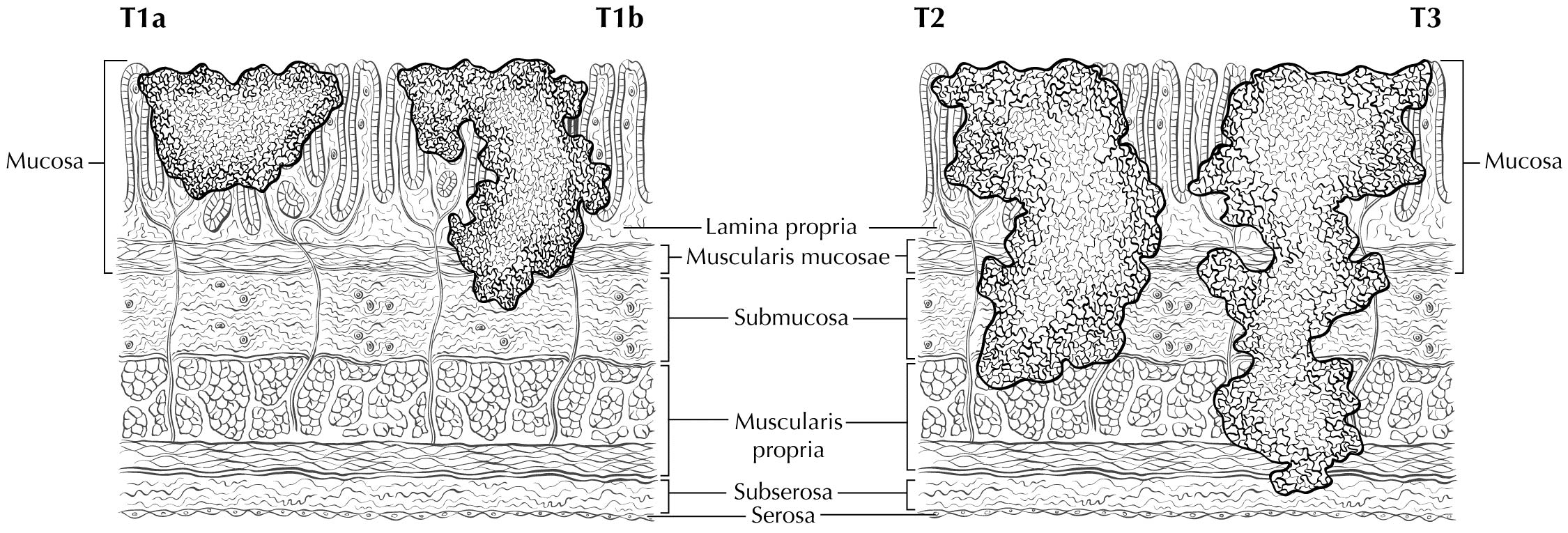
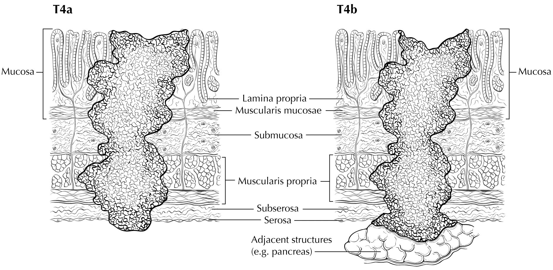
Staging of primary gastric adenocarcinoma depends on the depth of penetration of the primary tumor. The T1 designation is subdivided into T1a (invasion of the lamina propria or muscularis mucosae) and T1b (invasion of the submucosa). T2 is invasion of the muscularis propria, and T3 is invasion of the subserosal connective tissue without invasion of adjacent structures or the serosa (visceral peritoneum). T4 tumors penetrate the serosa (T4a) or invade adjacent structures/organs (T4b). EUS correlates highly with cT category. For tumors close to adjacent structures, radiologic imaging and EUS may help determine whether the adjacent anatomic structures are invaded (cT4a or cT4b).

Clinical N Category

Radiologic reports should document the number of enlarged nodes (with malignant features), and a multidisciplinary tumor board should review the images to document the number of nodes that appear malignant (see clinical staging methods in the next section). EUS also may be useful in identifying enlarged or malignant-appearing lymph nodes to determine cN category and may provide an opportunity to perform FNA or biopsy for cytologic assessment).

M Category for Clinical Staging

Imaging-based detection of organ metastasis (including peritoneal) is assigned cM1. This may be confirmed with tissue diagnosis, after which pM1 is assigned for the clinical stage. Confirmation of peritoneal metastasis by diagnostic laparoscopy or peritoneal washings performed as part of the staging workup is considered positive metastasis. Specifically, gross evidence of metastasis seen during diagnostic laparoscopy is cTcNcM1, whereas positive washings obtained during diagnostic laparoscopy without evidence of gross metastasis is considered cTcNpM1, which is clinical stage IV and pathological stage IV. See the detailed discussion in Chapter 1.

Endoscopy and Imaging

EUS and computed tomography (CT) of the chest, abdomen, and pelvis with oral and intravenous contrast are the initial imaging modalities used to determine clinical primary tumor (cT), node (cN), and metastasis (cM) categories. Positron emission tomography (PET)/CT with fluorine-18 (18F)-fluoro-2-deoxy-D-glucose (FDG) and magnetic resonance (MR) imaging may be used to further refine cN category and cM category in locoregionally advanced cTcN.

Endoscopic Evaluations

Clinical assessment of depth of invasion and nodal involvement, as well as some limited areas of distant disease, may be facilitated by the use of EUS. Gastric cancer staging is best performed by using commercially available ultrasound endoscopes with multifrequency (5-, 7.5-, 10-, and 12-MHz) radial transducers. Sonographic evaluation is performed as the instrument is withdrawn, starting at the pylorus. Orienting the images in an anteroposterior axis permits careful assessment of anatomic land marks to allow correlation with the location of the tumor, nodes, and surrounding organs.

The individual layers of the gastrointestinal wall are visualized throughout the examination to correlate the extent of the tumor relative to the alternating bright and dark layers seen on ultrasound. On the basis of in vitro studies, the first two layers (bright and dark starting at the lumen) correspond to the superficial and deep mucosa, the third (bright) layer corresponds to the submucosa, the fourth (dark) layer to the muscularis propria, and the fifth (bright) layer to the adventitia or serosa.6

Alterations in the thickness of individual layers are easily and reproducibly identified, allowing one to determine the depth of tumor invasion. The presence of a mass in the stomach usually is diagnosed as a hypoechoic or dark thickening in one or more layers, or a loss of the usual layer pattern.7 The first bright layer, which represents a transition echo layer, rarely is lost or thickened. Thickening of the second, or inner dark, layer suggests a T1 tumor. Although at higher EUS frequencies of 10 or 12 MHz one should be able to distinguish tumors limited to the mucosa (T1a) from those extending into the submucosa (T1b), some studies have shown reduced accuracy with T1 staging.8-11 A dark thickening extending from the second to the third layer (mucosa and submucosa), but not reaching the fourth layer (muscularis propria), is evidence of a T1b tumor. A dark thickening extending to but not completely through the fourth layer with a preserved smooth outer echogenic border is associated with a T2 tumor. Complete loss of all the layers, associated with a smooth white outer layer representing the serosal surface, suggests penetration through the muscularis propria into the subserosal fat, consistent with a T3 tumor in the stomach. If there is extension of the dark wall thickening with loss of the serosal echogenic stripe, the tumor is staged T4a. The distinction between T3 and T4a by EUS may be very challenging, as the actual thickness of the subserosal fat may vary and the serosa is very thin and likely not seen clearly on EUS. Full transmural extension with loss of the echogenic stripe separating the stomach from surrounding structures, such as the aorta, pancreas, liver, or other adjacent structure, indicates a T4b tumor.

The lymphatic drainage areas routinely investigated are the lower periesophageal, right and left paracardial, lesser curve (gastrohepatic ligament), greater curve, and supra- and infrapyloric regions, and areas along major vessels, including the celiac axis, splenic and hepatic artery, porta hepatis, and splenic hilum. Opinions vary regarding the specific cutoff for what is considered malignant. The presence of hypoechoic, rounded, sharply demarcated structures greater than 10 mm on EUS may be considered diagnostic of malignant nodes.12 Histologic confirmation of nodal disease by EUS-guided FNA is strongly encouraged if it can be achieved without traversing an area of tumor.13 Although not necessary for the current prognostic staging, the number of nodes remains important for future prognostic efforts; therefore, we encourage careful counting and reporting of suspicious nodes along with interpretation of clinical T category.

Parts of the liver are seen readily with EUS with the scope positioned in the antrum and along the lesser curvature and cardia, allowing identification of liver metastases (M1), which may be confirmed by EUS-FNA.14 Similarly, the presence of ascites adjacent to the stomach raises suspicion for peritoneal metastases, if other causes of ascites are ruled out. However, this finding has not been shown to be a reliable indicator of M1 disease.15,16

Imaging

Primary Tumor (cT)

EUS may be ideal for cT categorization of the primary tumor. With advances in multidetector CT scanners and greater attention to improving gastric distention, together with the use of negative oral contrast, dynamic intravenous contrast enhancement, and multiplanar reformatting of images, the accuracy of cT category has improved.17-19 In this regard, CT of the chest, abdomen, and pelvis with intravenous and oral contrast is used to describe cT in terms of location and degree of local invasion. However, overall, CT of the chest, abdomen, and pelvis still has a limited role in the cT category, especially for cT1, cT2, and cT3 tumors, although limitations also exist in identifying invasion of adjacent structures (cT4) unless gross invasion is present.20,21 FDG PET/CT generally is not useful in cT categorization because of the normal increased background uptake of FDG in the gastric mucosa together with the general lack of FDG uptake by signet ring cells and /or poorly differentiated adenocarcinomas.22-24 Although MR imaging has better contrast resolution than CT and has been reported in small studies to be comparable to CT, overall the imaging evaluation of cT categorization is limited.

Regional Lymph Nodes (cN)

CT of the chest and abdomen with intravenous and oral contrast and FDG PET/CT imaging are used to describe locoregional (cN) lymph nodes. In clinical practice, locoregional nodes generally are suspicious for tumor involvement if round and /or greater than 10 mm in short axis diameter. The portocaval lymph node, however, is an exception to these criteria. This lymph node has an elongated shape with a long transverse diameter and small anterior posterior diameter, and relying on measurement alone for this node results in frequent false positive interpretations. CT and FDG PET/CT are not optimal in detecting locoregional nodal metastasis.25-30 Additionally, the diagnostic benefit of FDG PET/CT is especially limited in patients with an early T classification (pT1) because of the low prevalence of nodal and distant metastases and the high rate of false positive PET findings.31,32 Because the criteria for cN classification have not been rigorously defined in peer-reviewed literature, the current cN classification requires evaluation of the size, shape, and number of abnormal lymph nodes in determining the cN classification. The diagnostic benefit of FDG PET/CT in detecting nodal metastasis is of limited value because of the high rate of false negative PET findings due to its inability to detect microscopic disease and because of poor FDG uptake by signet ring cells and /or poorly differentiated adenocarcinomas.

Metastatic Disease (cM)

CT of the chest, abdomen, and pelvis with intravenous and oral contrast can detect distant metastasis (cM1). Because advanced gastric adenocarcinoma has a propensity to metastasize to the peritoneum, ovaries, and rectovesical pouch, pelvic CT imaging is useful in cM categorization. MR imaging is used less frequently as a primary imaging modality. The addition of FDG PET/CT imaging to conventional clinical staging improves the detection of metastases missed or not visualized on CT of the chest, abdomen, and pelvis.

Diagnostic Laparoscopy and Peritoneal Washing Evaluations

Diagnostic peritoneal staging laparoscopy is used to identify occult metastates not detected by imaging (or by EUS) and is recommended for all patients with tumor depth cT3 or greater, or in the presence of clinically suspicious nodes in the absence of distant metastases on imaging (CT or PET). It generally is performed as a separate surgical procedure and allows detection of gross metastases on the peritoneal surface and visceral organs as well as of microscopic malignant cells in the washings. Clinical studies have found positive peritoneal cytology to be an independent prognosticator,33,34 and this finding is considered pM1 for both the clinical and pathological classification M categories, resulting in clinical Stage IV and pathological Stage IV.

Peritoneal washing generally involves instilling ~200 mL of normal saline into the different quadrants of the abdominal cavity. The fluid is dispersed by gentle stirring of the area and then is aspirated from different portions of the body. Areas typically include the right and left subphrenic space and the pouch of Douglas. Ideally, greater than 50 mL of washings should be retrieved for cytologic assessment.

Residual Disease (R Classification)

Assessment of the R classification applies only to a surgically resected specimen. In addition to proximal and distal margins of resection, the status of the radial or circumferential margin of resection determines whether the tumor was excised completely. The R classification is based on a combination of intraoperative assessment by the surgeon and pathological evaluation of the resected specimen. R0 indicates no evidence of residual tumor. R1 indicates the presence of microscopic tumor at the margins, as defined by the College of American Pathologists (CAP); however, the Royal College of Pathologists (RCP) definition of R1 includes tumors within a 1-mm margin. Macroscopically visible tumor at margins is classified as R2. The presence of tumor cells at the inked radial margin constitutes a positive margin according to CAP criteria.

Early malignant lesions removed endoscopically by either an endoscopic submucosal dissection or an endoscopic mucosal resection should be assessed for pT designation along with the assessment of deep and lateral margins. The pathological assessment also should include the presence/absence of lymphovascular and /or perineural invasion, maximum tumor diameter, and the final histologic grade (including the presence of high-grade dysplasia). This information may assist in making future therapeutic decisions, including surveillance strategies.

Pathological Classification

Pathological staging depends on data acquired clinically, together with findings from subsequent surgical exploration and examination of resected tissue (specimen). Surgical resections that qualify for pathological staging include total, near-total, subtotal, partial, proximal, and distal gastrectomy and antrectomy.

Primary Tumor (pT)

Depth of tumor invasion should be based on examination of the surgical specimen after resection. Tumor distance from closest margin or involvement of margin should be documented. Histologic classification, as well as grade and presence of lymphovascular or perineural invasion, should be documented according to CAP guidelines. The specimen should be tested for the presence of Helicobacter pylori infection.

Regional Lymph Nodes (pN)

Pathological assessment of regional lymph nodes entails the removal and histologic examination of nodes to evaluate the total number removed, as well as how many contain tumor. The number of nodes, as well as the number of positive nodes, should be documented.

Metastatic tumor deposits in the subserosal fat adjacent to a gastric carcinoma, without evidence of residual lymph node tissue, are considered regional lymph node metastases for purposes of gastric cancer staging. Tumor deposits are defined as discrete tumor nodules within the lymph drainage area of the primary carcinoma without identifiable lymph node tissue or identifiable vascular or neural structure. Shape, contour, and size of the deposit are not considered in these designations.

Metastatic Disease (pM)

Pathologically confirmed metastatic tissue obtained from a site outside of what is considered local or regional for gastric cancer is considered pM1. This designation includes tumor identified in distant nodal stations in the surgical resection specimen and tissue samples obtained from other organs (including peritoneum) showing malignant cells in peritoneal washings or implants.

When recording pathological stage, clinical stage M category (cM) may be used for final pathological Stage IV, such as pTpNcM0-1.
Although grading systems for tumor response have not been established for this disease, response of tumor to preoperative (neoadjuvant) therapy should be reported using the post-neoadjuvant therapy classification staging system. The assessment of pathological response to neoadjuvant therapy involves both the gross and the microscopic examination of the resected surgical specimen. At the microscopic level, a positive treatment-related effect is observed as abolition of the malignant epithelium and replacement by dense fibrosis or fibroinflammation. The pathological response to treatment is determined by the amount of residual viable carcinoma in relation to areas of fibrosis or fibroinflammation within the gross lesion. This relationship may be expressed as the inverse percentage of a favorable treatment response. Thus, a 100% treatment response indicates fibrosis or fibroinflammation within an entire gross lesion, without microscopic evidence of carcinoma, whereas a 0% response represents an unaffected tumor in the absence of any fibrosis or fibroinflammation. The presence of residual tumor cells suggests an incomplete response. Acellular mucin is regarded as a form of positive treatment response, not as residual tumor. The ypT category of the residual carcinoma is based on the deepest focus of residual malignant epithelium of the gastric wall. Positive lymph nodes are defined as having at least one focus of residual tumor cells in the lymph nodes.35 The pathology report should include ypT and ypN based on the submitted specimen. If no further diagnostic tests are performed after neoadjuvant therapy, the M designation should remain the same as cM. If further diagnostic tests are performed after neoadjuvant therapy, then ypM also should reflect these new tests. Pathologically confirmed metastatic tissue obtained from a site outside of what is considered local or regional for gastric cancer after neoadjuant therapy is considered ypM1.

Prognostic Factors

Prognostic Factors Required for Stage Grouping

Beyond the factors used to assign T, N, or M categories, no additional prognostic factors are required for stage grouping.

Additional Factors Recommended for Clinical Care

Carcinoembryonic Antigen

Elevated carcinoembryonic antigen (CEA) levels are not shown to have independent prognostic value. Treatment decisions should not be altered based on baseline levels of CEA. However, monitoring of CEA levels may be useful in the surveillance period.

AJCC Level of Evidence: III

Cancer Antigen 19-9

Elevated cancer antigen (CA) 19-9 levels are not shown to have independent prognostic value. Treatment decisions should not be altered based on baseline levels of CA 19-9. However, monitoring of CA 19-9 levels may be useful in the surveillance period.

AJCC Level of Evidence: III

HER2

The biomarker HER2 is examined directly on tumor tissue. There are mixed reports regarding the prognostic value of this biomarker; it is not shown to be independently prognostic. If the tumor is HER2 positive, HER2-directed therapy should be considered.

AJCC Level of Evidence: III

Microsatellite Instabiity

Microsatellite instability (MSI) is examined directly on the tumor tissue. The level of evidence is based on a limited number of patients. Patients with high MSI (MSI-H) tend to have a better overall prognosis. However, the independent prognostic value of MSI-H is not yet established.

AJCC Level of Evidence: III

Genomic and Molecular Assessments

Genomic and molecular analyses of gastric cancer may provide insights into the underlying pathogenesis, as well as cand idate therapeutic approaches. Distinct patterns of genomic alterations may reflect the molecular etiologies of these cancers. Tumors with antecedent infection with the Epstein-Barr virus (EBV) harbor marked DNA methylation.36 Tumors with inactivation of key DNA mismatch repair proteins undergo hypermutation accompanied by MSI.36 Both MSI and EBV-positive gastric cancers have been associated with longer survival.37,38 Many gastric cancers also harbor high rates of genomic instability, with frequent chromosomal amplifications of genes encoding key growth-promoting factors, with the patterns of these alterations highly resembling those seen in esophageal adenocarcinoma.36,39 Histologic classes of gastric adenocarcinoma also may be associated with genomic features. Diffuse-type gastric cancers often are found to have quieter genomic copy number profiles36 or more mesenchymal gene expression signatures38 and to harbor unique gene mutations40 relative to intestinal tumors.

For patients with resectable disease, the presence of an epithelial-to-mesenchymal gene expression signature is associated with higher rates of relapse and subsequent peritoneal metastases.38 Profiling of specific genomic alterations also may guide the use of specific biologic or targeted agents. Currently, the use of trastuzumab in metastatic gastric cancer is guided by evaluation of expression and amplification of HER2/ERBB2.41 Other agents, directed against targets genomically activated in subsets of gastric cancer, also are being evaluated. Genomic profiling ultimately may provide guidance regarding the use of emerging immunotherapies, as MSI status was shown to be a new cand idate predictor of response to PD-1 inhibitor therapy in colorectal cancer.42

AJCC Level of Evidence: Not identified

Risk Assessment

Risk Assesment Models

The AJCC recently established guidelines that will be used to evaluate published statistical prediction models for the purpose of granting endorsement for clinical use.43 Although this is a monumental step toward the goal of precision medicine, this work was published only very recently. Therefore, the existing models that have been published or may be in clinical use have not yet been evaluated for this cancer site by the Precision Medicine Core of the AJCC. In the future, the statistical prediction models for this cancer site will be evaluated, and those that meet all AJCC criteria will be endorsed.

Recommendations

TNM Definitions

Clinical T (cT)

cT CategorycT Criteria
cTXPrimary tumor cannot be assessed
cT0No evidence of primary tumor
cTisCarcinoma in situ: intraepithelial tumor without invasion of the lamina propria, high-grade dysplasia
cT1Tumor invades the lamina propria, muscularis mucosae, or submucosa
cT1aTumor invades the lamina propria or muscularis mucosae
cT1bTumor invades the submucosa
cT2Tumor invades the muscularis propria
cT3Tumor penetrates the subserosal connective tissue without invasion of the visceral peritoneum or adjacent structures
cT4Tumor invades the serosa (visceral peritoneum) or adjacent structures
cT4aTumor invades the serosa (visceral peritoneum)
cT4bTumor invades adjacent structures/organs

A tumor may penetrate the muscularis propria with extension into the gastrocolic or gastrohepatic ligaments, or into the greater or lesser omentum, without perforation of the visceral peritoneum covering these structures. In this case, the tumor is classified as T3. If there is perforation of the visceral peritoneum covering the gastric ligaments or the omentum, the tumor should be classified as T4.

The adjacent structures of the stomach include the spleen, transverse colon, liver, diaphragm, pancreas, abdominal wall, adrenal gland , kidney, small intestine, and retroperitoneum.

Intramural extension to the duodenum or esophagus is not considered invasion of an adjacent structure, but is classified using the depth of the greatest invasion in any of these sites.

Pathological T (pT)

pT CategorypT Criteria
pTXPrimary tumor cannot be assessed
pT0No evidence of primary tumor
pTisCarcinoma in situ: intraepithelial tumor without invasion of the lamina propria, high-grade dysplasia
pT1Tumor invades the lamina propria, muscularis mucosae, or submucosa
pT1aTumor invades the lamina propria or muscularis mucosae
pT1bTumor invades the submucosa
pT2Tumor invades the muscularis propria
pT3Tumor penetrates the subserosal connective tissue without invasion of the visceral peritoneum or adjacent structures
pT4Tumor invades the serosa (visceral peritoneum) or adjacent structures
pT4aTumor invades the serosa (visceral peritoneum)
pT4bTumor invades adjacent structures/organs
cTXPrimary tumor cannot be assessed
cT0No evidence of primary tumor
cTisCarcinoma in situ: intraepithelial tumor without invasion of the lamina propria, high-grade dysplasia
cT1Tumor invades the lamina propria, muscularis mucosae, or submucosa
cT1aTumor invades the lamina propria or muscularis mucosae
cT1bTumor invades the submucosa
cT2Tumor invades the muscularis propria
cT3Tumor penetrates the subserosal connective tissue without invasion of the visceral peritoneum or adjacent structures
cT4Tumor invades the serosa (visceral peritoneum) or adjacent structures
cT4aTumor invades the serosa (visceral peritoneum)
cT4bTumor invades adjacent structures/organs

A tumor may penetrate the muscularis propria with extension into the gastrocolic or gastrohepatic ligaments, or into the greater or lesser omentum, without perforation of the visceral peritoneum covering these structures. In this case, the tumor is classified as T3. If there is perforation of the visceral peritoneum covering the gastric ligaments or the omentum, the tumor should be classified as T4.

The adjacent structures of the stomach include the spleen, transverse colon, liver, diaphragm, pancreas, abdominal wall, adrenal gland , kidney, small intestine, and retroperitoneum.

Intramural extension to the duodenum or esophagus is not considered invasion of an adjacent structure, but is classified using the depth of the greatest invasion in any of these sites.

Neoadjuvant Clinical T (yT)

ycT CategoryycT Criteria
ycTXPrimary tumor cannot be assessed
ycT0No evidence of primary tumor
ycTisCarcinoma in situ: intraepithelial tumor without invasion of the lamina propria, high-grade dysplasia
ycT1Tumor invades the lamina propria, muscularis mucosae, or submucosa
ycT1aTumor invades the lamina propria or muscularis mucosae
ycT1bTumor invades the submucosa
ycT2Tumor invades the muscularis propria
ycT3Tumor penetrates the subserosal connective tissue without invasion of the visceral peritoneum or adjacent structures
ycT4Tumor invades the serosa (visceral peritoneum) or adjacent structures
ycT4aTumor invades the serosa (visceral peritoneum)
ycT4bTumor invades adjacent structures/organs

A tumor may penetrate the muscularis propria with extension into the gastrocolic or gastrohepatic ligaments, or into the greater or lesser omentum, without perforation of the visceral peritoneum covering these structures. In this case, the tumor is classified as T3. If there is perforation of the visceral peritoneum covering the gastric ligaments or the omentum, the tumor should be classified as T4.

The adjacent structures of the stomach include the spleen, transverse colon, liver, diaphragm, pancreas, abdominal wall, adrenal gland , kidney, small intestine, and retroperitoneum.

Intramural extension to the duodenum or esophagus is not considered invasion of an adjacent structure, but is classified using the depth of the greatest invasion in any of these sites.

Neoadjuvant Pathological T (yT)

ypT CategoryypT Criteria
ypTXPrimary tumor cannot be assessed
ypT0No evidence of primary tumor
ypTisCarcinoma in situ: intraepithelial tumor without invasion of the lamina propria, high-grade dysplasia
ypT1Tumor invades the lamina propria, muscularis mucosae, or submucosa
ypT1aTumor invades the lamina propria or muscularis mucosae
ypT1bTumor invades the submucosa
ypT2Tumor invades the muscularis propria
ypT3Tumor penetrates the subserosal connective tissue without invasion of the visceral peritoneum or adjacent structures
ypT4Tumor invades the serosa (visceral peritoneum) or adjacent structures
ypT4aTumor invades the serosa (visceral peritoneum)
ypT4bTumor invades adjacent structures/organs

A tumor may penetrate the muscularis propria with extension into the gastrocolic or gastrohepatic ligaments, or into the greater or lesser omentum, without perforation of the visceral peritoneum covering these structures. In this case, the tumor is classified as T3. If there is perforation of the visceral peritoneum covering the gastric ligaments or the omentum, the tumor should be classified as T4.

The adjacent structures of the stomach include the spleen, transverse colon, liver, diaphragm, pancreas, abdominal wall, adrenal gland , kidney, small intestine, and retroperitoneum.

Intramural extension to the duodenum or esophagus is not considered invasion of an adjacent structure, but is classified using the depth of the greatest invasion in any of these sites.

Definition of Regional Lymph Node (N)

Clinical N (cN)
cN CategorycN Criteria
cNXRegional lymph node(s) cannot be assessed
cN0No regional lymph node metastasis
cN1Metastasis in one or two regional lymph nodes
cN2Metastasis in three to six regional lymph nodes
cN3Metastasis in seven or more regional lymph nodes
cN3aMetastasis in seven to 15 regional lymph nodes
cN3bMetastasis in 16 or more regional lymph nodes
Pathological N (pN)
pN CategorypN Criteria
pNXRegional lymph node(s) cannot be assessed
pN0No regional lymph node metastasis
pN1Metastasis in one or two regional lymph nodes
pN2Metastasis in three to six regional lymph nodes
pN3Metastasis in seven or more regional lymph nodes
pN3aMetastasis in seven to 15 regional lymph nodes
pN3bMetastasis in 16 or more regional lymph nodes
cNXRegional lymph node(s) cannot be assessed
cN0No regional lymph node metastasis
cN1Metastasis in one or two regional lymph nodes
cN2Metastasis in three to six regional lymph nodes
cN3Metastasis in seven or more regional lymph nodes
cN3aMetastasis in seven to 15 regional lymph nodes
cN3bMetastasis in 16 or more regional lymph nodes
Neoadjuvant Clinical N (pY)
ycN CategoryycN Criteria
ycNXRegional lymph node(s) cannot be assessed
ycN0No regional lymph node metastasis
ycN1Metastasis in one or two regional lymph nodes
ycN2Metastasis in three to six regional lymph nodes
ycN3Metastasis in seven or more regional lymph nodes
ycN3aMetastasis in seven to 15 regional lymph nodes
ycN3bMetastasis in 16 or more regional lymph nodes
Neoadjuvant Pathological N (pY)
ypN CategoryypN Criteria
ypNXRegional lymph node(s) cannot be assessed
ypN0No regional lymph node metastasis
ypN1Metastasis in one or two regional lymph nodes
ypN2Metastasis in three to six regional lymph nodes
ypN3Metastasis in seven or more regional lymph nodes
ypN3aMetastasis in seven to 15 regional lymph nodes
ypN3bMetastasis in 16 or more regional lymph nodes

Definition of Distant Metastasis (M)- Clinical M (cN)

cM CategorycM Criteria
cM0No distant metastasis
cM1Distant metastasis
pM1Microscopic evidence of distant metastasis

Definition of Distant Metastasis (M)- Pathological M (pN)

pM CategorypM Criteria
cM0No distant metastasis
cM1Distant metastasis
pM1Microscopic evidence of distant metastasis

Definition of Distant Metastasis (M)- Neoadjuvant Clinical M (pY)

ycM CategoryycM Criteria
cM0No distant metastasis
cM1Distant metastasis
pM1Microscopic evidence of distant metastasis

Definition of Distant Metastasis (M)- Neoadjuvant Pathological M (pY)

ypM CategoryypM Criteria
cM0No distant metastasis
cM1Distant metastasis
pM1Microscopic evidence of distant metastasis

Stage Prognostic

Clinical

When T is…and N is…and M is…Then the Clinical Prognostic Stage Group is…
cTiscN0cM00
cT1cN0cM0I
cT1acN0cM0I
cT1bcN0cM0I
cT2cN0cM0I
cT1cN1cM0IIA
cT1cN2cM0IIA
cT1cN3cM0IIA
cT1cN3acM0IIA
cT1cN3bcM0IIA
cT1acN1cM0IIA
cT1acN2cM0IIA
cT1acN3cM0IIA
cT1acN3acM0IIA
cT1acN3bcM0IIA
cT1bcN1cM0IIA
cT1bcN2cM0IIA
cT1bcN3cM0IIA
cT1bcN3acM0IIA
cT1bcN3bcM0IIA
cT2cN1cM0IIA
cT2cN2cM0IIA
cT2cN3cM0IIA
cT2cN3acM0IIA
cT2cN3bcM0IIA
cT3cN0cM0IIB
cT4acN0cM0IIB
cT3cN1cM0III
cT3cN2cM0III
cT3cN3cM0III
cT3cN3acM0III
cT3cN3bcM0III
cT4acN1cM0III
cT4acN2cM0III
cT4acN3cM0III
cT4acN3acM0III
cT4acN3bcM0III
cT4bcNXcM0IVA
cT4bcN0cM0IVA
cT4bcN1cM0IVA
cT4bcN2cM0IVA
cT4bcN3cM0IVA
cT4bcN3acM0IVA
cT4bcN3bcM0IVA
cTXcNXcM1IVB
cTXcN0cM1IVB
cTXcN1cM1IVB
cTXcN2cM1IVB
cTXcN3cM1IVB
cTXcN3acM1IVB
cTXcN3bcM1IVB
cT0cNXcM1IVB
cT0cN0cM1IVB
cT0cN1cM1IVB
cT0cN2cM1IVB
cT0cN3cM1IVB
cT0cN3acM1IVB
cT0cN3bcM1IVB
cT1cNXcM1IVB
cT1cN0cM1IVB
cT1cN1cM1IVB
cT1cN2cM1IVB
cT1cN3cM1IVB
cT1cN3acM1IVB
cT1cN3bcM1IVB
cT1acNXcM1IVB
cT1acN0cM1IVB
cT1acN1cM1IVB
cT1acN2cM1IVB
cT1acN3cM1IVB
cT1acN3acM1IVB
cT1acN3bcM1IVB
cT1bcNXcM1IVB
cT1bcN0cM1IVB
cT1bcN1cM1IVB
cT1bcN2cM1IVB
cT1bcN3cM1IVB
cT1bcN3acM1IVB
cT1bcN3bcM1IVB
cT2cNXcM1IVB
cT2cN0cM1IVB
cT2cN1cM1IVB
cT2cN2cM1IVB
cT2cN3cM1IVB
cT2cN3acM1IVB
cT2cN3bcM1IVB
cT3cNXcM1IVB
cT3cN0cM1IVB
cT3cN1cM1IVB
cT3cN2cM1IVB
cT3cN3cM1IVB
cT3cN3acM1IVB
cT3cN3bcM1IVB
cT4cNXcM1IVB
cT4cN0cM1IVB
cT4cN1cM1IVB
cT4cN2cM1IVB
cT4cN3cM1IVB
cT4cN3acM1IVB
cT4cN3bcM1IVB
cT4acNXcM1IVB
cT4acN0cM1IVB
cT4acN1cM1IVB
cT4acN2cM1IVB
cT4acN3cM1IVB
cT4acN3acM1IVB
cT4acN3bcM1IVB
cT4bcNXcM1IVB
cT4bcN0cM1IVB
cT4bcN1cM1IVB
cT4bcN2cM1IVB
cT4bcN3cM1IVB
cT4bcN3acM1IVB
cT4bcN3bcM1IVB
cTXcNXpM1IVB
cTXcN0pM1IVB
cTXcN1pM1IVB
cTXcN2pM1IVB
cTXcN3pM1IVB
cTXcN3apM1IVB
cTXcN3bpM1IVB
cT0cNXpM1IVB
cT0cN0pM1IVB
cT0cN1pM1IVB
cT0cN2pM1IVB
cT0cN3pM1IVB
cT0cN3apM1IVB
cT0cN3bpM1IVB
cT1cNXpM1IVB
cT1cN0pM1IVB
cT1cN1pM1IVB
cT1cN2pM1IVB
cT1cN3pM1IVB
cT1cN3apM1IVB
cT1cN3bpM1IVB
cT1acNXpM1IVB
cT1acN0pM1IVB
cT1acN1pM1IVB
cT1acN2pM1IVB
cT1acN3pM1IVB
cT1acN3apM1IVB
cT1acN3bpM1IVB
cT1bcNXpM1IVB
cT1bcN0pM1IVB
cT1bcN1pM1IVB
cT1bcN2pM1IVB
cT1bcN3pM1IVB
cT1bcN3apM1IVB
cT1bcN3bpM1IVB
cT2cNXpM1IVB
cT2cN0pM1IVB
cT2cN1pM1IVB
cT2cN2pM1IVB
cT2cN3pM1IVB
cT2cN3apM1IVB
cT2cN3bpM1IVB
cT3cNXpM1IVB
cT3cN0pM1IVB
cT3cN1pM1IVB
cT3cN2pM1IVB
cT3cN3pM1IVB
cT3cN3apM1IVB
cT3cN3bpM1IVB
cT4cNXpM1IVB
cT4cN0pM1IVB
cT4cN1pM1IVB
cT4cN2pM1IVB
cT4cN3pM1IVB
cT4cN3apM1IVB
cT4cN3bpM1IVB
cT4acNXpM1IVB
cT4acN0pM1IVB
cT4acN1pM1IVB
cT4acN2pM1IVB
cT4acN3pM1IVB
cT4acN3apM1IVB
cT4acN3bpM1IVB
cT4bcNXpM1IVB
cT4bcN0pM1IVB
cT4bcN1pM1IVB
cT4bcN2pM1IVB
cT4bcN3pM1IVB
cT4bcN3apM1IVB
cT4bcN3bpM1IVB

Pathological

When T is…and N is…and M is…Then the Pathological Prognostic Stage Group is…
pTispN0cM00
pT1pN0cM0IA
pT1apN0cM0IA
pT1pN1cM0IB
pT1apN1cM0IB
pT1bpN1cM0IB
pT2pN0cM0IB
pT1pN2cM0IIA
pT1apN2cM0IIA
pT1bpN2cM0IIA
pT2pN1cM0IIA
pT3pN0cM0IIA
pT1pN3acM0IIB
pT1apN3acM0IIB
pT1bpN3acM0IIB
pT2pN2cM0IIB
pT3pN1cM0IIB
pT4apN0cM0IIB
pT2pN3acM0IIIA
pT3pN2cM0IIIA
pT4apN1cM0IIIA
pT4apN2cM0IIIA
pT4bpN0cM0IIIA
pT1pN3bcM0IIIB
pT1apN3bcM0IIIB
pT1bpN3bcM0IIIB
pT2pN3bcM0IIIB
pT3pN3acM0IIIB
pT4apN3acM0IIIB
pT4bpN1cM0IIIB
pT4bpN2cM0IIIB
pT3pN3bcM0IIIC
pT4apN3bcM0IIIC
pT4bpN3acM0IIIC
pT4bpN3bcM0IIIC
pT4bpN3cM0IIIC
pTXpNXcM1IV
pTXpN0cM1IV
pTXpN1cM1IV
pTXpN2cM1IV
pTXpN3cM1IV
pTXpN3acM1IV
pTXpN3bcM1IV
pT0pNXcM1IV
pT0pN0cM1IV
pT0pN1cM1IV
pT0pN2cM1IV
pT0pN3cM1IV
pT0pN3acM1IV
pT0pN3bcM1IV
pT1pNXcM1IV
pT1pN0cM1IV
pT1pN1cM1IV
pT1pN2cM1IV
pT1pN3cM1IV
pT1pN3acM1IV
pT1pN3bcM1IV
pT1apNXcM1IV
pT1apN0cM1IV
pT1apN1cM1IV
pT1apN2cM1IV
pT1apN3cM1IV
pT1apN3acM1IV
pT1apN3bcM1IV
pT1bpNXcM1IV
pT1bpN0cM1IV
pT1bpN1cM1IV
pT1bpN2cM1IV
pT1bpN3cM1IV
pT1bpN3acM1IV
pT1bpN3bcM1IV
pT2pNXcM1IV
pT2pN0cM1IV
pT2pN1cM1IV
pT2pN2cM1IV
pT2pN3cM1IV
pT2pN3acM1IV
pT2pN3bcM1IV
pT3pNXcM1IV
pT3pN0cM1IV
pT3pN1cM1IV
pT3pN2cM1IV
pT3pN3cM1IV
pT3pN3acM1IV
pT3pN3bcM1IV
pT4pNXcM1IV
pT4pN0cM1IV
pT4pN1cM1IV
pT4pN2cM1IV
pT4pN3cM1IV
pT4pN3acM1IV
pT4pN3bcM1IV
pT4apNXcM1IV
pT4apN0cM1IV
pT4apN1cM1IV
pT4apN2cM1IV
pT4apN3cM1IV
pT4apN3acM1IV
pT4apN3bcM1IV
pT4bpNXcM1IV
pT4bpN0cM1IV
pT4bpN1cM1IV
pT4bpN2cM1IV
pT4bpN3cM1IV
pT4bpN3acM1IV
pT4bpN3bcM1IV
pTXpNXpM1IV
pTXpN0pM1IV
pTXpN1pM1IV
pTXpN2pM1IV
pTXpN3pM1IV
pTXpN3apM1IV
pTXpN3bpM1IV
pT0pNXpM1IV
pT0pN0pM1IV
pT0pN1pM1IV
pT0pN2pM1IV
pT0pN3pM1IV
pT0pN3apM1IV
pT0pN3bpM1IV
pT1pNXpM1IV
pT1pN0pM1IV
pT1pN1pM1IV
pT1pN2pM1IV
pT1pN3pM1IV
pT1pN3apM1IV
pT1pN3bpM1IV
pT1apNXpM1IV
pT1apN0pM1IV
pT1apN1pM1IV
pT1apN2pM1IV
pT1apN3pM1IV
pT1apN3apM1IV
pT1apN3bpM1IV
pT1bpNXpM1IV
pT1bpN0pM1IV
pT1bpN1pM1IV
pT1bpN2pM1IV
pT1bpN3pM1IV
pT1bpN3apM1IV
pT1bpN3bpM1IV
pT2pNXpM1IV
pT2pN0pM1IV
pT2pN1pM1IV
pT2pN2pM1IV
pT2pN3pM1IV
pT2pN3apM1IV
pT2pN3bpM1IV
pT3pNXpM1IV
pT3pN0pM1IV
pT3pN1pM1IV
pT3pN2pM1IV
pT3pN3pM1IV
pT3pN3apM1IV
pT3pN3bpM1IV
pT4pNXpM1IV
pT4pN0pM1IV
pT4pN1pM1IV
pT4pN2pM1IV
pT4pN3pM1IV
pT4pN3apM1IV
pT4pN3bpM1IV
pT4apNXpM1IV
pT4apN0pM1IV
pT4apN1pM1IV
pT4apN2pM1IV
pT4apN3pM1IV
pT4apN3apM1IV
pT4apN3bpM1IV
pT4bpNXpM1IV
pT4bpN0pM1IV
pT4bpN1pM1IV
pT4bpN2pM1IV
pT4bpN3pM1IV
pT4bpN3apM1IV
pT4bpN3bpM1IV
cTXcNXpM1IV
cTXcN0pM1IV
cTXcN1pM1IV
cTXcN2pM1IV
cTXcN3pM1IV
cTXcN3apM1IV
cTXcN3bpM1IV
cT0cNXpM1IV
cT0cN0pM1IV
cT0cN1pM1IV
cT0cN2pM1IV
cT0cN3pM1IV
cT0cN3apM1IV
cT0cN3bpM1IV
cT1cNXpM1IV
cT1cN0pM1IV
cT1cN1pM1IV
cT1cN2pM1IV
cT1cN3pM1IV
cT1cN3apM1IV
cT1cN3bpM1IV
cT1acNXpM1IV
cT1acN0pM1IV
cT1acN1pM1IV
cT1acN2pM1IV
cT1acN3pM1IV
cT1acN3apM1IV
cT1acN3bpM1IV
cT1bcNXpM1IV
cT1bcN0pM1IV
cT1bcN1pM1IV
cT1bcN2pM1IV
cT1bcN3pM1IV
cT1bcN3apM1IV
cT1bcN3bpM1IV
cT2cNXpM1IV
cT2cN0pM1IV
cT2cN1pM1IV
cT2cN2pM1IV
cT2cN3pM1IV
cT2cN3apM1IV
cT2cN3bpM1IV
cT3cNXpM1IV
cT3cN0pM1IV
cT3cN1pM1IV
cT3cN2pM1IV
cT3cN3pM1IV
cT3cN3apM1IV
cT3cN3bpM1IV
cT4cNXpM1IV
cT4cN0pM1IV
cT4cN1pM1IV
cT4cN2pM1IV
cT4cN3pM1IV
cT4cN3apM1IV
cT4cN3bpM1IV
cT4acNXpM1IV
cT4acN0pM1IV
cT4acN1pM1IV
cT4acN2pM1IV
cT4acN3pM1IV
cT4acN3apM1IV
cT4acN3bpM1IV
cT4bcNXpM1IV
cT4bcN0pM1IV
cT4bcN1pM1IV
cT4bcN2pM1IV
cT4bcN3pM1IV
cT4bcN3apM1IV
cT4bcN3bpM1IV

Neoadjuvant Clinical

There is no Postneoadjuvant Clinical Therapy (ycTNM) stage group available at this time.

Neoadjuvant Pathological

When T is…and N is…and M is…Then the Postneoadjuvant Pathological Stage Group is…
ypT1ypN0cM0I
ypT1aypN0cM0I
ypT1bypN0cM0I
ypT2ypN0cM0I
ypT1ypN1cM0I
ypT1aypN1cM0I
ypT1bypN1cM0I
ypT3ypN0cM0II
ypT2ypN1cM0II
ypT1ypN2cM0II
ypT1aypN2cM0II
ypT1bypN2cM0II
ypT4aypN0cM0II
ypT3ypN1cM0II
ypT2ypN2cM0II
ypT1ypN3cM0II
ypT1ypN3acM0II
ypT1ypN3bcM0II
ypT1aypN3cM0II
ypT1aypN3acM0II
ypT1aypN3bcM0II
ypT1bypN3cM0II
ypT1bypN3acM0II
ypT1bypN3bcM0II
ypT4aypN1cM0III
ypT4ypN1cM0III
ypT3ypN2cM0III
ypT2ypN3cM0III
ypT2ypN3acM0III
ypT2ypN3bcM0III
ypT4bypN0cM0III
ypT4bypN1cM0III
ypT4aypN2cM0III
ypT4ypN2cM0III
ypT3ypN3cM0III
ypT3ypN3acM0III
ypT3ypN3bcM0III
ypT4bypN2cM0III
ypT4aypN3cM0III
ypT4aypN3acM0III
ypT4aypN3bcM0III
ypT4ypN3cM0III
ypT4ypN3acM0III
ypT4ypN3bcM0III
ypT4bypN3cM0III
ypT4bypN3acM0III
ypT4bypN3bcM0III
ypTXypNXcM1IV
ypTXypN0cM1IV
ypTXypN1cM1IV
ypTXypN2cM1IV
ypTXypN3cM1IV
ypTXypN3acM1IV
ypTXypN3bcM1IV
ypT0ypNXcM1IV
ypT0ypN0cM1IV
ypT0ypN1cM1IV
ypT0ypN2cM1IV
ypT0ypN3cM1IV
ypT0ypN3acM1IV
ypT0ypN3bcM1IV
ypT1ypNXcM1IV
ypT1ypN0cM1IV
ypT1ypN1cM1IV
ypT1ypN2cM1IV
ypT1ypN3cM1IV
ypT1ypN3acM1IV
ypT1ypN3bcM1IV
ypT1aypNXcM1IV
ypT1aypN0cM1IV
ypT1aypN1cM1IV
ypT1aypN2cM1IV
ypT1aypN3cM1IV
ypT1aypN3acM1IV
ypT1aypN3bcM1IV
ypT1bypNXcM1IV
ypT1bypN0cM1IV
ypT1bypN1cM1IV
ypT1bypN2cM1IV
ypT1bypN3cM1IV
ypT1bypN3acM1IV
ypT1bypN3bcM1IV
ypT2ypNXcM1IV
ypT2ypN0cM1IV
ypT2ypN1cM1IV
ypT2ypN2cM1IV
ypT2ypN3cM1IV
ypT2ypN3acM1IV
ypT2ypN3bcM1IV
ypT3ypNXcM1IV
ypT3ypN0cM1IV
ypT3ypN1cM1IV
ypT3ypN2cM1IV
ypT3ypN3cM1IV
ypT3ypN3acM1IV
ypT3ypN3bcM1IV
ypT4ypNXcM1IV
ypT4ypN0cM1IV
ypT4ypN1cM1IV
ypT4ypN2cM1IV
ypT4ypN3cM1IV
ypT4ypN3acM1IV
ypT4ypN3bcM1IV
ypT4aypNXcM1IV
ypT4aypN0cM1IV
ypT4aypN1cM1IV
ypT4aypN2cM1IV
ypT4aypN3cM1IV
ypT4aypN3acM1IV
ypT4aypN3bcM1IV
ypT4bypNXcM1IV
ypT4bypN0cM1IV
ypT4bypN1cM1IV
ypT4bypN2cM1IV
ypT4bypN3cM1IV
ypT4bypN3acM1IV
ypT4bypN3bcM1IV
ypTXypNXpM1IV
ypTXypN0pM1IV
ypTXypN1pM1IV
ypTXypN2pM1IV
ypTXypN3pM1IV
ypTXypN3apM1IV
ypTXypN3bpM1IV
ypT0ypNXpM1IV
ypT0ypN0pM1IV
ypT0ypN1pM1IV
ypT0ypN2pM1IV
ypT0ypN3pM1IV
ypT0ypN3apM1IV
ypT0ypN3bpM1IV
ypT1ypNXpM1IV
ypT1ypN0pM1IV
ypT1ypN1pM1IV
ypT1ypN2pM1IV
ypT1ypN3pM1IV
ypT1ypN3apM1IV
ypT1ypN3bpM1IV
ypT1aypNXpM1IV
ypT1aypN0pM1IV
ypT1aypN1pM1IV
ypT1aypN2pM1IV
ypT1aypN3pM1IV
ypT1aypN3apM1IV
ypT1aypN3bpM1IV
ypT1bypNXpM1IV
ypT1bypN0pM1IV
ypT1bypN1pM1IV
ypT1bypN2pM1IV
ypT1bypN3pM1IV
ypT1bypN3apM1IV
ypT1bypN3bpM1IV
ypT2ypNXpM1IV
ypT2ypN0pM1IV
ypT2ypN1pM1IV
ypT2ypN2pM1IV
ypT2ypN3pM1IV
ypT2ypN3apM1IV
ypT2ypN3bpM1IV
ypT3ypNXpM1IV
ypT3ypN0pM1IV
ypT3ypN1pM1IV
ypT3ypN2pM1IV
ypT3ypN3pM1IV
ypT3ypN3apM1IV
ypT3ypN3bpM1IV
ypT4ypNXpM1IV
ypT4ypN0pM1IV
ypT4ypN1pM1IV
ypT4ypN2pM1IV
ypT4ypN3pM1IV
ypT4ypN3apM1IV
ypT4ypN3bpM1IV
ypT4aypNXpM1IV
ypT4aypN0pM1IV
ypT4aypN1pM1IV
ypT4aypN2pM1IV
ypT4aypN3pM1IV
ypT4aypN3apM1IV
ypT4aypN3bpM1IV
ypT4bypNXpM1IV
ypT4bypN0pM1IV
ypT4bypN1pM1IV
ypT4bypN2pM1IV
ypT4bypN3pM1IV
ypT4bypN3apM1IV
ypT4bypN3bpM1IV

Registry Data

Registry Data Collection Variables

  1. Tumor location (needed because C16.0 is both cardia and EGJ)
  2. Serum CEA
  3. Serum CA 19-9
  4. Clinical staging modalities (endoscopy and biopsy, EUS, EUS-FNA, CT, PET/CT)
  5. Tumor length
  6. Depth of invasion
  7. Number of suspicious malignant lymph nodes on baseline radiologic images
  8. Number of suspicious malignant lymph nodes by EUS assessment
  9. Location of suspicious nodes (clinical)
  10. Location of malignant nodes (pathological)
  11. Number of tumor deposits
  12. Lymphovascular invasion
  13. Neural invasion
  14. Extranodal extension
  15. HER2 status (positive or negative)
  16. MSI
  17. Surgical margin (negative, microscopic, macroscopic)
  18. Sites of metastasis, if applicable
  19. Type of surgery

Histopathologic type

The staging system applies to all primary carcinomas that arise in the stomach, but it excludes sarcomas, lymphomas, and neuroendocrine tumors. Adenocarcinoma is the most common histologic type, whereas other histologic types are observed less frequently.

Adenocarcinomas may be divided into general subtypes. In addition, the histologic terms intestinal, diffuse, and mixed may be applied. Mixed gland ular/neuroendocrine carcinomas should be staged using the gastric carcinoma staging system described in this chapter, not the staging system for well-differentiated neuroendocrine tumors of the stomach.

Histologic grade

HISTOLOGIC GRADE (G)

GG Definition
GXGrade cannot be assessed
G1Well differentiated
G2Moderately differentiated
G3Poorly differentiated, undifferentiated

Survival

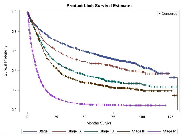
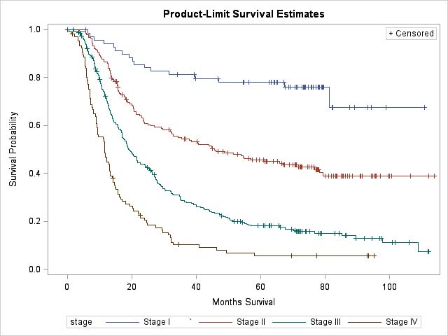
17.4 Clinical stage (cTNM) and overall survival in patients diagnosed with gastric cancer, stratified by clinical stage groupings, based on NCDB data (2004-2008; median follow-up, 12 months; n = 7,306).

17.1 Clinical stage and 1-, 3-, and 5-year and median overall survivals in patients diagnosed with gastric cancer, stratified by clinical stage groupings, based on NCDB data

Clinical stage groupPatients, n1-y Survival, %3-y Survival, %5-y Survival, %Median survival, mo
I (T1/2, N0)1,14880.664.956.784.93
IIA (T1/2, N+)29674.253.747.346.06
IIB (T3/T4a, N0)78368.941.433.123.82
III (T3/T4a, N+)1,42766.433.125.919.12
IV (T4b & M+)3,38228.37.85.06.24

17.5 Clinical stage and overall survival in patients with gastric cancer who received curative or palliative surgery, stratified by clinical stage groupings, based on Shizuoka Cancer Center data (2002-2015; median follow-up, 47 months; n = 4,091).

17.2 Clinical stage and 1-, 3-, and 5-year and median overall survivals in patients with gastric cancer who received curative or palliative surgery, stratified by clinical stage groupings, based on Shizuoka Cancer Center data

Clinical stage groupPatients, n1-y Survival, %3-y Survival, %5-y Survival, %Median survival
I (T1/2, N0)2,31898.995.090.2Not reached
IIA (T1/2, N+)16196.883.675.2Not reached
IIB (T3/T4a, N0)56687.867.759.398.73 mo
III (T3/T4a, N+)75882.955.143.445.07 mo
IV (T4b & M+)28851.722.114.113.3 mo

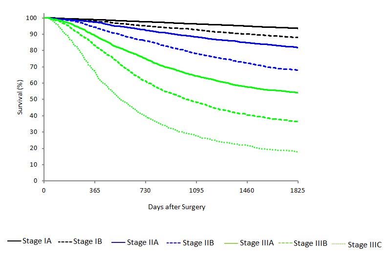
17.6 Pathological stage (pTNM) and overall survival in gastric cancer patients who underwent surgical resection with adequate lymphadenectomy (D2) without prior chemotherapy or radiation therapy, stratified by pathological stage groupings, based on IGCA data (2000-2004; only patients with complete 5-year follow-up were included, n = 25,411).

17.3 Pathological stage and 1-, 3-, and 5-year and median overall survivals in patients with gastric cancer who received curative surgery, stratified by pathological stage groupings, based on IGCA data3

Pathological stage groupPatients, n1-y Survival, %3-y Survival, %5-y Survival, %Median survival
IA10,6069996.3093.60Not reached
IB2,6069892.8088Not reached
IIA2,29197.4088.3081.80Not reached
IIB2,48194.3078.2068Not reached
IIIA3,0448964.4054.20Not reached
IIIB2,21883.1048.2036.2032.8 mo
IIIC1,35066.8027.7017.9018.5 mo

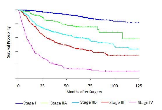
17.7 Post-neoadjuvant therapy stage (ypTNM) and overall survival in patients who underwent surgical resection and were given chemotherapy and /or radiation therapy before surgery, stratified by ypStage groupings, based on NCDB data (2004-2008; median follow-up, 23 months; n = 683).

17.4 Post-neoadjuvant therapy stage (ypTNM) and 1-, 3-, and 5-year and median overall survivals in patients with gastric cancer, stratified by ypStage groupings, based on NCDB data

Posttreatment stage groupPatients, n1-y Survival, %3-y Survival, %5-y Survival, %Median survival, mo
I7094.381.476.5117.8
II19586.754.846.346.0
III30171.728.818.319.2
IV11746.710.25.711.6

Illustrations

17.8 T1a is defined as tumor that invades the lamina propria. T1b is defined as tumor that invades the submucosa. T2 is defined as tumor that invades the muscularis propria, whereas T3 is defined as tumor that extends through the muscularis propria into the subserosal tissue.

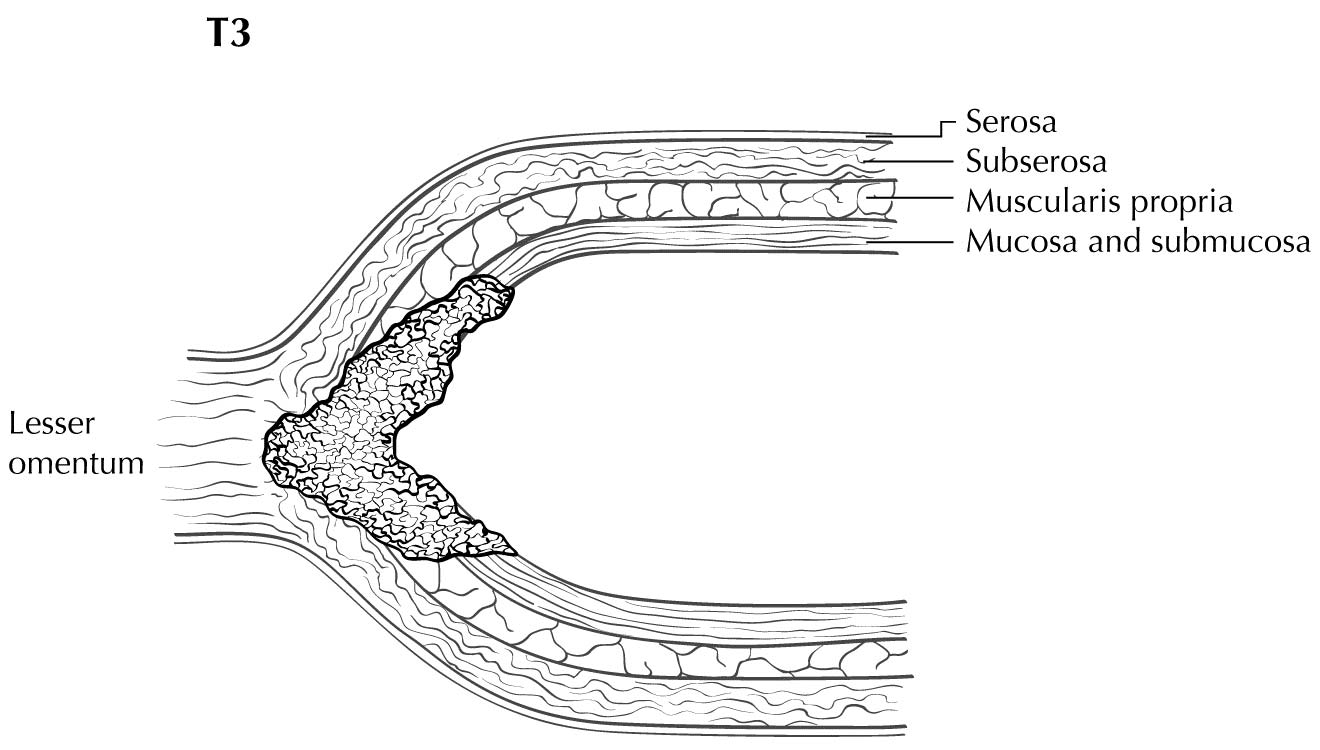
17.9 T3 is defined as tumor that invades the subserosa, shown here invading the lesser omentum without involvement of the serosa (visceral peritoneum).

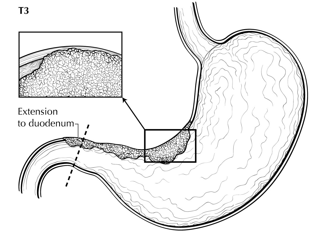
17.10 Distal extension to duodenum does not affect the T3 category.

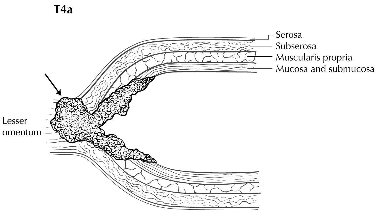
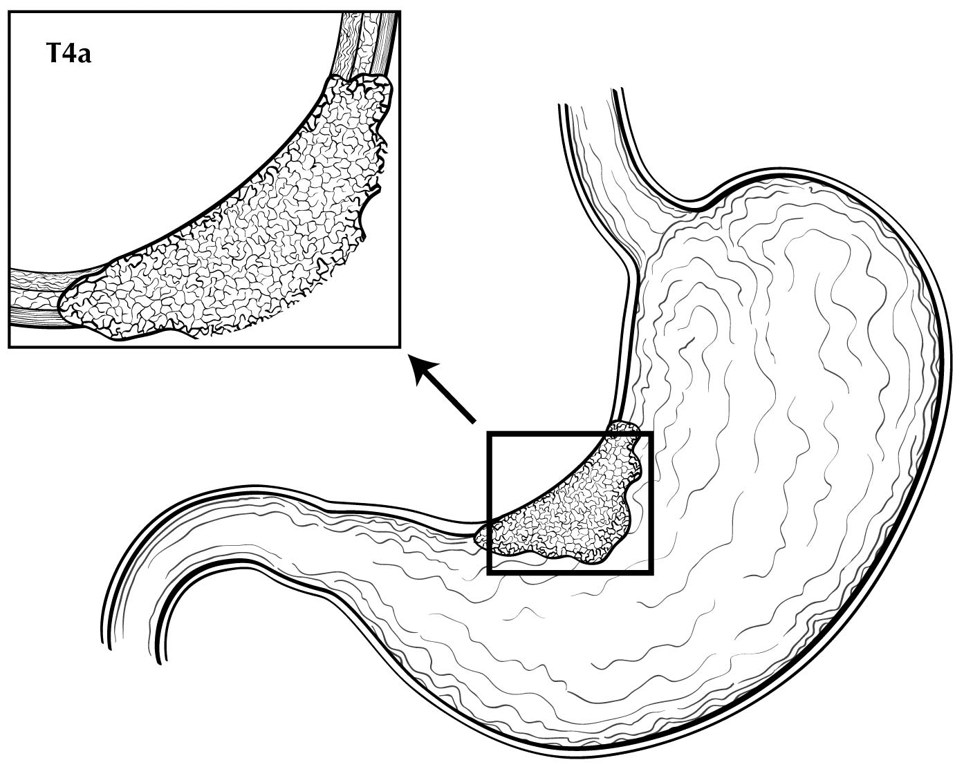
17.11 T4a is defined as tumor that penetrates the serosa (visceral peritoneum) without invasion of adjacent structures.

17.12 T4a is defined as tumor that penetrates the serosa (visceral peritoneum) without invasion of adjacent structures.

17.13 T4a is defined as tumor that penetrates the serosa (visceral peritoneum) without invasion of adjacent structures, whereas T4b is defined as tumor that radially invades adjacent structures, shown here invading the pancreas.

Bibliography

  1. Torre LA, Bray F, Siegel RL, Ferlay J, Lortet-Tieulent J, Jemal A. Global cancer statistics, 2012. CA: a cancer journal for clinicians. Mar 2015;65(2):87-108.
  2. Ajani JA, Bentrem DJ, Besh S, et al. Gastric cancer, version 2.2013: featured updates to the NCCN Guidelines. Journal of the National Comprehensive Cancer Network : JNCCN. May 1 2013;11(5):531-546.
  3. Sano T, Coit DG, Kim HH, et al. Proposal of a new stage grouping of gastric cancer for TNM classification: International Gastric Cancer Association staging project. Gastric cancer : official journal of the International Gastric Cancer Association and the Japanese Gastric Cancer Association. Feb 20 2016.
  4. Choi AH, Kim J, Chao J. Perioperative chemotherapy for resectable gastric cancer: MAGIC and beyond. World journal of gastroenterology : WJG. Jun 28 2015;21(24):7343-7348.
  5. Association JGC. Japanese gastric cancer treatment guidelines 2010 (ver. 3). Gastric cancer : official journal of the International Gastric Cancer Association and the Japanese Gastric Cancer Association. 2011;14(2):113-123.
  6. Kimmey MB, Martin RW, Haggitt RC, Wang KY, Franklin DW, Silverstein FE. Histologic correlates of gastrointestinal ultrasound images. Gastroenterology. Feb 1989;96(2 Pt 1):433-441.
  7. Botet JF, Lightdale CJ, Zauber AG, et al. Preoperative staging of gastric cancer: comparison of endoscopic US and dynamic CT. Radiology. Nov 1991;181(2):426-432.
  8. Barbour AP, Rizk NP, Gerdes H, et al. Endoscopic ultrasound predicts outcomes for patients with adenocarcinoma of the gastroesophageal junction. Journal of the American College of Surgeons. Oct 2007;205(4):593-601.
  9. Blackshaw G, Lewis WG, Hopper AN, et al. Prospective comparison of endosonography, computed tomography, and histopathological stage of junctional oesophagogastric cancer. Clin Radiol. Oct 2008;63(10):1092-1098.
  10. Murata Y, Napoleon B, Odegaard S. High-frequency endoscopic ultrasonography in the evaluation of superficial esophageal cancer. Endoscopy. May 2003;35(5):429-435; discussion 436.
  11. Mocellin S, Pasquali S. Diagnostic accuracy of endoscopic ultrasonography (EUS) for the preoperative locoregional staging of primary gastric cancer. Cochrane Database Syst Rev. 2015;2:CD009944.
  12. Catalano MF, Sivak MV, Jr., Rice T, Gragg LA, Van Dam J. Endosonographic features predictive of lymph node metastasis. Gastrointestinal endoscopy. Jul-Aug 1994;40(4):442-446.
  13. Puli SR, Reddy JB, Bechtold ML, Antillon D, Ibdah JA, Antillon MR. Staging accuracy of esophageal cancer by endoscopic ultrasound: a meta-analysis and systematic review. World journal of gastroenterology : WJG. Mar 14 2008;14(10):1479-1490.
  14. tenBerge J, Hoffman BJ, Hawes RH, et al. EUS-guided fine needle aspiration of the liver: indications, yield, and safety based on an international survey of 167 cases. Gastrointestinal endoscopy. Jun 2002;55(7):859-862.
  15. Lee YT, Ng EK, Hung LC, et al. Accuracy of endoscopic ultrasonography in diagnosing ascites and predicting peritoneal metastases in gastric cancer patients. Gut. Nov 2005;54(11):1541-1545.
  16. Sultan J, Robinson S, Hayes N, Griffin SM, Richardson DL, Preston SR. Endoscopic ultrasonography-detected low-volume ascites as a predictor of inoperability for oesophagogastric cancer. The British journal of surgery. Sep 2008;95(9):1127-1130.
  17. Chen CY, Hsu JS, Wu DC, et al. Gastric cancer: preoperative local staging with 3D multi-detector row CT--correlation with surgical and histopathologic results. Radiology. Feb 2007;242(2):472-482.
  18. Park HS, Lee JM, Kim SH, et al. Three-dimensional MDCT for preoperative local staging of gastric cancer using gas and water distention methods: a retrospective cohort study. AJR. American journal of roentgenology. Dec 2010;195(6):1316-1323.
  19. Anzidei M, Napoli A, Zaccagna F, et al. Diagnostic performance of 64-MDCT and 1.5-T MRI with high-resolution sequences in the T staging of gastric cancer: a comparative analysis with histopathology. Radiol Med. Oct 2009;114(7):1065-1079.
  20. Kim YH, Lee KH, Park SH, et al. Staging of T3 and T4 gastric carcinoma with multidetector CT: added value of multiplanar reformations for prediction of adjacent organ invasion. Radiology. Mar 2009;250(3):767-775.
  21. Fujikawa H, Yoshikawa T, Hasegawa S, et al. Diagnostic value of computed tomography for staging of clinical T1 gastric cancer. Annals of surgical oncology. Sep 2014;21(9):3002-3007.
  22. Yun M, Lim JS, Noh SH, et al. Lymph node staging of gastric cancer using (18)F-FDG PET: a comparison study with CT. Journal of nuclear medicine : official publication, Society of Nuclear Medicine. Oct 2005;46(10):1582-1588.
  23. Chen J, Cheong JH, Yun MJ, et al. Improvement in preoperative staging of gastric adenocarcinoma with positron emission tomography. Cancer. Jun 1 2005;103(11):2383-2390.
  24. Namikawa T, Okabayshi T, Nogami M, Ogawa Y, Kobayashi M, Hanazaki K. Assessment of (18)F-fluorodeoxyglucose positron emission tomography combined with computed tomography in the preoperative management of patients with gastric cancer. International journal of clinical oncology. Aug 2014;19(4):649-655.
  25. Kato H, Kuwano H, Nakajima M, et al. Comparison between positron emission tomography and computed tomography in the use of the assessment of esophageal carcinoma. Cancer. Feb 15 2002;94(4):921-928.
  26. Lowe VJ, Booya F, Fletcher JG, et al. Comparison of positron emission tomography, computed tomography, and endoscopic ultrasound in the initial staging of patients with esophageal cancer. Molecular imaging and biology : MIB : the official publication of the Academy of Molecular Imaging. Nov-Dec 2005;7(6):422-430.
  27. Takizawa K, Matsuda T, Kozu T, et al. Lymph node staging in esophageal squamous cell carcinoma: a comparative study of endoscopic ultrasonography versus computed tomography. J Gastroenterol Hepatol. Oct 2009;24(10):1687-1691.
  28. Choi J, Kim SG, Kim JS, Jung HC, Song IS. Comparison of endoscopic ultrasonography (EUS), positron emission tomography (PET), and computed tomography (CT) in the preoperative locoregional staging of resectable esophageal cancer. Surg Endosc. Jun 2010;24(6):1380-1386.
  29. Walker AJ, Spier BJ, Perlman SB, et al. Integrated PET/CT fusion imaging and endoscopic ultrasound in the pre-operative staging and evaluation of esophageal cancer. Molecular imaging and biology : MIB : the official publication of the Academy of Molecular Imaging. Feb 2011;13(1):166-171.
  30. Findlay JM, Bradley KM, Maile EJ, et al. Pragmatic staging of oesophageal cancer using decision theory involving selective endoscopic ultrasonography, PET and laparoscopy. The British journal of surgery. Nov 2015;102(12):1488-1499.
  31. Cuellar SL, Carter BW, Macapinlac HA, et al. Clinical staging of patients with early esophageal adenocarcinoma: does FDG-PET/CT have a role? J Thorac Oncol. Aug 2014;9(8):1202-1206.
  32. Little SG, Rice TW, Bybel B, et al. Is FDG-PET indicated for superficial esophageal cancer? European journal of cardio-thoracic surgery : official journal of the European Association for Cardio-thoracic Surgery. May 2007;31(5):791-796.
  33. Shiozaki H, Elimova E, Slack RS, et al. Prognosis of gastric adenocarcinoma patients with various burdens of peritoneal metastases. Journal of surgical oncology. 2016;113(1):29-35.
  34. Mezhir JJ, Shah MA, Jacks LM, Brennan MF, Coit DG, Strong VE. Positive peritoneal cytology in patients with gastric cancer: natural history and outcome of 291 patients. Annals of surgical oncology. Dec 2010;17(12):3173-3180.
  35. Mansour JC, Tang L, Shah M, et al. Does graded histologic response after neoadjuvant chemotherapy predict survival for completely resected gastric cancer? Annals of surgical oncology. 2007;14(12):3412-3418.
  36. Network CGAR. Comprehensive molecular characterization of gastric adenocarcinoma. Nature. 2014;513(7517):202-209.
  37. van Beek J, zur Hausen A, Klein Kranenbarg E, et al. EBV-positive gastric adenocarcinomas: a distinct clinicopathologic entity with a low frequency of lymph node involvement. J Clin Oncol. Feb 15 2004;22(4):664-670.
  38. Cristescu R, Lee J, Nebozhyn M, et al. Molecular analysis of gastric cancer identifies subtypes associated with distinct clinical outcomes. Nature medicine. May 2015;21(5):449-456.
  39. Dulak AM, Stojanov P, Peng S, et al. Exome and whole-genome sequencing of esophageal adenocarcinoma identifies recurrent driver events and mutational complexity. Nature genetics. May 2013;45(5):478-486.
  40. Kakiuchi M, Nishizawa T, Ueda H, et al. Recurrent gain-of-function mutations of RHOA in diffuse-type gastric carcinoma. Nature genetics. Jun 2014;46(6):583-587.
  41. Bang YJ, Van Cutsem E, Feyereislova A, et al. Trastuzumab in combination with chemotherapy versus chemotherapy alone for treatment of HER2-positive advanced gastric or gastro-oesophageal junction cancer (ToGA): a phase 3, open-label, rand omised controlled trial. Lancet. Aug 28 2010;376(9742):687-697.
  42. Le DT, Uram JN, Wang H, et al. PD-1 Blockade in Tumors with Mismatch-Repair Deficiency. N Engl J Med. Jun 25 2015;372(26):2509-2520.
  43. Kattan MW, Hess KR, Amin MB, et al. American Joint Committee on Cancer acceptance criteria for inclusion of risk models for individualized prognosis in the practice of precision medicine. CA: a cancer journal for clinicians. Jan 19 2016.