section name header

Disease Prologue

Summary

Cancers Staged Using This Staging System

Adenocarcinomas, high-grade neuroendocrine carcinomas, and squamous carcinomas of the colon and rectum are covered by this staging system.

Cancers Not Staged Using This Staging System

These histopathologic types of cancer…Are staged according to the classification for…and can be found in chapter…
Appendiceal carcinomasAppendix - carcinoma19
Anal carcinomasAnus21
Well-differentiated neuroendocrine tumors (carcinoids)Well-differentiated neuroendocrine tumors of the colon and rectum33

Summary of Changes

ChangeDetails of ChangeLevel of Evidence
Definition of Distant Metastasis (M)Introduced M1c, which details peritoneal carcinomatosis as a poor prognostic factorI
Definition of Regional Lymph Node (N)Clarified the definition of tumor depositsII
Additional Factors Recommended for Clinical CareLymphovascular invasion: reintroduced the L and V elements to better identify lymphatic and vessel invasionI
Additional Factors Recommended for Clinical CareMicrosatellite instability (MSI): clarified the importance of MSI as a prognostic and predictive factorI
Additional Factors Recommended for Clinical CareIdentified KRAS, NRAS, and BRAF mutations as critical prognostic factors that are also predictiveI and II

ICD-O-3 Topography Codes

CodeDescription
C18.0Cecum
C18.2Ascending colon
C18.3Hepatic flexure of colon
C18.4Transverse colon
C18.5Splenic flexure of colon
C18.6Descending colon
C18.7Sigmoid colon
C18.8Overlapping lesion of colon
C18.9Colon, NOS
C19.9Rectosigmoid junction
C20.9Rectum, NOS

WHO Classification of Tumors

This list includes histology codes and preferred terms from the WHO Classification of Tumors and the International Classification of Diseases for Oncology (ICD-O). Most of the terms in this list represent malignant behavior. For cancer reporting purposes, behavior codes /3 (denoting malignant neoplasms), /2 (denoting in situ neoplasms), and in some cases /1 (denoting neoplasms with uncertain and unknown behavior) may be appended to the 4-digit histology codes to create a complete morphology code.

CodeDescription
8000Neoplasm, malignant
8010Carcinoma, NOS
8010Carcinoma in situ, NOS
8013Large cell neuroendocrine carcinoma (NEC)
8020Undifferentiated carcinoma
8041Small cell neuroendocrine carcinoma (NEC)
8070Squamous cell carcinoma
8140Adenocarcinoma in situ
8140Adenocarcinoma
8213Serrated adenocarcinoma
8246Neuroendocrine carcinoma (NEC)
8265Micropapillary carcinoma
8480Mucinous adenocarcinoma
8481Mucin-producing adenocarcinoma
8490Signet ring cell carcinoma
8510Medullary carcinoma
8560Adenosquamous carcinoma

Histology is not ideal for clinical use in patient care, as it describes an unspecified or outdated diagnosis. Data collectors may use this code only if there is not enough information in the medical record to document a more specific diagnosis.

Bosman FT, Carneiro F, Hruban RH, Theise ND, eds. World Health Organization Classification of Tumours of the Digestive System. Lyon: IARC; 2010. Used with permission.

International Agency for Research on Cancer, World Health Organization. International Classification of Diseases for Oncology. ICD-O-3-Online.http://codes.iarc.fr/home. Accessed September 29, 2017. Used with permission.

Introduction

Adenocarcinoma of the colon and rectum is the second most lethal cancer in the United States; its treatment is determined primarily by TNM staging. The advent of detailed molecular characterization of colorectal carcinoma has led to better understand ing of not only the etiology of the malignancy but also how the disease responds to stage-specific treatment. The molecular characteristics provide a set of prognostic factors that likely will become more important in the future. In addition, histopathologic analysis of primary carcinomas, as well as the development of immune checkpoint inhibitors, has led to an increasing appreciation of the role of host immunity in improving survival. This approach involves the current development of a histologic prognostic and predictive scoring system, called Immunoscore, that may improve TNM staging after it is validated. It also involves early trials of inhibitors of checkpoint proteins such as PD-1, PD-L1, and CTL4. These recent molecular and immune findings portend exciting future methods for treating colorectal carcinoma.

The AJCC Cancer Staging Manual, 8th Edition is very similar to the AJCC Cancer Staging Manual, 7th Edition. The colorectal disease team of the Lower GI Expert Panel has attempted to clarify the issues that have perplexed some experts in the last several editions. This has has led to the recommendation that small vessel and large venous involvement be collected as registry data items in addition to tumor deposits. We also present data that validate the division of T4 colon or rectal cancer primaries into T4a and T4b categories in a dataset independent from the one used in the 7th Edition. We have strengthened the evidence for collecting molecular data such as microsatellite instability (MSI) status and BRAF mutations as prognostic factors, as well as mutations in BRAF, KRAS, and NRAS as predictive factors.

Anatomy

Primary Site(s)

The large intestine (colon and rectum) extends from the terminal ileum to the anal canal. Excluding the vermiform appendix and rectum, the colon is divided into four parts: the right or ascending colon, the middle or transverse colon, the left or descending colon, and the sigmoid colon. The sigmoid colon is continuous with the rectum, which terminates at the anal canal (Figures 20.1 and 20.2).

20.1 Anatomic subsites of the colon.

20.2 Anatomic subsites of the rectum.

The ascending colon begins with the cecum, a 6- to 9-cm pouch that arises as the proximal segment of the right colon at the end of the terminal ileum. It is covered with a visceral peritoneum (serosa). The ascending colon continues from the cecum and measures about 15 to 20 cm in length. The posterior surface of the ascending (and descending) colon lacks peritoneum and thus is in direct contact with the retroperitoneum. In contrast, the anterior and lateral surfaces of the ascending (and descending) colon have serosa and are intraperitoneal. The ascending colon ends at the hepatic flexure, which transitions the ascending colon into the transverse colon, passing just inferior to the liver and anterior to the duodenum.

The transverse colon is entirely intraperitoneal, about 18 to 22 cm long, and supported on a mesentery that is attached to the pancreas. Anteriorly, its serosa is continuous with the gastrocolic ligament. The transverse colon ends at the splenic flexure, which transitions into the descending colon.

The descending colon passes inferior to the spleen and anterior to the tail of the pancreas. As noted earlier, the posterior aspect of the descending colon lacks serosa and is in direct contact with the retroperitoneum, whereas the lateral and anterior surfaces have serosa and are intraperitoneal. The descending colon measures about 10 to 15 cm in length.

The sigmoid colon is completely intraperitoneal, once again with a mesentery that develops at the medial border of the left psoas major muscle and extends to the rectum. The transition from sigmoid colon to rectum is marked by the fusion of the taenia of the sigmoid colon to the circumferential longitudinal muscle of the rectum. The sigmoid colon is approximately 15 to 20 cm long.

The proximal rectum is defined by the fusion of the taenia, which typically occurs at the level of the sacral promontory. The distal boundary of the rectal reservoir or ampulla is the puborectalis ring, which is palpable as the anorectal ring on digital rectal examination. The rectal mucosa extends below this ring into the functional anal canal to the dentate line. This feature is critical to understand ing how rectal cancer may occur within the functional (“surgical”) anal canal. The rectum is approximately 12 to 16 cm in length. It is covered by peritoneum in front and on both sides in its upper third and only on the anterior wall in its middle third. The peritoneum is reflected laterally from the rectum to form the perirectal fossa and , anteriorly, the uterine or rectovesical fold. Depending on body habitus and gender, this fossa may be widely variable, and may extend to the pelvic floor.

In general, the lower third of the rectum (the reservoir or ampulla) does not have a peritoneal covering. This extraperitoneal rectum is encircled by a variably thick fatty sheath containing perirectal lymph nodes and enveloped circumferentially by the fascia propria, with separation posteriorly from the sacrum by Waldeyer's fascia, from the pelvic sidewalls by the pelvic parietal fascia, and anteriorly from the prostate or vagina by Denonvilliers' fascia. The mesorectum tapers distally so that no fatty sheath surrounds the rectal wall at the puborectalis sling. The rectum has semilunar transverse rectal folds, also known as the valves of Houston. Most commonly, there are three folds, although two or four may be present. These boundaries are described further in the evaluation of total mesorectal excision (TME) specimens.

The mucosa of the colon and rectum comprises a single layer of epithelial cells arranged in invaginations called the crypts of Lieberkühn, separated by the lamina propria, a loose connective tissue stroma investing the crypts. The base of the mucosa is separated from the submucosa by a thin but distinct muscle layer, the muscularis mucosae. The thicker, separate, and deeper layer of smooth muscle is the muscularis propria. The area between the two muscle layers is the submucosa. The layer of connective tissue beyond the muscularis propria is the pericolorectal connective tissue, which alternatively may be called subserosal tissue when covered by a peritoneal lining, or adventia, in areas lacking peritoneal lining. Histologically, the colorectal mucosa extends to the dentate line, which is the superior boundary of the anal mucosa.

Two definitions of the anal canal exist, one based on function (the “surgical” anal canal) and the other on embryologic development. The functional anal canal is defined by the anal sphincter, which measures 3 to 5 cm in length and whose boundaries are the puborectalis sling cephalad and the intersphincteric groove (the anal verge) caudad on digital rectal examination. The superior border of the embryologic anal canal is the dentate line, which is visible but not palpable and coincides with the midpoint of the functional anal canal, approximately 1 to 2 cm distal to the puborectalis sling (Figure 20.3). The embryologic anal canal shares the same distal boundary as the functional anal canal: the anal verge. The common practice of reporting rectal tumor level relative to the anal verge and the variable length of the sphincter complex contribute to challenges in relating the measurement of the distal extent of the rectal tumor to the probability of sphincter preservation. Distinguishing the origin of distal rectal carcinomas from anal carcinomas is sometimes difficult, because the rectal mucosa may extend within 1 to 2 cm of the anal verge (Figure 20.3).

20.3 The anal canal extends from the proximal aspect of the external sphincter to the anal verge at the intersphincteric groove.

Regional Lymph Nodes

Regional nodes are located 1) along the course of the major vessels supplying the colon and rectum, 2) along the vascular arcades of the marginal artery, and 3) adjacent to the colon—that is, along the mesocolic borders of the colon. Specifically, the regional lymph nodes are termed pericolic and perirectal/mesorectal and also are found along the ileocolic, right colic, middle colic, left colic, inferior mesenteric, superior rectal (hemorrhoidal), and internal iliac arteries (Figure 20.4).

The regional lymph nodes for each segment of the large bowel are designated as follows:

SegmentRegional lymph nodes
CecumPericolic, ileocolic, right colic
Ascending colonPericolic, ileocolic, right colic, right branch of the middle colic
Hepatic flexurePericolic, ileocolic, right colic, middle colic
Transverse colonPericolic, middle colic
Splenic flexurePericolic, middle colic, left colic
Descending colonPericolic, left colic, sigmoid, inferior mesenteric
Sigmoid colonPericolic, sigmoid, superior rectal (hemorrhoidal), inferior mesenteric
RectosigmoidPericolic, sigmoid, superior rectal (hemorrhoidal), inferior mesenteric
RectumMesorectal, superior rectal (hemorrhoidal), inferior mesenteric, internal iliac, inferior rectal (hemorrhoidal)

20.4 The regional lymph nodes of the colon and rectum.

Metastatic Sites

Although carcinomas of the colon and rectum can metastasize to almost any organ, the liver and lungs are most commonly affected. Seeding of other segments of the colon, small intestine, or peritoneum also may occur.

Classification Rules

Clinical Classification

Clinical assessment is based on medical history, physical examination, radiology, and endoscopy with biopsy. Radiologic examinations designed to demonstrate the presence of extrarectal or extracolonic metastasis may include chest radiographs, computed tomography (CT; abdomen, pelvis, chest), magnetic resonance (MR) imaging, positron emission tomography (PET), or fused PET/CT scans. Clinical stage (cTNM) then may be assigned. Pathological stage (pTNM) is assigned based on the resection specimen. Preoperative measurement of carcinoembryonic antigen (CEA) is recommended, as CEA level reflects the likelihood that subclinical or clinical liver or lung metastases are present. In the event of recurrence or synchronous metastases, it now is recommended that the status of the genes KRAS, NRAS, and BRAF be evaluated and MSI or mismatch repair (MMR) be measured.

Primary Site(s)

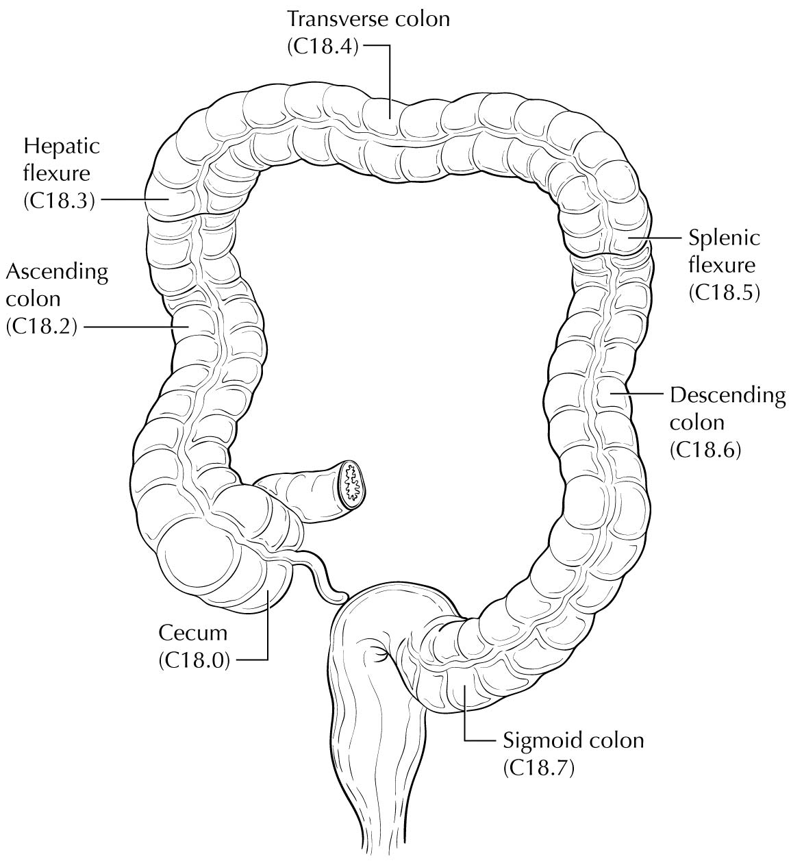
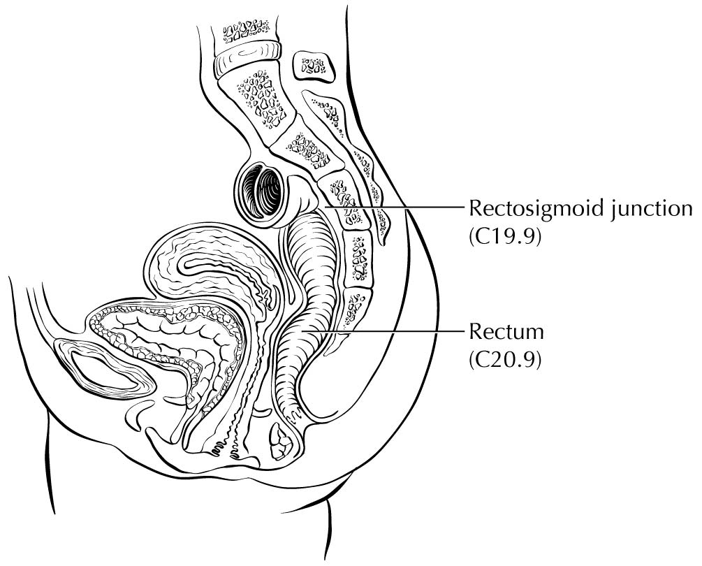
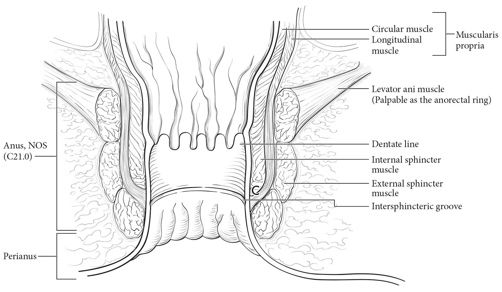
Carcinoma arising at the ileocecal valve should be classified as colonic cancer. For staging purposes, adenocarcinomas should be classified as rectal cancers if proximal to the dentate line or anorectal ring on digital examination. Squamous carcinomas should be staged as anal canal cancers if they are distal to the dentate line or the anorectal ring. However, there are instances of rectal squamous carcinomas and anal adenocarcinomas in this area. The former may need to be treated according to anal squamous carcinoma regimens, whereas the anal adenocarcinomas may require surgery in addition to chemotherapy and radiation. For rectal cancers that extend beyond the dentate line, as for anal canal cancers, the superficial inguinal lymph nodes are among the regional nodal groups at risk for metastatic spread and are included in cN/pN analysis.

Carcinomas that arise in the colon or rectum spread by direct invasion into the mucosa, submucosa, muscularis propria, and subserosal tissue (or adventitia) of the bowel wall, and each level of penetration is annotated by a T category. Primary tumors also spread by invading lymphatics and blood vessels to form metastases in lymph nodes or distant sites; this is annotated by the N and M categories, respectively. In addition, carcinomas may spread and grow in the adventitia as discrete nodules of cells called tumor deposits. These characteristics, along with further description of the T categories, are described in detail later in the chapter.

For patients with rectal cancer, the pelvic extent of disease (cT and cN categories), combined with the status of extrapelvic metastasis (cM) and patient symptoms, determines whether preoperative adjuvant treatment is appropriate. The primary imaging modalities to assess the pelvic extent of disease are endoscopic ultrasound (EUS) and pelvic MR imaging. To improve the accuracy of nodal staging, EUS may be augmented with fine-needle aspiration of lymph nodes suspicious for metastasis, but microscopic evidence of tumor by such a procedure is part of the clinical TNM (cTNM) staging. It is especially important that patients who will receive preoperative adjuvant treatment or neoadjuvant therapy be assigned a pretreatment clinical stage based on disease extent before beginning treatment (cTNM).1-4 Pathological stage is assigned if the patient undergoes resection, and a modified pathological stage is generated if the patient undergoes neoadjuvant therapy (ypTNM).

For carcinomas of the colon or rectum, the number of metastatic sites involved is an important prognostic factor and is reflected in the subdivision of M1, as described in greater detail later in the chapter. Metastases to both ovaries or both lobes of the lungs are considered involvement of a single site by themselves. Peritoneal carcinomatosis with or without blood-borne metastasis to visceral organs has a worse prognosis.

Imaging

As stated elsewhere in this chapter, several imaging studies may be performed in newly diagnosed colon or rectal carcinoma patients. The National Comprehensive Cancer Network (NCCN) guidelines for evaluation of colon1 or rectal3 carcinoma cases recommend that a CT scan with intravenous and oral contrast be performed on the chest, abdomen, and pelvis. If the CT scan cannot be performed because of contrast sensitivity or the images are not adequate, then MR imaging with contrast may be performed with a noncontrast CT scan.1 PET/CT is recommended only if there is an equivocal finding on a contrast-enhanced CT scan or in the case of sensitivity to CT contrast. If synchronous metastases or distant metastases appear later and resection seems possible, then PET/CT should be considered to further delineate the extent of disease.1,3

Pathological Classification

Most cancers of the colon and many cancers of the rectum are pathologically staged after microscopic examination of the resected specimen (pTNM) resulting from surgical exploration of the abdomen and cancer-directed surgical resection.

Primary Tumor

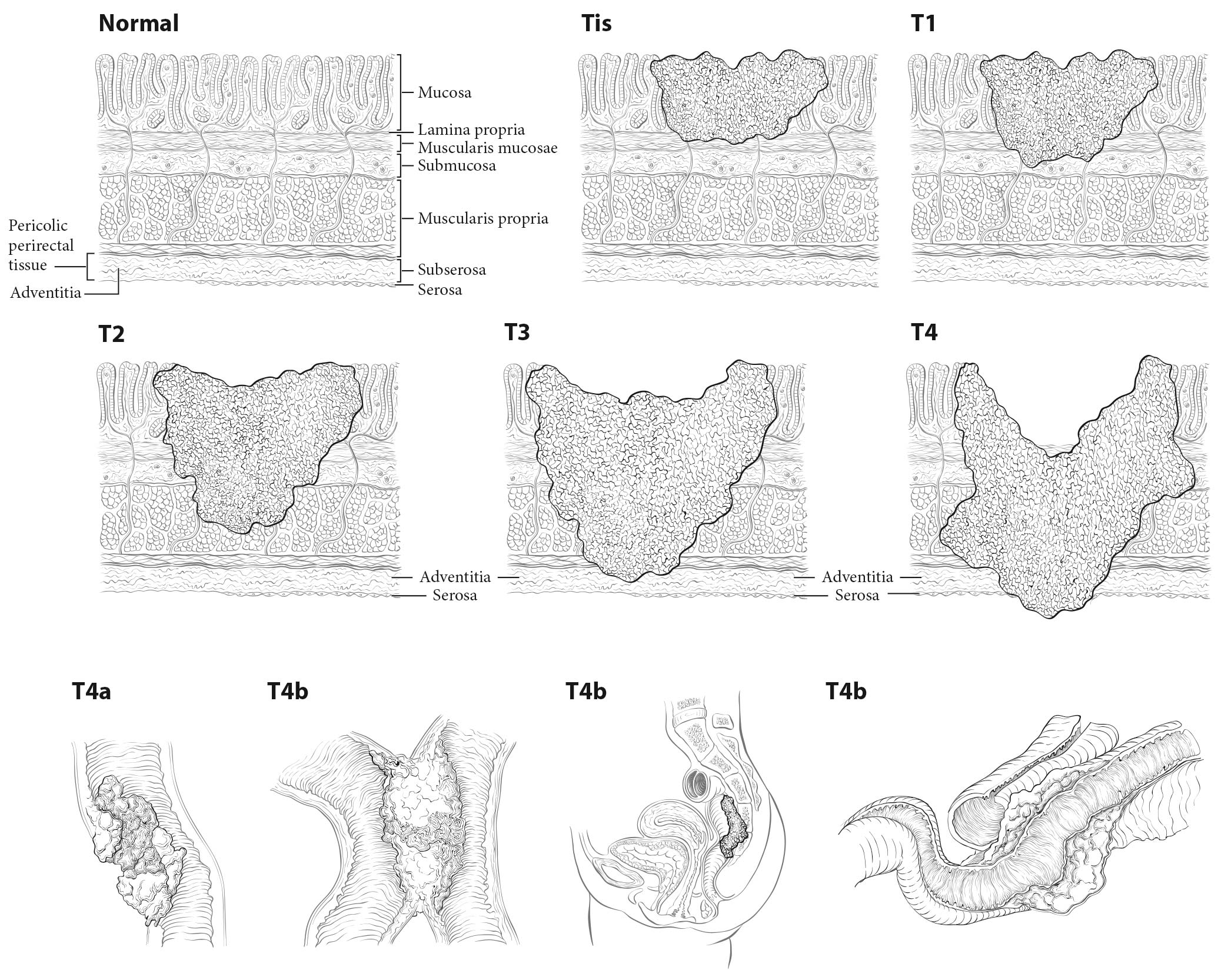
Tis and T1. Regarding the colorectum, pathologists apply the term high-grade dysplasia to lesions that are confined to the epithelial layer of crypts and lack invasion through the basement membrane into the lamina propria. The term intraepithelial carcinoma is synonymous with high-grade dysplasia but rarely is used to apply to the colorectum. High-grade dysplasia should not be assigned to the Tis category or recorded in cancer registries, because these lesions lack potential for tumor spread. However, Tis is assigned to lesions confined to the mucosa in which cancer cells invade into the lamina propria and may involve but not penetrate through the muscularis mucosa. (These lesions are more correctly termed intramucosal carcinoma.) Although invasion through the basement membrane in all gastrointestinal sites is considered invasive, in colorectal tumors, invasion of the lamina propria without penetration through the muscularis mucosa (intramucosal carcinoma) is designated Tis, as it is associated with a negligible risk for metastasis. Because there is potential for missing deeper invasion because of sampling, such lesions should be recorded in the cancer registry. The term invasive adenocarcinoma is used for colorectal cancer if the tumor extends through the muscularis mucosae into the submucosa or beyond (Figure 20.5).

20.5 T1-T3 as defined in Definition of Primary Tumor (T). T4 is a tumor that penetrates or perforates the visceral peritoneum in the parts of the colon or rectum covered only by peritoneum (T4a) or that invades an adjacent structure or organ (T4b).

Carcinoma in a Polyp. These lesions are classified according to the pT definitions adopted for colorectal carcinomas. For instance, invasive carcinoma limited to the muscularis mucosae and /or lamina propria is classified as pTis, whereas tumor that has invaded through the muscularis mucosae and has entered the submucosa of the polyp head or stalk is classified as pT1. pTis in a polyp resected with a clear margin during endoscopy is a Stage 0 carcinoma with nodal and metastatic status unknown, but with a sufficiently low probability of nodal involvement that node resection is not justified. The probability of metastasis is similarly low. Haggitt levels and submucosal depth of invasion categories may be used to classify polyps for their malignant potential, but reporting of these parameters is optional.5-10 Guidelines from several organizations1,3,4,11,12 and authors8,12,13 recommend surgical resection for polyps that contain high-grade carcinoma, have invasive carcinoma at or less than 1 mm from the resection margin, or have lymphatic/venous vessel invasion.

T1, T2, and T3. As in previous AJCC editions, these tumors are defined as involvement of the submucosa, penetration through the submucosa into but not through the muscularis propria, and penetration through the muscularis propria, respectively.14,15

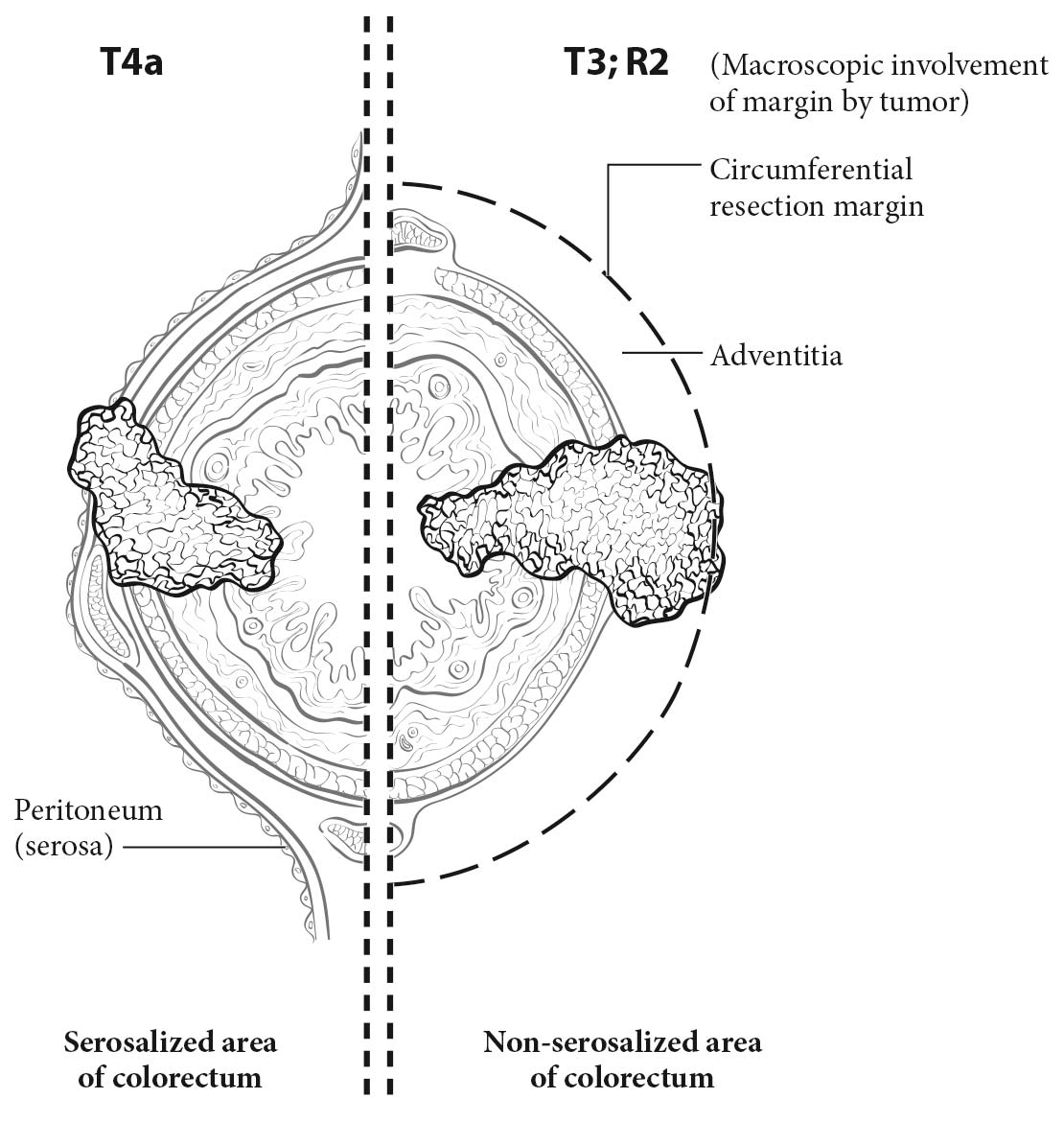
T4. Tumors that involve the serosal surface (visceral peritoneum) or directly invade adjacent organs or structures are assigned to the T4 category. For both colon and rectum, T4 is divided into two categories (T4a and T4b) based on different outcomes shown in expand ed datasets16,17 (Figures 20.6 and 20.7). T4a tumors are characterized by involvement of the serosal surface (visceral peritoneum) by direct tumor extension. Tumors with perforation in which the tumor cells are continuous with the serosal surface through inflammation also are considered T4a. The significance of tumors that are <1 mm from the serosal surface and accompanied by serosal reaction is unclear, with some18 but not all studies19 indicating a higher risk for peritoneal relapse. Multiple-level sections and /or additional tissue blocks of the tumor should be examined in these cases to detect serosal surface involvement. If the latter is not present after additional evaluation, the tumor should be assigned to the pT3 category. In portions of the colorectum that are not peritonealized (e.g., posterior aspects of the ascending and descending colon, lower portion of the rectum), the T4a category is not applicable.

Treatment of Primary Colorectal Carcinomas. Colon carcinomas are resected according to guidelines by several organizations.1-4 The stand ard of care for primary treatment of rectal carcinoma should include sharp dissection with an intact fascia propria of the mesorectum. The extent of mesorectal excision may be total (TME) or partial, as in the setting of proximal tumors located more than 5 cm from the distal extent of the mesorectum (tumor-specific mesorectal excision [TSME]). For the purpose of discussion throughout the rest of this chapter, TME is used to indicate either TME or TSME. The rate of local recurrence is inversely proportional to the completeness of the excision and the distance from the tumor to the circumferential resection margin (CRM).20-22 Thus, macroscopic assessment of the quality and completeness of the excision of the mesorectum and the state of the fascia propria should be reported for rectal cancers. As suggested by Parfitt and Driman,23 the College of American Pathologists (CAP) guidelines support a three-tiered evaluation system of a) complete, b) nearly complete, and c) incomplete (Table 20.1). The site-specific factor of the CRM is related to, but separate from, the grading of the TME specimen, as TME grading includes an assessment of the mesorectum for bulk, presence of surgical defects, degree of coning, and perforation. The CRM is described further elsewhere in the chapter and applies to the status of the nonserosal margin closest to the deepest point of penetration by the primary cancer throughout the colorectum.11 As indicated in the current CAP guidelines11 and in a systematic review,24 patients with an incomplete mesorectum after rectal resection have a worse outcome than those with a complete mesorectum. For those with a negative CRM, the quality of the TME specimen is especially important, because the recurrence rate is higher in those with incomplete specimens.

20.1 Grading of quality and completeness of the mesorectum in a total mesorectal excision

MesorectumDefectsConingCRM*
CompleteIntact, smoothNot deeper than 5 mmNoneSmooth, regular
Nearly completeModerate bulk, irregularNo visible muscularis propriaModerateIrregular
IncompleteLittle bulkDown to muscularis propriaModerate/markedIrregular

Both the specimen as a whole (fresh) and cross-sectional slices (fixed) are examined to make an adequate interpretation (Adapted from Parfitt and Driman23 with permission).

*CRM, circumferential resection margin.

Tumor Regression after Neoadjuvant Preoperative Radiotherapy and /or Chemotherapy for Rectal Carcinoma

The pathological response to preoperative radiotherapy and /or chemotherapy is an important prognostic factor for rectal carcinoma. This response is assessed by the pathologist and is reported with the prefix y before a pT and pN. Both a large institutional study25 and a rand omized phase III trial26 demonstrated that complete eradication of the tumor, as detected by microscopic examination of the resected specimen, is associated with a better prognosis than an incomplete or poor response to neoadjuvant therapy. Failure of the tumor to respond to neoadjuvant treatment is an adverse prognostic factor. Parallel associations are observed in breast, esophageal, and pancreatic carcinomas. The US Food and Drug Administration (FDA) has approved the use of complete pathological response to neoadjuvant therapy as such a strong positive prognostic factor in breast cancer that it may be used as an end point for evaluating drug responses. In addition, measures of tumor response to therapy such as minimal residual disease in leukemias after induction therapy and necrosis after neoadjuvant treatment of osteosarcomas have similar prognostic impact.

According to the CAP guidelines for recording tumor regression, the resection specimen must be evaluated and recorded by the pathologist (see CAP's “Protocol for the Examination of Specimens from Patients with Carcinomas of the Colon and Rectum”11,12), because neoadjuvant chemoradiation in rectal cancer often is associated with significant tumor response and downstaging. Therefore, specimens from patients receiving neoadjuvant chemoradiation should be examined thoroughly at the primary tumor site, in regional nodes, and for peritumoral satellite nodules or deposits in the remainder of the specimen. It is especially important to evaluate the resection specimen for lymph node metastases, because nodal metastasis is an important poor prognostic factor.26

Lymph Nodes

In the assessment of pN, the number of lymph nodes sampled should be recorded. The number of nodes removed and retrieved from an operative specimen has been reported to correlate with improved survival, possibly because of increased accuracy in staging. For nodal sampling to be accurate, it is important to obtain and examine at least 12 lymph nodes in radical colon and rectum resections in patients who undergo surgery for cure. In cases in which tumor is resected for palliation, or in patients who have received preoperative radiation or chemoradiation, fewer lymph nodes may be present. In all cases, however, it is essential that the total number of regional lymph nodes recovered from the resection specimen be recorded, because that number is prognostically important. A pN0 determination is assigned if all nodes are histologically negative, even if fewer than the recommended number of nodes have been analyzed.

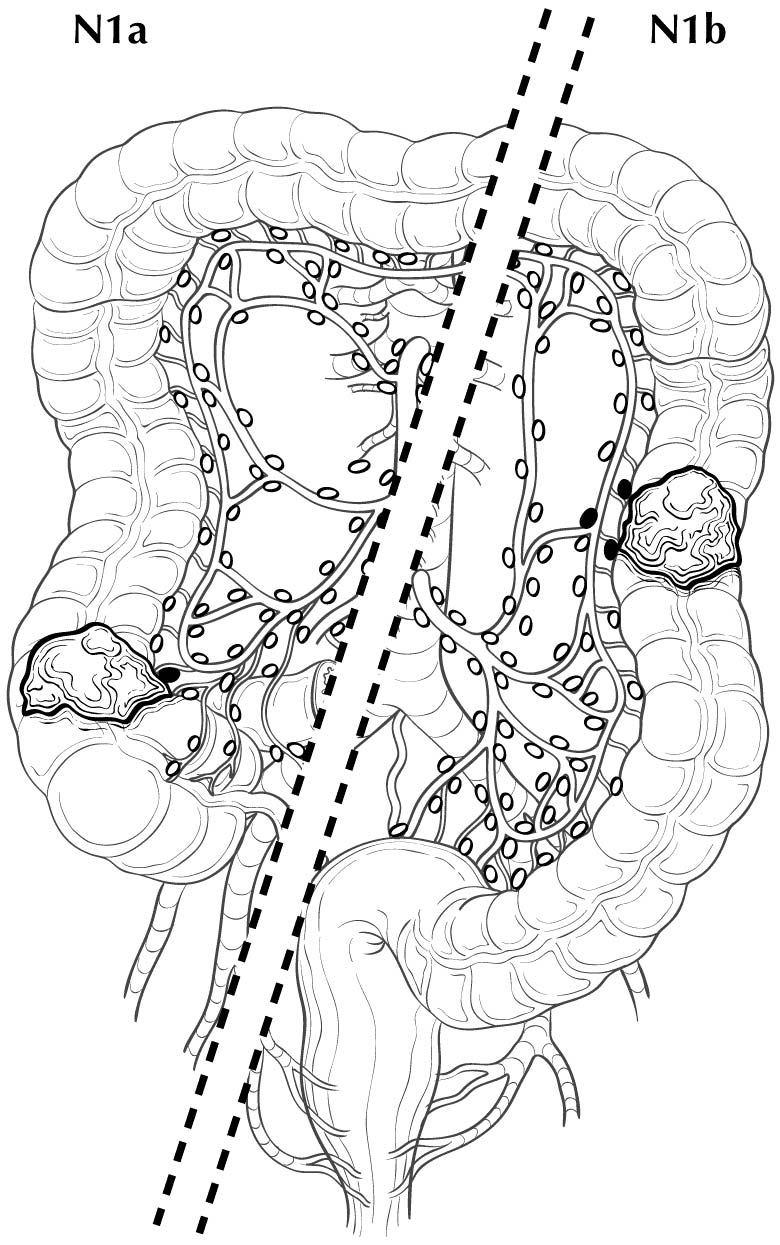
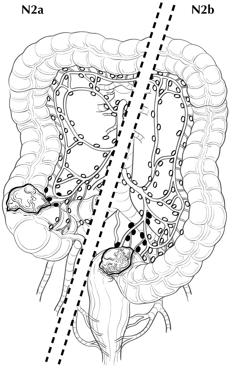
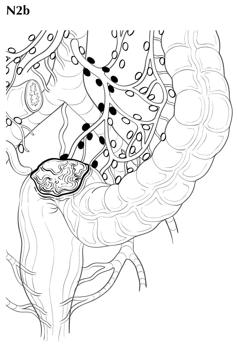
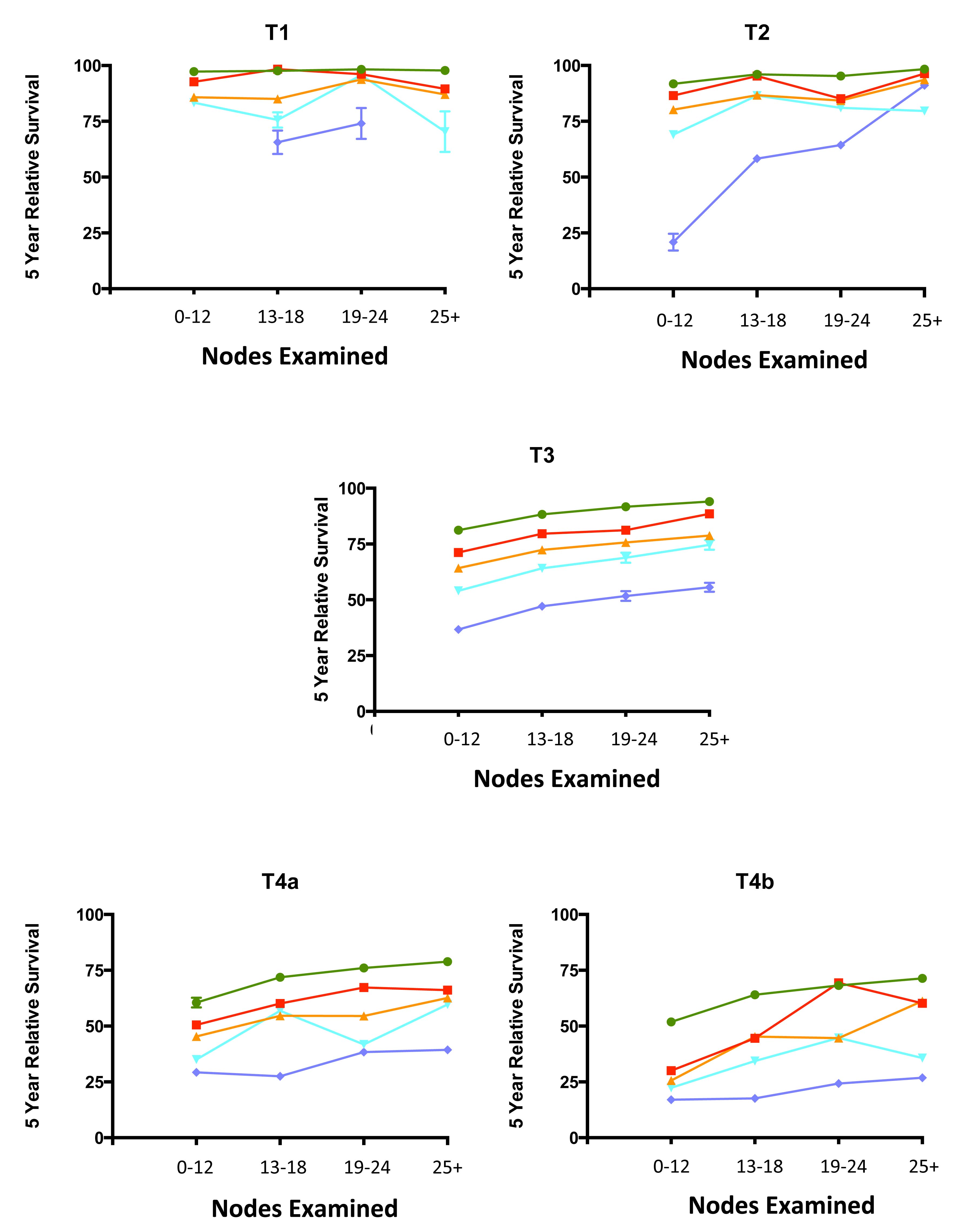
Regional lymph nodes are classified as N1 or N2 according to the number involved by metastatic tumor. Involvement of one to three nodes by metastasis is pN1; involvement of four or more nodes by tumor metastasis is pN2. The number of nodes involved with metastasis influences outcome in both the N1 and N2 groups (Figures 20.5 and 20.6). pN1 is subdivided further into pN1a (metastasis in one regional lymph node) and pN1b (metastasis in two or three regional lymph nodes), and pN2 is subdivided into pN2a (metastasis in four to six regional lymph nodes) and pN2b (metastasis in seven or more regional lymph nodes), because each subgroup represents roughly half the population of N1 or N2, and the subgroups with fewer positive nodes have better survival than those with more positive nodes within the N1 and N2 categories16,17 (Figures 20.6 and 20.7). Lymph nodes outside the regional drainage area of the primary tumor should be categorized as distant metastases; for example, external iliac or common iliac node involvement in a rectosigmoid carcinoma would be M1a.

Controversy exists regarding whether isolated tumor cells or micrometastases in regional nodes are prognostically important. The literature is contradictory because some authors have defined isolated tumor cells as not only individual tumor cells in the subcapsular or marginal sinus but clumps of up to 20 tumor cells.27Micrometastases have been defined as clusters of 10 to 20 tumor cells or clumps of tumor on cut section that measure >=0.2 mm in diameter. These cell clusters indicate that tumor cells have entered a node and replicated and are not merely isolated dormant cells. A recent meta-analysis28 demonstrated that micrometastases, defined as clusters of tumor cells greater than 0.2 mm in diameter, are a significant poor prognostic factor. Lymph nodes harboring such clusters therefore should be considered positive. Although these micrometastases may be designated as N1mi, it may be better to consider these as stand ard positive nodes with the corresponding number, as pathologists likely have considered these to be positive nodes in the past.

In a recent multicenter prospective trial, nearly 200 patients with Stage I or II disease had negative nodes based on stand ard hematoxylin and eosin (H&E) staining but were found to have tumor cells <0.2 mm in diameter that stained positively with a pan-keratin antibody (N0i+). These patients had a 10% decrease in overall survival compared with patients with N0 disease.29 This decrease in survival occurred in patients with T3-T4 primary tumors but not in those with T1 or T2 primary tumors.29 Further research into N0(i+) detected by pan-keratin staining in otherwise negative H&E-stained lymph nodes may be warranted, especially in patients with T3 or T4 primary carcinomas.

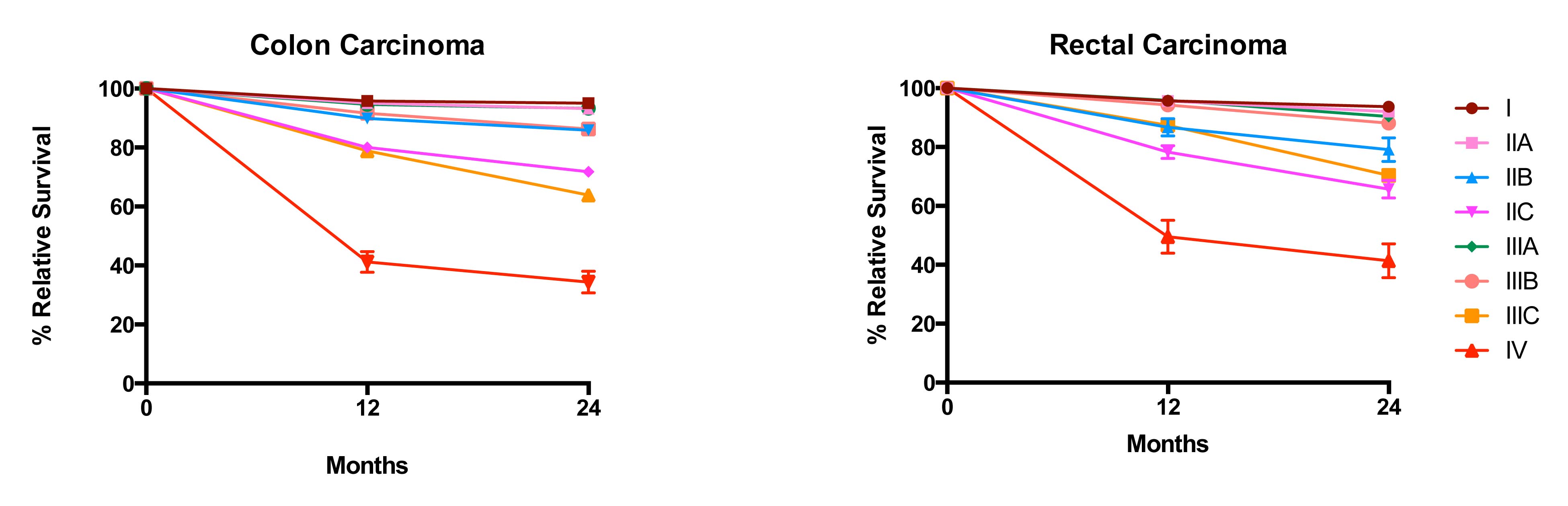
20.6 Impact of positive nodes, the total number of nodes examined, and the depth of primary tumor invasion in a population-based cohort of rectal carcinoma. Data from 30,202 patients diagnosed with rectal carcinoma between 2004 and 2010 and included in the population-based Surveillance, Epidemiology, and End Results (SEER) database were analyzed for 5-year relative survival. Each T category is presented separately. The N categories are color coded as follows: green, N0 (all nodes negative); red, N1a (one positive node); orange, N1b (two or three positive nodes); light blue, N2a (four to six positive nodes); and dark blue, N2b (seven or more positive nodes). The N1c category is not represented because there is not yet a mature cohort in SEER with 5-year relative survival. The lines for N category levels spread across the numbers of nodes examined. All patients were free of clinical metastases by surgical and clinical staging; that is, they were in stage groups I to III. Results are mean ± SEM of 5-year relative survival except for T4a and T4b, for which the number of patients is small and the error is large. The results suggest that the examination of more nodes for a given N category is associated with increasing survival. N0(i+) and N0mi are not included because they were not recorded previously for colorectal carcinoma.

20.7 Impact of positive nodes, the total number of nodes examined, and the depth of primary tumor invasion in a population-based cohort of colon carcinoma. Data from 144,744 patients diagnosed with colon carcinoma between 2004 and 2011 and included in the population-based SEER database were analyzed for 5-year relative survival. Each T category is presented separately. The N categories are color-coded as follows: green, N0 (all nodes negative); red, N1a (one positive node); orange, N1b (two or three positive nodes); light blue, N2a (four to six positive nodes); and dark blue, N2b (seven or more positive nodes). N1c category is not represented because there is not yet a mature cohort in SEER with 5-year relative survival. The lines for N category levels spread across the numbers of nodes examined. All patients were free of clinical metastases by surgical and clinical staging; that is, they were in stage groups I to III. Results are mean ± SEM of 5-year relative survival. Results suggest that the examination of more nodes for a given N category is associated with increasing survival. N0(i+) and N0mi are not included because they were not recorded previously for colorectal carcinoma.

N1c—Tumor Deposits

Tumor deposits are defined as discrete tumor nodules within the lymph drainage area of the primary carcinoma without identifiable lymph node tissue or identifiable vascular or neural structure. The shape, contour, and size of the deposit are not considered in these designations.

If the vessel wall or its remnant is identifiable on H&E, elastin, or any other stain, the lesion should be classified as lymphovascular invasion (LVI) present (a CAP-required data element). Further documentation should subclassify LVI as small vessel invasion (“L” positive for either lymphatic or small venule involvement) or venous invasion (“V” positive for tumor within an endothelial-lined space that contains red cells or is surrounded by smooth muscle [adapted from the RCPath Colorectal Cancer Dataset 2014]).30 These definitions are similar to those for large vessel invasion on page 122 of the AJCC Cancer Staging Manual, 6th Edition,14 and LVI is a new data item to be collected. If neural structures are identifiable, the lesion should be classified as perineural invasion.

One to four individual tumor deposits or five or more deposits without involvement of lymphatic, venous, or neural structures within the lymph drainage area of the primary carcinoma should be recorded. In the evaluation of tumors pretreated with radiation and /or chemotherapy, it is important for the pathologist to assess whether tumor nodules represent tumor deposits as defined earlier or discontinuous eradication of the original tumor so that he or she can record the appropriate ypT and ypN categories.

As reported by Quirke, Nagtagaal, and others,31-35 tumor deposits are associated with poor overall survival. A recent population-based study36 demonstrated that approximately 10% of primary colon or rectal carcinomas have tumor deposits and that 2.5% of colon and 3.3% of rectal cases have tumor deposits with otherwise histologically negative lymph nodes. The strength of tumor deposits as a negative prognostic factor in the absence of any nodal metastases resulted in the introduction of the N1c category.

In cases with tumor deposits but no identified lymph node metastases, the N1c category is used and is applicable to all T categories. The presence of tumor deposits does not change the primary tumor T category, but does change the node status (N) to N1c if all regional lymph nodes are pathologically negative. The number of tumor deposits is notadded to the number of positive regional lymph nodes if one or more lymph nodes contain cancer.

Metastasis

Metastasis to only one site/solid organ (e.g., liver, lung, ovaries, nonregional lymph node) should be recorded as M1a. Multiple metastases within only one organ, even if the organ is paired (e.g., the ovaries or lungs), is still M1a disease. Metastases to multiple sites or solid organs distant from the primary site is M1b, excluding peritoneal carcinomatosis. Peritoneal carcinomatosis with or without blood-borne metastasis to visceral organs is designated as M1c, because recent studies suggest that this occurs in 1-4% of patients37,38 and that the prognosis for peritoneal disease is worse than that for visceral metastases to one or more solid organs.39,40 The pathologist should not assign pM0 because M0 is a global designation referring to the absence of detectable metastasis anywhere in the body.

Anastomotic Recurrence

If the tumor recurs at the site of surgery, it is anatomically assigned to the proximal segment of the anastomosis (unless that segment is the small intestine, in which case the colonic or rectal segment should be designated as appropriate) and restaged by the TNM classification. The rprefix is used for the recurrent tumor stage (rTNM).

Colorectal Carcinoma Found at Death

The aprefix is used for cancer discovered as an incidental finding during autopsy and not suspected before death.

Molecular Advances That Will Lead to Future Markers and Therapies

In the past few years, there has been an explosion in the understand ing of the molecular pathology of colorectal carcinoma. The Cancer Genome Atlas (TCGA) project provided a reference against which alterations that occur in cancer may be compared. The TCGA study in primary colorectal carcinomas41 identified several different pathways that may lead to colorectal carcinoma, including chromosomal instability characterized by truncating mutations in the APCgene; MSI due to loss of function by somatic mutation or promoter hypermethylation of at least one of the DNA MMR genes (MLH1, MSH2, MSH6, PMS2) with or without POLE mutations and /or the presence of hypermutation; and the CpG island methylator phenotype (CIMP) pathway caused by epigenetic alterations. The various pathways provide several molecular alterations that will create not only prognostic and predictive markers but also opportunities for therapy to be developed in coming years. As stated, hypermutated carcinomas show frequent deficiency in DNA MMR as well as specific mutations associated with this subset.42 Their high rate of somatic mutation may form neoantigens that may induce an antitumor immune response, leading to a better prognosis.43 The emergence of the Immunoscore project, which quantitates host immune infiltrating cells in colorectal carcinoma,44 may be stand ardized in the future. Currently, however, its lack of stand ardization prevents it from being added to TNM staging.

Presently, the only major molecular alterations validated as significant markers with level I evidence are high levels of MSI/defective MMR (MSH-H/dMMR; good prognosis) and mutation in the BRAF gene (poor prognosis). Predictive markers are mutations in KRAS, the related NRAS, and BRAF genes that cause resistance to therapies using monoclonal antibodies to epidermal growth factor receptor (EGFR)45 and possibly to vascular endothelial growth factor A (VEGFA)46 in advanced colorectal carcinoma. The prognostic effects of mutations in the RAS genes appear to be small; this is discussed further in the site-specific factor section. Mutations in the gene PIK3CA47 also may be prognostic and may predict lack of response to therapy against EGFR in advanced colorectal carcinoma, but their level of evidence currently is low.

Prognostic Factors

Prognostic Factors Required for Stage Grouping

Beyond the factors used to assign T, N, or M categories, no additional prognostic factors are required for stage grouping.

Additional Factors Recommended for Clinical Care

Factors Important to Consider in Making Decisions about Treatment

Eight prognostic factors now are judged to be clinically significant in colorectal carcinoma and should be considered when physicians and patients are deciding on what treatments to use. These factors, which have varying degrees of usefulness depending on disease stage, are as follows:

  1. Serum CEA levels in patients who are to undergo surgery for potential cure (e.g., patients with Stage I-III colorectal carcinoma or Stage IV patients undergoing metastectomy) and changes in CEA as a response marker during chemotherapy for Stage IV disease. Data must be recorded as XXXX.X ng/mL
  2. Tumor regression score in rectal carcinoma, which quantitates the pathological response to neoadjuvant therapy (similar to the pathological complete response measurement in breast cancer, which is FDA approved as a marker for drug development)
  3. Circumferential resection margin (CRM), measured in millimeters from the edge of the tumor to the nearest dissected margin of the surgical resection in rectal cancer and retroperitoneal regions of the colon
  4. Lymphovascular invasion (LVI) in all colorectal specimens, to identify small vessel or venous invasion
  5. Perineural invasion (PNI), which provides histologic evidence of invasion of nerves or perineural spaces by the primary tumor and may have a negative prognostic impact similar to that of LVI
  6. Microsatellite instability (MSI), which not only is a prognostic factor but also predicts lack of response to 5-fluorouracil (5-FU) chemotherapy
  7. KRAS and NRAS mutation status
  8. BRAF mutation, which along with KRAS and NRAS mutation is important because mutation in these genes is associated with lack of response to treatment with monoclonal antibodies directed against the epidermal growth factor receptor (EGFR) in patients with Stage IV colorectal carcinoma

Tumor Deposits

A tumor deposit is a discrete nodule of cancer in pericolic/perirectal fat or in adjacent mesentery (mesocolic or rectal fat) within the lymph drainage area of the primary carcinoma, without identifiable lymph node tissue or identifiable vascular structure. If the vessel wall or its remnant is identifiable on H&E, elastic, or any other stain, it should be classified as vascular (venous) invasion. Similarly, if neural structures are identifiable, the lesion should be classified as perineural invasion.

If tumor deposits are present in the absence of any regional nodes involved with carcinoma, then the nodal classification is N1c, regardless of T category, and the tumor deposits should be recorded on the staging form. Evidence indicates that tumor deposits are equivalent to positive nodes as a negative prognostic factor and that adjuvant therapy is warranted in patients whose stage group would otherwise be I or II. The level of evidence supporting this marker is I.1,3

Tumor deposits are identified during pathological review of the resection specimen, as described in Pathological Classification. Venous invasion (V+) with lymphatic invasion (L+) or perineural invasion or spread (PNI) should be separated from tumor deposits based on histologic review and should not be categorized as N1c. If tumor deposits are observed in lesions that otherwise would be classified as pN0, then the primary tumor classification is not changed, but the nodule is recorded as a tumor deposit and categorized as N1c. The number of tumor deposits should be recorded on the staging form as one to four individual tumor deposits or five or more tumor deposits. If preoperative or neoadjuvant therapy has been administered, it is important to consider whether the potential tumor nodule near a partially responding tumor represents residual primary carcinoma or a true tumor deposit. Nagtegaal and Quirke32 provide guidance in evaluating potential tumor deposits after preoperative therapy. Currently, the level of evidence for tumor deposits as a factor is II, but it must be included in data collection because it is required for staging node-negative carcinoma of the colon or rectum.

AJCC Level of Evidence: I, II

Serum CEA Levels

Carcinoembryonic antigen (CEA), now known as CEACAM5, belongs to a 35-member family of related molecules that are part of the immunoglobulin supergene family.48 Produced by epithelial cells that line the gut,49 this 185-kDa glycoprotein is produced by almost all adenocarcinomas from all sites, as well as by many squamous cell carcinomas of the lung and other sites. CEA is cleared from the blood only by cells in the liver and lung50; therefore, it is an important marker for the presence of subclinical hepatic or pulmonary metastases, even before such lesions are detectable by current imaging modalities.51 Although CEA has been considered a glycoprotein without specific function, research shows that it can promote metastasis in human xenograft models52 through increased cell adhesion,53-55 induction of cytokines that promote cancer cell survival,56 inhibition of inflammatory responses,57 and inhibition of programmed cell death, or apoptosis.58,59 These functions of CEA are important for its ability to cause treatment resistance. The level of evidence supporting CEA as a prognostic marker is I.1,51

The ability of CEA to increase cell-cell adhesion, decrease reactive oxygen and nitrogen radical formation, and block apoptosis makes it a significant molecule for metastasis and response to therapy.

CEA levels may be measured in blood, plasma, or serum, primarily by an enzyme-linked immunosorbent assay (ELISA)-based method in a laboratory accredited by the Clinical Laboratory Improvement Amendments (CLIA). It is recommended that a preoperative level be obtained before potentially curative resection surgery (e.g., for clinical Stage I-III disease), then every 3 to 6 months for 2 years and annually thereafter until 5 years after first treatment. CEA levels also may be used as a response marker for treatment of Stage IV disease when measured at monthly intervals.

AJCC Level of Evidence: I

Tumor Regression Score

The pathological response to preoperative radiotherapy (rectal cancer), chemoradiation (rectal cancer), or chemotherapy (colon or rectal cancer) is an important prognostic factor. This response is assessed by the pathologist and is reported as a score based on resection specimens from patients who have undergone preoperative radiotherapy and /or chemotherapy.

Strong observational evidence exists that complete eradication of the tumor, as detected by pathological examination of the resected specimen, is associated with a better prognosis. Moreover, the degree of response appears to be associated with the degree of improvement in prognosis. Patients with minimal or no residual disease after therapy have a better prognosis than those with gross residual disease. Failure of the tumor to respond to neoadjuvant treatment is an adverse prognostic factor. Parallel associations are observed in breast and pancreatic carcinomas. The FDA has approved the use of complete pathological response to neoadjuvant therapy as such a strong positive prognostic factor in breast cancer that it may be used as an end point for evaluating drug responses. In addition, similar measures of tumor response to therapy have similar prognostic impacts, including minimal residual disease in leukemias after induction therapy and necrosis after neoadjuvant treatment of osteosarcomas. The level of evidence is II.

The pathologist must evaluate and record the resection specimen according to the CAP guidelines for recording tumor regression (see CAP's “Protocol for the Examination of Specimens from Patients with Carcinomas of the Colon and Rectum”11,12). Neoadjuvant chemoradiation in rectal cancer is often associated with significant tumor response, including the potential for sterilization of involved mesorectal lymph nodes. Therefore, specimens from patients receiving neoadjuvant chemoradiation should be examined thoroughly at the primary tumor site, in regional nodes, and for peritumoral satellite nodules or deposits in the remainder of the specimen. Sterilizing the primary tumor site does not assure sterilization of the regional lymph nodes. Although several different scoring systems for tumor regression have been advocated, a four-point tumor regression score is used to assess response; this system is similar to that of Ryan et al.,11,12,60 except the score for complete absence of viable tumor is recorded as 0 (Table 20.2).

Acellular pools of mucin in specimens from patients receiving neoadjuvant therapy are considered to represent completely eradicated tumor and are not used to assign pT category or counted as positive lymph nodes.

20.2 Modified Ryan scheme for tumor regression score. Adapted from Ryan et al11,12,60with permission.

DescriptionTumor regression score
No viable cancer cells (complete response)0
Single cells or rare small groups of cancer cells (near-complete response)1
Residual cancer with evident tumor regression, but more than single cells or rare small groups of cancer cells (partial response)2
Extensive residual cancer with no evident tumor regression (poor or no response)3
AJCC Level of Evidence: II

Circumferential Resection Margins (CRM)

The CRM is the distance in millimeters between the deepest point of tumor invasion in the primary cancer and the margin of resection in the retroperitoneum or mesentery (Figure 20.8). The longer this distance is, the better the prognosis for rectal carcinomas and for colon carcinomas arising in areas of the colon that partially lack a peritoneal lining. The CRM is produced by surgical resection of pericolic or perirectal fibroadipose tissue or pelvic structures. The term does not apply to the anatomic serosa of the colon or rectum that is peritonealized.

Strong evidence exists that CRM status is one of the most important determinants of local control in colon and rectal cancers.61 Distance to the CRM is an important prognostic indicator, and tumors with a CRM distance of 0 to 1 mm are at high risk for recurrence. In addition, involvement of the CRM is associated with decreased survival. If the CRM distance is 1 cm or more, the chance of local recurrence is significantly reduced and survival is improved. AJCC Level of Evidence: I

The circumferential surface of surgical resection specimens from the ascending colon, descending colon, or upper rectum is only partially peritonealized, and the demarcation between the peritonealized surface and the nonperitonealized surface (corresponding to the CRM) of such specimens is not always easily appreciated on pathological examination. The nonperitonealized resection margin may occur on an adjacent structure beyond the fascia propria that has been resected en bloc to achieve a clear CRM. Therefore, the surgeon is encouraged to mark the peritoneal reflection and /or the area of deepest tumor penetration adjacent to a nonperitonealized surface with a clip or suture or to specifically orient the specimen for the pathologist so that the CRM may be identified and evaluated accurately.

For mid- and distal rectal cancers (below the peritoneal reflection), the entire surface of the resection specimen (anterior, posterior, medial, and lateral) is a CRM. For proximal rectal or retroperitoneal colon cancers (ascending and descending colon and cecum), surgically dissected margins include those that lie in a retroperitoneal or subperitoneal location (Figure 20.8). For segments of the colon covered completely by visceral peritoneum (transverse, sigmoid, sometimes cecum), the only radial margin dissected surgically is the mesenteric margin, unless the cancer adheres to or invades an adjacent organ or structure. Therefore, for cancers of the cecum, transverse colon, or sigmoid colon that extend to the cut edge of the mesentery, assignment of a positive CRM is appropriate.

For rectal cancer, the quality of the surgical technique is a key factor in the success of surgical outcomes relative to local recurrence and possibly long-term survival. Numerous nonrand omized studies demonstrated that total mesorectal excision (TME) with adequate surgical clearance around the penetrating edge of the tumor decreases the rate of local relapse. The TME technique entails precise sharp dissection within the areolar plane of loose connective tissue outside (lateral to) the visceral mesorectal fascia in order to remove the rectum. With this approach, all mesorectal soft tissues encasing the rectum, including the mesentery and all mesorectal and mesenteric lymph nodes, are removed en bloc. Thus, the circumferential surface (CRM) of TME resection specimens is the mesorectal fascia. The CRM of extended resections beyond the mesorectal fascial plane corresponds to the resection plane of the extended resection. Rectal resection performed by less precise techniques may be associated with incomplete excision of the mesorectum.

It is critical that the analysis of the surgical specimen follow the CAP guidelines that refer to examination of the TME specimen. In addition, it is essential that the distance between the closest leading edge of the tumor and the CRM (known as the surgical clearance) be measured pathologically and recorded in millimeters in the CRM field on the staging form. A margin greater than 1 mm is considered a negative margin. Because surgical clearance of 1 mm or less is associated with a significantly increased risk of local recurrence, margins less than 1 mm should be classified as positive61 (Figure 20.8).

20.8 Depiction of T4a lesions and the importance of the circumferential margin.

AJCC Level of Evidence: I

Lymphovascular Invasion (LVI)

Invasion of either small or large vessels by the primary tumor is an important poor prognostic factor. Small vessel invasion is involvement by tumor of thin-walled structures lined by endothelium, without an identifiable smooth muscle layer or elastic lamina. These thin-walled structures include lymphatics, capillaries, and postcapillary venules. Large vessel invasion is defined by tumor involving endothelium-lined spaces that have an elastic lamina and /or smooth muscle layer. Circumscribed tumor nodules surrounded by an elastic lamina on H&E or elastic stain also are considered venous invasion and may be extramural (beyond the muscularis propria) or intramural (submucosa or muscularis propria).

LVI has been recognized as a category I factor since 1999 (summarized with recommendation by Compton et al.62). Although formal meta-analyses have not been performed, the depth of data in the literature indicates that involvement of either small or large vessels is a significant sign of poor prognosis. As such, it should be considered a prognostic factor with level I evidence. Small vessel invasion is associated with lymph node metastasis, and several studies found it to be an independent indicator of adverse outcome.63-65 Multivariate analysis demonstrated that extramural venous invasion was an independent adverse prognostic factor in multiple studies and is a risk factor for liver metastasis.66-68 The significance of intramural venous invasion is less clear.

Analysis of resected specimens and indications for using special stains should follow CAP guidelines.11,12

AJCC Level of Evidence: I

Perineural Invasion (PNI)

Invasion of the nerves within or adjacent to the primary tumor by colorectal carcinoma is a negative prognostic factor that may be as important as invasion of lymphatics or blood vessels. However, it often is overlooked and may be present in as many as 20% of primary colonic or rectal carcinomas.69

Carcinoma invasion of peripheral nerves, including perineural spaces within the regional drainage area of the primary tumor, is an adverse prognostic factor, as identified in multiple institutional studies,69-71 and indicates an especially aggressive carcinoma. The level of evidence for this factor is I.

If present, PNI usually is apparent on stand ard H&E staining of formalin-fixed tissues. Its presence in any field is sufficient to warrant a positive or present designation on a pathology report, and this should be so designated on the staging form.

AJCC Level of Evidence: I

Microsatellite Instability (MSI)

One form of genetic instability is manifested by changes in the length of repeated single- to six-nucleotide sequences (known as DNA microsatellite sequences), which are caused by a functional defect in DNA MMR.72 High levels of MSI (MSI-H) occur in about 15% of colorectal carcinomas and are associated with right-sided colon carcinomas, frequently with poorly differentiated and mucinous histology but good prognosis.73 This MSI is a hallmark of hereditary nonpolyposis colorectal carcinoma (HNPCC), or Lynch syndrome.74 The vast majority of MSI tumors are sporadic because of epigenetic inactivation of the MLH1 gene, whereas the others occur in patients with a mutation in a DNA MMR gene. Frequently, the mutation in the latter group is in the germline and confers Lynch syndrome. Thus, new patients with MSI tumors should be screened for Lynch syndrome in accordance with current NCCN guidelines.1,3

MSI-H is important because it not only is a good prognostic factor, but it also predicts a poor response to 5-FU chemotherapy.75 However, the addition of oxaliplatin (in the FOLFOX regimens) negates the adverse effects of MSI-H.76,77 Recent data suggest that mutation in BRAF is associated with MSI-H tumors and that colon carcinomas with both gene alterations have a significantly, although modestly, worse prognosis in Stage III and IV colon carcinomas.78 MSI-H also is associated with Lynch syndrome/ HNPCC.79

MSI-H is identified by DNA polymerase chain reaction (PCR) amplification of known DNA microsatellites to show that the length of the microsatellites is greater in tumor than in normal colon. In addition, immunohistochemistry may be used to detect loss of expression of DNA MMR proteins, including MLH1, MSH2, MSH6, and PMS2. The most common cause of MSI-H is sporadic nonfamilial loss of expression of MLH1 due to somatic promoter hypermethylation. MSI-H results in hypermutated colorectal cancers with large numbers of mutated genes, especially in those that contain microsatellites.

The NCCN3,4 and Spanish Society of Pathology80 recommend testing for MSI in patients younger than 70 years, especially those with high-grade right-sided colon carcinomas, mucinous histology, or Crohn's disease-like peritumoral lymphoid follicles, which are features of MSI-H cancers. Alternatively, several reports indicate that the sensitivity and specificity of identifying MSI-H tumors increases if all tumors are analyzed,81 as opposed to following either the Bethesda guidelines82 or the MSI-like morphology described here. All these considerations make MSI a level I factor to be collected.

AJCC Level of Evidence: I

KRAS and NRAS Mutation

KRAS and NRAS are important signaling intermediates in the growth receptor pathway, which controls cell proliferation and survival. These genes are activated when EGFR binds EGF or similar growth factors and then activate the RAF or PIK3CA proteins. Both KRAS and NRAS may be constitutively activated through mutation during colorectal carcinogenesis so that they continuously stimulate cell proliferation and prevent cell death. Activating mutations are most likely to occur in codons 12 and 13, but codons 61, 146, and , less frequently, other codons in KRAS also may be mutated.47 Similarly, codons in NRAS may have activating mutations. KRAS may be activated by somatic mutation in up to 40% of colorectal carcinomas and NRAS in about 7%.47 Activation of either RAS gene is a modestly poor prognostic factor in Stage III and IV disease.45,83,84 More importantly, RAS activation predicts a poor response to monoclonal anti-EGFR antibody therapy in advanced colorectal carcinoma.45 KRAS mutation also may predict a poor response to anti-VEGF therapy in advanced colorectal carcinoma.46 The level of evidence for the poor predictive effect of KRAS mutation is I in advanced colorectal carcinoma and II as a prognostic factor in Stage II-IV disease. The level of evidence for NRAS mutation as a prognostic factor currently is II.

FDA-approved kits now are available for detecting KRAS mutations. There is no consensus regarding the level of mutation necessary to suspect a tumor of containing an activated RAS gene; however, the level often is considered to be around 5% of the amount of KRAS gene expression, as detected by PCR or a sequencing method.

AJCC Level of Evidence: II

BRAF Mutation

The BRAF oncoprotein is a serine-threonine kinase that transmits cell growth and proliferation signals from KRAS or NRAS to other enzymes, leading to cell proliferation and growth. An activating point mutation at BRAF V600E may be detected in 6-10% of colorectal carcinomas,41 which constitutively stimulate these other enzymes to promote continuous cell growth. This stimulation abrogates the ability of EGFR inhibitors to block cell proliferation and growth.

Multiple studies in Stage IV colorectal cancers and more recent data in Stage III disease indicate that the BRAF V600E mutation is associated with significantly worse prognosis, including survival after tumor recurrence.85 MSI status and BRAF mutation are prognostic factors that interact significantly. Although BRAF mutation is associated with poor prognosis, the presence of MSI may attenuate its adverse impact. MSI is an established good prognostic factor.73-76 The concurrent presence of a BRAF mutation may attenuate survival slightly.85 MSI without BRAF is a good prognostic factor, whereas MSI-H with BRAF mutation portends slightly worse survival.83,84 Both patient groups have better survival than those with microsatellite stability (MSS) without BRAF mutation, who in turn have better survival than those with MSS tumors with BRAF mutation.83-87 BRAF V600E mutation in colorectal carcinomas blocks the effect of anti-EGFR antibodies on disease progression in Stage IV colorectal carcinoma.50 The level of evidence for these effects of BRAF is I (meta-analysis) for blocking the effect of anti-EGFR antibody therapy and II for the prognostic effect on survival.

BRAF V600E mutation may be determined by an assay approved by the FDA for detecting the mutation in melanoma. In addition, laboratory-developed tests that involve stand ard genotyping or next-generation sequencing may be used to measure the level of this mutation in colorectal carcinoma specimens. As with other somatic mutations, the cutoff or threshold for assigning a positive result is under consideration, but a common stand ard is that 5% of the alleles or gene expression should be mutated for a test to be considered positive. Immunohistochemistry for mutated BRAF V600E protein is not recommended for use in colorectal carcinomas, because it is not as sensitive and concordant with genomic sequencing as it is in melanomas.88

AJCC Level of Evidence: I, II

Immunoscore and Immune Response Markers

Galon et al.89,90 reported that measuring the lymphoid infiltrate of tumors and comparing the numbers of CD4+ and CD8+ T cells and other lymphoid subsets in the advancing edge and center of the tumor was strongly prognostic in colon cancers and stronger than TNM staging. Limitations of this provocative work, however, include failure to identify the MSI subset and the use of retrospective case series. Importantly, a consortium is seeking to stand ardize this immunohistochemical analysis using primary colorectal carcinomas.91,92 These results are forthcoming and will stand ardized within the next few years; if this method is validated and it outperforms MSI, it has the potential for inclusion in TNM.

Other important immune response markers are the immune checkpoint proteins and inhibitors PD-1 and PD-L1, CTLA4, and IDO. These molecules also are targets for specific therapies. Recent data suggest that blocking these checkpoint inhibitors may enhance endogenous immune responses to cancer, leading to enhanced survival.90 Finally, it is unclear what role cytokines and other soluble factors released by host cells as well as tumors have in the tumor-host relationship. Current research may well identify a few as being sufficiently critical to be modifiers of the TNM stage groups.

AJCC Level of Evidence: Not identified

Gene Expression Alterations

While risk assessment tools may be useful, current medical decision-making is increasingly being placed on the molecular variants that drive cancers, gene expression alterations caused by those variants or the activation of specific pathways within a patient's cancer since all of these may be targets for therapy. It will take time for the utility of these molecular classifiers in colorectal carcinoma to reach the level of evidence necessary for FDA approval or for inclusion within guidelines but healthcare professionals should know that a 12-gene signature is now at least level II evidence as a prognostic factor in stage II-III colon or rectal carcinoma.93-96

AJCC Level of Evidence: II

Risk Assessment

Risk Assesment Models

Prognostic models will continue to play an important role in 21st century medicine for several reasons.97 First, by identifying which factors predict outcomes, clinicians gain insight into the biology and natural history of the disease. Second, treatment strategies may be optimized based on the outcome risks of the individual patient. Third, because of the heterogeneity of disease in most cancers, prognostic models will play a critical role in the design, conduct, and analysis of clinical trials in oncology.97 If developed and validated appropriately, these models will become part of routine patient care, decision making, and trial design and conduct.

The AJCC Precision Medicine Core (PMC) developed and published criteria for critical evaluation of prognostic tool quality,98 which are presented and discussed in Chapter 4. Although developed independently by the PMC, the AJCC quality criteria corresponded fully to the recently developed Cochrane CHARMS tool for critical appraisal in systematic reviews of prediction modeling studies.99

Existing prognostic models for colon and rectum cancer meeting all the AJCC inclusion/exclusion criteria and meriting AJCC endorsement are presented in this section. A full list of the evaluated models and their adherence to the quality criteria is available at www.cancerstaging.org.

The PMC performed a systematic search of published literature for prognostic models/tools in colon and rectum cancer from January 2011 to December 2015. The search strategy is provided in Chapter 4. The PMC defined prognostic model as a multivariable model in which factors predict a clinical outcome that will occur in the future. Each tool identified was compared against the quality criteria developed by the PMC as guidelines for AJCC recommendation for prognostication models (see Chapter 4).

Twenty-nine prognostication tools100-128 for colon or colorectal cancer were identified: 14 for patients with resected liver metastases,104-107,110,113-118,124,128 two for patients with unresectable liver metastases,100,122 four in the adjuvant (Stage I/II/III) setting,102,120,126,127 seven for patients with metastatic disease,101,103,109,111,112,119,121 one for patients with resected pulmonary metastases,108 one for patients with locally advanced rectal cancer,125 and one across all disease stages.123

Of the 14 models for patients with resected liver metastases, none met all the predefined criteria. Most were excluded because their development used single-institution series lacking sufficient external validation,105,106,110,114-117 made predictions generated from data not reflecting current clinical stand ards of treatment,104,107,108,113,118,128 or lacked external validation.110,124Both tools for patients with unresectable liver metastases were excluded because they were from single-institution series and were too small to be reliably generalizable.100,122 Of the tools for patients with metastatic disease, Elias et al. (2014)103 lacked external validation; Peng et al. (2014)119 and Shitara et al. (2011)121 were from a single institution or the patient set was too small to be generalizable; and Chibaudel et al. (2011),101 Kato et al. (2005),109 Kobayashi et al. (2013),111 and Kohne et al. (2002)112 were felt to be based on datasets not reflective of current treatment paradigms for patients with metastatic disease (although the Kohne tool112 was of very high quality).

Among the four models for patients in the adjuvant setting, two met all inclusion criteria.120,126 We note that the Numeracy model102 was excluded because it was replaced by ACCENT.120 Weiser et al. (2008)127 was excluded because it predicted only recurrence, although it met all other criteria. The model in Stojadinovic et al. (2013)123 was considered very promising; however, it lacked sufficient detail for it to be implemented in practice. The final model, which predicted outcomes in patients with locally advanced rectal cancer,110 met all criteria and is endorsed by the committee for this somewhat limited treatment setting.

Twenty-nine models for prognostication in colon or colorectal cancer were identified, but only three models, two for adjuvant disease120,126 and one for local advanced rectal cancer,125 met all predefined AJCC inclusion and exclusion criteria and therefore are endorsed by the AJCC.120,125,126 Table 20.3 presents the models meeting the AJCC quality criteria. The two models in the adjuvant setting were developed using very different datasets. Renfro et al. (2014)120 was based on a large collection of completed rand omized clinical trials, whereas Weiser et al. (2011)126 was built using SEER data. However, both models were externally validated. The third endorsed tool, by Valentini et al. (2011)125 in locally advanced rectal cancer, also was developed using data from completed clinical trials.

In the interest of precision medicine and informed individualized care for patients, the AJCC supports the appropriate use of both high-value patient classifiers (prognostic factors) and prognostication tools (risk calculators). Both are valuable. Prognostication tools (i.e., risk calculators) provide individualized probability estimates, whereas patient classifiers group patients into ordered risk strata (either directly or based on cut-points for individual probability estimates). The TNM staging system is an example of such a classification tool, yielding at the least granular level ordered classes (I, II, III, IV) of increasingly poor prognosis. Strata based on prognostic factors (e.g., a gene signature) are other examples.

While such stratification is useful, it also limited by the number of categories that are manageable, by the complexity of combining information from multiple predictors to form discrete ordered categories in a transparent manner, and by the inherent variability of prognosis of patients in a given risk class. Risk calculators, in contrast, are designed to deliver a more precise estimate of outcome for an individual patient through computational integration of a variety of patient-specific data elements.

Prognostic tools for colon and rectum cancer meeting all AJCC quality criteria
Approved Prognostic ToolWeb AddressFactors Included in the Model
ACCENT-based web calculators to predict recurrence and overall survival in Stage III colon cancer120http://www.mayoclinic.org/medical-professionals/cancer-prediction-tools/colon-cancerAge, sex, race, BMI, performance status (PS), T category, lymph node ratio, grade, treatment group, location
Predicting survival after curative colectomy for cancer: indvidualizing colon cancer staging126https://www.mskcc.org/nomograms/colorectal/overall-survival-probabilityT category, N category, age, sex, tumor differentiation/grade, number of regional lymph nodes evaluated, number of regional lymph nodes positive
Nomograms predicting local recurrence, distant metastases, and overall survival for patients with locally advanced rectal cancer on the basis of European rand omized clinical trials125http://www.predictcancer.org/Main.php?page=RectumFollowUpModelAge, sex, T category, radiotherapy dose, concomitant chemotherapy, surgical procedure, pT category, pN category, adjuvant chemotherapy

Recommendations

The Colorectal Committee of the Lower GI Expert Panel recommends that all T, N, and M categories, as well as the factors required and recommended for collection, be included as appropriate for clinical trials.

TNM Definitions

Clinical T (cT)

cT CategorycT Criteria
cTXPrimary tumor cannot be assessed
cT0No evidence of primary tumor
cTisCarcinoma in situ, intramucosal carcinoma (involvement of lamina propria with no extension through muscularis mucosae)
cT1Tumor invades the submucosa (through the muscularis mucosa but not into the muscularis propria)
cT2Tumor invades the muscularis propria
cT3Tumor invades through the muscularis propria into pericolorectal tissues
cT4Tumor invades the visceral peritoneum or invades or adheres to adjacent organ or structure
cT4aTumor invades through the visceral peritoneum (including gross perforation of the bowel through tumor and continuous invasion of tumor through areas of inflammation to the surface of the visceral peritoneum)
cT4bTumor directly invades or adheres to adjacent organs or structures

Direct invasion in T4 includes invasion of other organs or other segments of the colorectum as a result of direct extension through the serosa, as confirmed on microscopic examination (for example, invasion of the sigmoid colon by a carcinoma of the cecum) or, for cancers in a retroperitoneal or subperitoneal location, direct invasion of other organs or structures by virtue of extension beyond the muscularis propria (i.e., respectively, a tumor on the posterior wall of the descending colon invading the left kidney or lateral abdominal wall; or a mid or distal rectal cancer with invasion of prostate, seminal vesicles, cervix, or vagina).

Tumor that is adherent to other organs or structures, grossly, is classified cT4b. However, if no tumor is present in the adhesion, microscopically, the classification should be pT1-4a depending on the anatomical depth of wall invasion. The V and L classification should be used to identify the presence or absence of vascular or lymphatic invasion whereas the PN prognostic factor should be used for perineural invasion.

Pathological T (pT)

pT CategorypT Criteria
pTXPrimary tumor cannot be assessed
pT0No evidence of primary tumor
pTisCarcinoma in situ, intramucosal carcinoma (involvement of lamina propria with no extension through muscularis mucosae)
pT1Tumor invades the submucosa (through the muscularis mucosa but not into the muscularis propria)
pT2Tumor invades the muscularis propria
pT3Tumor invades through the muscularis propria into pericolorectal tissues
pT4Tumor invades the visceral peritoneum or invades or adheres to adjacent organ or structure
pT4aTumor invades through the visceral peritoneum (including gross perforation of the bowel through tumor and continuous invasion of tumor through areas of inflammation to the surface of the visceral peritoneum)
pT4bTumor directly invades or adheres to adjacent organs or structures
cTXPrimary tumor cannot be assessed
cT0No evidence of primary tumor
cTisCarcinoma in situ, intramucosal carcinoma (involvement of lamina propria with no extension through muscularis mucosae)
cT1Tumor invades the submucosa (through the muscularis mucosa but not into the muscularis propria)
cT2Tumor invades the muscularis propria
cT3Tumor invades through the muscularis propria into pericolorectal tissues
cT4Tumor invades the visceral peritoneum or invades or adheres to adjacent organ or structure
cT4aTumor invades through the visceral peritoneum (including gross perforation of the bowel through tumor and continuous invasion of tumor through areas of inflammation to the surface of the visceral peritoneum)
cT4bTumor directly invades or adheres to adjacent organs or structures

Direct invasion in T4 includes invasion of other organs or other segments of the colorectum as a result of direct extension through the serosa, as confirmed on microscopic examination (for example, invasion of the sigmoid colon by a carcinoma of the cecum) or, for cancers in a retroperitoneal or subperitoneal location, direct invasion of other organs or structures by virtue of extension beyond the muscularis propria (i.e., respectively, a tumor on the posterior wall of the descending colon invading the left kidney or lateral abdominal wall; or a mid or distal rectal cancer with invasion of prostate, seminal vesicles, cervix, or vagina).

Tumor that is adherent to other organs or structures, grossly, is classified cT4b. However, if no tumor is present in the adhesion, microscopically, the classification should be pT1-4a depending on the anatomical depth of wall invasion. The V and L classification should be used to identify the presence or absence of vascular or lymphatic invasion whereas the PN prognostic factor should be used for perineural invasion.

Neoadjuvant Clinical T (yT)

ycT CategoryycT Criteria
ycTXPrimary tumor cannot be assessed
ycT0No evidence of primary tumor
ycTisCarcinoma in situ, intramucosal carcinoma (involvement of lamina propria with no extension through muscularis mucosae)
ycT1Tumor invades the submucosa (through the muscularis mucosa but not into the muscularis propria)
ycT2Tumor invades the muscularis propria
ycT3Tumor invades through the muscularis propria into pericolorectal tissues
ycT4Tumor invades the visceral peritoneum or invades or adheres to adjacent organ or structure
ycT4aTumor invades through the visceral peritoneum (including gross perforation of the bowel through tumor and continuous invasion of tumor through areas of inflammation to the surface of the visceral peritoneum)
ycT4bTumor directly invades or adheres to adjacent organs or structures

Direct invasion in T4 includes invasion of other organs or other segments of the colorectum as a result of direct extension through the serosa, as confirmed on microscopic examination (for example, invasion of the sigmoid colon by a carcinoma of the cecum) or, for cancers in a retroperitoneal or subperitoneal location, direct invasion of other organs or structures by virtue of extension beyond the muscularis propria (i.e., respectively, a tumor on the posterior wall of the descending colon invading the left kidney or lateral abdominal wall; or a mid or distal rectal cancer with invasion of prostate, seminal vesicles, cervix, or vagina).

Tumor that is adherent to other organs or structures, grossly, is classified cT4b. However, if no tumor is present in the adhesion, microscopically, the classification should be pT1-4a depending on the anatomical depth of wall invasion. The V and L classification should be used to identify the presence or absence of vascular or lymphatic invasion whereas the PN prognostic factor should be used for perineural invasion.

Neoadjuvant Pathological T (yT)

ypT CategoryypT Criteria
ypTXPrimary tumor cannot be assessed
ypT0No evidence of primary tumor
ypTisCarcinoma in situ, intramucosal carcinoma (involvement of lamina propria with no extension through muscularis mucosae)
ypT1Tumor invades the submucosa (through the muscularis mucosa but not into the muscularis propria)
ypT2Tumor invades the muscularis propria
ypT3Tumor invades through the muscularis propria into pericolorectal tissues
ypT4Tumor invades the visceral peritoneum or invades or adheres to adjacent organ or structure
ypT4aTumor invades through the visceral peritoneum (including gross perforation of the bowel through tumor and continuous invasion of tumor through areas of inflammation to the surface of the visceral peritoneum)
ypT4bTumor directly invades or adheres to adjacent organs or structures

Direct invasion in T4 includes invasion of other organs or other segments of the colorectum as a result of direct extension through the serosa, as confirmed on microscopic examination (for example, invasion of the sigmoid colon by a carcinoma of the cecum) or, for cancers in a retroperitoneal or subperitoneal location, direct invasion of other organs or structures by virtue of extension beyond the muscularis propria (i.e., respectively, a tumor on the posterior wall of the descending colon invading the left kidney or lateral abdominal wall; or a mid or distal rectal cancer with invasion of prostate, seminal vesicles, cervix, or vagina).

Tumor that is adherent to other organs or structures, grossly, is classified cT4b. However, if no tumor is present in the adhesion, microscopically, the classification should be pT1-4a depending on the anatomical depth of wall invasion. The V and L classification should be used to identify the presence or absence of vascular or lymphatic invasion whereas the PN prognostic factor should be used for perineural invasion.

Definition of Regional Lymph Node (N)

Clinical N (cN)
cN CategorycN Criteria
cNXRegional lymph nodes cannot be assessed
cN0No regional lymph node metastasis
cN1One to three regional lymph nodes are positive (tumor in lymph nodes measuring greater than or equal to 0.2 mm), or any number of tumor deposits are present and all identifiable lymph nodes are negative
cN1aOne regional lymph node is positive
cN1bTwo or three regional lymph nodes are positive
cN1cNo regional lymph nodes are positive, but there are tumor deposits in the subserosa, mesentery or nonperitonealized pericolic, or perirectal/mesorectal tissues.
cN2Four or more regional nodes are positive
cN2aFour to six regional lymph nodes are positive
cN2bSeven or more regional lymph nodes are positive
Pathological N (pN)
pN CategorypN Criteria
pNXRegional lymph nodes cannot be assessed
pN0No regional lymph node metastasis
pN1One to three regional lymph nodes are positive (tumor in lymph nodes measuring greater than or equal to 0.2 mm), or any number of tumor deposits are present and all identifiable lymph nodes are negative
pN1aOne regional lymph node is positive
pN1bTwo or three regional lymph nodes are positive
pN1cNo regional lymph nodes are positive, but there are tumor deposits in the subserosa, mesentery or nonperitonealized pericolic, or perirectal/mesorectal tissues.
pN2Four or more regional nodes are positive
pN2aFour to six regional lymph nodes are positive
pN2bSeven or more regional lymph nodes are positive
cNXRegional lymph nodes cannot be assessed
cN0No regional lymph node metastasis
cN1One to three regional lymph nodes are positive (tumor in lymph nodes measuring greater than or equal to 0.2 mm), or any number of tumor deposits are present and all identifiable lymph nodes are negative
cN1aOne regional lymph node is positive
cN1bTwo or three regional lymph nodes are positive
cN1cNo regional lymph nodes are positive, but there are tumor deposits in the subserosa, mesentery or nonperitonealized pericolic, or perirectal/mesorectal tissues.
cN2Four or more regional nodes are positive
cN2aFour to six regional lymph nodes are positive
cN2bSeven or more regional lymph nodes are positive
Neoadjuvant Clinical N (pY)
ycN CategoryycN Criteria
ycNXRegional lymph nodes cannot be assessed
ycN0No regional lymph node metastasis
ycN1One to three regional lymph nodes are positive (tumor in lymph nodes measuring greater than or equal to 0.2 mm), or any number of tumor deposits are present and all identifiable lymph nodes are negative
ycN1aOne regional lymph node is positive
ycN1bTwo or three regional lymph nodes are positive
ycN1cNo regional lymph nodes are positive, but there are tumor deposits in the subserosa, mesentery or nonperitonealized pericolic, or perirectal/mesorectal tissues.
ycN2Four or more regional nodes are positive
ycN2aFour to six regional lymph nodes are positive
ycN2bSeven or more regional lymph nodes are positive
Neoadjuvant Pathological N (pY)
ypN CategoryypN Criteria
ypNXRegional lymph nodes cannot be assessed
ypN0No regional lymph node metastasis
ypN1One to three regional lymph nodes are positive (tumor in lymph nodes measuring greater than or equal to 0.2 mm), or any number of tumor deposits are present and all identifiable lymph nodes are negative
ypN1aOne regional lymph node is positive
ypN1bTwo or three regional lymph nodes are positive
ypN1cNo regional lymph nodes are positive, but there are tumor deposits in the subserosa, mesentery or nonperitonealized pericolic, or perirectal/mesorectal tissues.
ypN2Four or more regional nodes are positive
ypN2aFour to six regional lymph nodes are positive
ypN2bSeven or more regional lymph nodes are positive

Definition of Distant Metastasis (M)- Clinical M (cN)

cM CategorycM Criteria
cM0No distant metastasis by imaging, etc.; no evidence of tumor in distant sites or organs (This category is not assigned by pathologists.)
cM1Metastasis to one or more distant sites or organs or peritoneal metastasis is identified
cM1aMetastasis to one site or organ is identified without peritoneal metastasis
cM1bMetastasis to two or more sites or organs is identified without peritoneal metastasis
cM1cMetastasis to the peritoneal surface is identified alone or with other site or organ metastases
pM1Microscopic evidence of metastasis to one or more distant sites or organs or peritoneal metastasis
pM1aMicroscopic evidence of metastasis to one site or organ is identified without peritoneal metastasis
pM1bMicroscopic evidence of metastasis to two or more sites or organs is identified without peritoneal metastasis
pM1cMicroscopic evidence of metastasis to the peritoneal surface is identified alone or with other site or organ metastases

Definition of Distant Metastasis (M)- Pathological M (pN)

pM CategorypM Criteria
cM0No distant metastasis by imaging, etc.; no evidence of tumor in distant sites or organs (This category is not assigned by pathologists.)
cM1Metastasis to one or more distant sites or organs or peritoneal metastasis is identified
cM1aMetastasis to one site or organ is identified without peritoneal metastasis
cM1bMetastasis to two or more sites or organs is identified without peritoneal metastasis
cM1cMetastasis to the peritoneal surface is identified alone or with other site or organ metastases
pM1Microscopic evidence of metastasis to one or more distant sites or organs or peritoneal metastasis
pM1aMicroscopic evidence of metastasis to one site or organ is identified without peritoneal metastasis
pM1bMicroscopic evidence of metastasis to two or more sites or organs is identified without peritoneal metastasis
pM1cMicroscopic evidence of metastasis to the peritoneal surface is identified alone or with other site or organ metastases

Definition of Distant Metastasis (M)- Neoadjuvant Clinical M (pY)

ycM CategoryycM Criteria
cM0No distant metastasis by imaging, etc.; no evidence of tumor in distant sites or organs (This category is not assigned by pathologists.)
cM1Metastasis to one or more distant sites or organs or peritoneal metastasis is identified
cM1aMetastasis to one site or organ is identified without peritoneal metastasis
cM1bMetastasis to two or more sites or organs is identified without peritoneal metastasis
cM1cMetastasis to the peritoneal surface is identified alone or with other site or organ metastases
pM1Microscopic evidence of metastasis to one or more distant sites or organs or peritoneal metastasis
pM1aMicroscopic evidence of metastasis to one site or organ is identified without peritoneal metastasis
pM1bMicroscopic evidence of metastasis to two or more sites or organs is identified without peritoneal metastasis
pM1cMicroscopic evidence of metastasis to the peritoneal surface is identified alone or with other site or organ metastases

Definition of Distant Metastasis (M)- Neoadjuvant Pathological M (pY)

ypM CategoryypM Criteria
cM0No distant metastasis by imaging, etc.; no evidence of tumor in distant sites or organs (This category is not assigned by pathologists.)
cM1Metastasis to one or more distant sites or organs or peritoneal metastasis is identified
cM1aMetastasis to one site or organ is identified without peritoneal metastasis
cM1bMetastasis to two or more sites or organs is identified without peritoneal metastasis
cM1cMetastasis to the peritoneal surface is identified alone or with other site or organ metastases
pM1Microscopic evidence of metastasis to one or more distant sites or organs or peritoneal metastasis
pM1aMicroscopic evidence of metastasis to one site or organ is identified without peritoneal metastasis
pM1bMicroscopic evidence of metastasis to two or more sites or organs is identified without peritoneal metastasis
pM1cMicroscopic evidence of metastasis to the peritoneal surface is identified alone or with other site or organ metastases

Stage Prognostic

Clinical

When T is…and N is…and M is…Then the Clinical Prognostic Stage Group is…
cTiscN0cM00
cT1cN0cM0I
cT2cN0cM0I
cT3cN0cM0IIA
cT4acN0cM0IIB
cT4bcN0cM0IIC
cT1cN1cM0IIIA
cT1cN1acM0IIIA
cT1cN1bcM0IIIA
cT1cN1ccM0IIIA
cT1cN2acM0IIIA
cT2cN1cM0IIIA
cT2cN1acM0IIIA
cT2cN1bcM0IIIA
cT2cN1ccM0IIIA
cT1cN2bcM0IIIB
cT2cN2cM0IIIB
cT2cN2acM0IIIB
cT2cN2bcM0IIIB
cT3cN1cM0IIIB
cT3cN1acM0IIIB
cT3cN1bcM0IIIB
cT3cN1ccM0IIIB
cT3cN2acM0IIIB
cT4acN1cM0IIIB
cT4acN1acM0IIIB
cT4acN1bcM0IIIB
cT4acN1ccM0IIIB
cT3cN2bcM0IIIC
cT4cN2cM0IIIC
cT4cN2acM0IIIC
cT4cN2bcM0IIIC
cT4acN2cM0IIIC
cT4acN2acM0IIIC
cT4acN2bcM0IIIC
cT4bcN1cM0IIIC
cT4bcN1acM0IIIC
cT4bcN1bcM0IIIC
cT4bcN1ccM0IIIC
cT4bcN2cM0IIIC
cT4bcN2acM0IIIC
cT4bcN2bcM0IIIC
cTXcNXcM1aIVA
cTXcNXpM1aIVA
cTXcN0cM1aIVA
cTXcN0pM1aIVA
cTXcN1cM1aIVA
cTXcN1pM1aIVA
cTXcN1acM1aIVA
cTXcN1apM1aIVA
cTXcN1bcM1aIVA
cTXcN1bpM1aIVA
cTXcN1ccM1aIVA
cTXcN1cpM1aIVA
cTXcN2cM1aIVA
cTXcN2pM1aIVA
cTXcN2acM1aIVA
cTXcN2apM1aIVA
cTXcN2bcM1aIVA
cTXcN2bpM1aIVA
cT0cNXcM1aIVA
cT0cNXpM1aIVA
cT0cN0cM1aIVA
cT0cN0pM1aIVA
cT0cN1cM1aIVA
cT0cN1pM1aIVA
cT0cN1acM1aIVA
cT0cN1apM1aIVA
cT0cN1bcM1aIVA
cT0cN1bpM1aIVA
cT0cN1ccM1aIVA
cT0cN1cpM1aIVA
cT0cN2cM1aIVA
cT0cN2pM1aIVA
cT0cN2acM1aIVA
cT0cN2apM1aIVA
cT0cN2bcM1aIVA
cT0cN2bpM1aIVA
cT1cNXcM1aIVA
cT1cNXpM1aIVA
cT1cN0cM1aIVA
cT1cN0pM1aIVA
cT1cN1cM1aIVA
cT1cN1pM1aIVA
cT1cN1acM1aIVA
cT1cN1apM1aIVA
cT1cN1bcM1aIVA
cT1cN1bpM1aIVA
cT1cN1ccM1aIVA
cT1cN1cpM1aIVA
cT1cN2cM1aIVA
cT1cN2pM1aIVA
cT1cN2acM1aIVA
cT1cN2apM1aIVA
cT1cN2bcM1aIVA
cT1cN2bpM1aIVA
cT2cNXcM1aIVA
cT2cNXpM1aIVA
cT2cN0cM1aIVA
cT2cN0pM1aIVA
cT2cN1cM1aIVA
cT2cN1pM1aIVA
cT2cN1acM1aIVA
cT2cN1apM1aIVA
cT2cN1bcM1aIVA
cT2cN1bpM1aIVA
cT2cN1ccM1aIVA
cT2cN1cpM1aIVA
cT2cN2cM1aIVA
cT2cN2pM1aIVA
cT2cN2acM1aIVA
cT2cN2apM1aIVA
cT2cN2bcM1aIVA
cT2cN2bpM1aIVA
cT3cNXcM1aIVA
cT3cNXpM1aIVA
cT3cN0cM1aIVA
cT3cN0pM1aIVA
cT3cN1cM1aIVA
cT3cN1pM1aIVA
cT3cN1acM1aIVA
cT3cN1apM1aIVA
cT3cN1bcM1aIVA
cT3cN1bpM1aIVA
cT3cN1ccM1aIVA
cT3cN1cpM1aIVA
cT3cN2cM1aIVA
cT3cN2pM1aIVA
cT3cN2acM1aIVA
cT3cN2apM1aIVA
cT3cN2bcM1aIVA
cT3cN2bpM1aIVA
cT4cNXcM1aIVA
cT4cNXpM1aIVA
cT4cN0cM1aIVA
cT4cN0pM1aIVA
cT4cN1cM1aIVA
cT4cN1pM1aIVA
cT4cN1acM1aIVA
cT4cN1apM1aIVA
cT4cN1bcM1aIVA
cT4cN1bpM1aIVA
cT4cN1ccM1aIVA
cT4cN1cpM1aIVA
cT4cN2cM1aIVA
cT4cN2pM1aIVA
cT4cN2acM1aIVA
cT4cN2apM1aIVA
cT4cN2bcM1aIVA
cT4cN2bpM1aIVA
cT4acNXcM1aIVA
cT4acNXpM1aIVA
cT4acN0cM1aIVA
cT4acN0pM1aIVA
cT4acN1cM1aIVA
cT4acN1pM1aIVA
cT4acN1acM1aIVA
cT4acN1apM1aIVA
cT4acN1bcM1aIVA
cT4acN1bpM1aIVA
cT4acN1ccM1aIVA
cT4acN1cpM1aIVA
cT4acN2cM1aIVA
cT4acN2pM1aIVA
cT4acN2acM1aIVA
cT4acN2apM1aIVA
cT4acN2bcM1aIVA
cT4acN2bpM1aIVA
cT4bcNXcM1aIVA
cT4bcNXpM1aIVA
cT4bcN0cM1aIVA
cT4bcN0pM1aIVA
cT4bcN1cM1aIVA
cT4bcN1pM1aIVA
cT4bcN1acM1aIVA
cT4bcN1apM1aIVA
cT4bcN1bcM1aIVA
cT4bcN1bpM1aIVA
cT4bcN1ccM1aIVA
cT4bcN1cpM1aIVA
cT4bcN2cM1aIVA
cT4bcN2pM1aIVA
cT4bcN2acM1aIVA
cT4bcN2apM1aIVA
cT4bcN2bcM1aIVA
cT4bcN2bpM1aIVA
cTXcNXcM1bIVB
cTXcNXpM1bIVB
cTXcN0cM1bIVB
cTXcN0pM1bIVB
cTXcN1cM1bIVB
cTXcN1pM1bIVB
cTXcN1acM1bIVB
cTXcN1apM1bIVB
cTXcN1bcM1bIVB
cTXcN1bpM1bIVB
cTXcN1ccM1bIVB
cTXcN1cpM1bIVB
cTXcN2cM1bIVB
cTXcN2pM1bIVB
cTXcN2acM1bIVB
cTXcN2apM1bIVB
cTXcN2bcM1bIVB
cTXcN2bpM1bIVB
cT0cNXcM1bIVB
cT0cNXpM1bIVB
cT0cN0cM1bIVB
cT0cN0pM1bIVB
cT0cN1cM1bIVB
cT0cN1pM1bIVB
cT0cN1acM1bIVB
cT0cN1apM1bIVB
cT0cN1bcM1bIVB
cT0cN1bpM1bIVB
cT0cN1ccM1bIVB
cT0cN1cpM1bIVB
cT0cN2cM1bIVB
cT0cN2pM1bIVB
cT0cN2acM1bIVB
cT0cN2apM1bIVB
cT0cN2bcM1bIVB
cT0cN2bpM1bIVB
cT1cNXcM1bIVB
cT1cNXpM1bIVB
cT1cN0cM1bIVB
cT1cN0pM1bIVB
cT1cN1cM1bIVB
cT1cN1pM1bIVB
cT1cN1acM1bIVB
cT1cN1apM1bIVB
cT1cN1bcM1bIVB
cT1cN1bpM1bIVB
cT1cN1ccM1bIVB
cT1cN1cpM1bIVB
cT1cN2cM1bIVB
cT1cN2pM1bIVB
cT1cN2acM1bIVB
cT1cN2apM1bIVB
cT1cN2bcM1bIVB
cT1cN2bpM1bIVB
cT2cNXcM1bIVB
cT2cNXpM1bIVB
cT2cN0cM1bIVB
cT2cN0pM1bIVB
cT2cN1cM1bIVB
cT2cN1pM1bIVB
cT2cN1acM1bIVB
cT2cN1apM1bIVB
cT2cN1bcM1bIVB
cT2cN1bpM1bIVB
cT2cN1ccM1bIVB
cT2cN1cpM1bIVB
cT2cN2cM1bIVB
cT2cN2pM1bIVB
cT2cN2acM1bIVB
cT2cN2apM1bIVB
cT2cN2bcM1bIVB
cT2cN2bpM1bIVB
cT3cNXcM1bIVB
cT3cNXpM1bIVB
cT3cN0cM1bIVB
cT3cN0pM1bIVB
cT3cN1cM1bIVB
cT3cN1pM1bIVB
cT3cN1acM1bIVB
cT3cN1apM1bIVB
cT3cN1bcM1bIVB
cT3cN1bpM1bIVB
cT3cN1ccM1bIVB
cT3cN1cpM1bIVB
cT3cN2cM1bIVB
cT3cN2pM1bIVB
cT3cN2acM1bIVB
cT3cN2apM1bIVB
cT3cN2bcM1bIVB
cT3cN2bpM1bIVB
cT4cNXcM1bIVB
cT4cNXpM1bIVB
cT4cN0cM1bIVB
cT4cN0pM1bIVB
cT4cN1cM1bIVB
cT4cN1pM1bIVB
cT4cN1acM1bIVB
cT4cN1apM1bIVB
cT4cN1bcM1bIVB
cT4cN1bpM1bIVB
cT4cN1ccM1bIVB
cT4cN1cpM1bIVB
cT4cN2cM1bIVB
cT4cN2pM1bIVB
cT4cN2acM1bIVB
cT4cN2apM1bIVB
cT4cN2bcM1bIVB
cT4cN2bpM1bIVB
cT4acNXcM1bIVB
cT4acNXpM1bIVB
cT4acN0cM1bIVB
cT4acN0pM1bIVB
cT4acN1cM1bIVB
cT4acN1pM1bIVB
cT4acN1acM1bIVB
cT4acN1apM1bIVB
cT4acN1bcM1bIVB
cT4acN1bpM1bIVB
cT4acN1ccM1bIVB
cT4acN1cpM1bIVB
cT4acN2cM1bIVB
cT4acN2pM1bIVB
cT4acN2acM1bIVB
cT4acN2apM1bIVB
cT4acN2bcM1bIVB
cT4acN2bpM1bIVB
cT4bcNXcM1bIVB
cT4bcNXpM1bIVB
cT4bcN0cM1bIVB
cT4bcN0pM1bIVB
cT4bcN1cM1bIVB
cT4bcN1pM1bIVB
cT4bcN1acM1bIVB
cT4bcN1apM1bIVB
cT4bcN1bcM1bIVB
cT4bcN1bpM1bIVB
cT4bcN1ccM1bIVB
cT4bcN1cpM1bIVB
cT4bcN2cM1bIVB
cT4bcN2pM1bIVB
cT4bcN2acM1bIVB
cT4bcN2apM1bIVB
cT4bcN2bcM1bIVB
cT4bcN2bpM1bIVB
cTXcNXcM1cIVC
cTXcNXpM1cIVC
cTXcN0cM1cIVC
cTXcN0pM1cIVC
cTXcN1cM1cIVC
cTXcN1pM1cIVC
cTXcN1acM1cIVC
cTXcN1apM1cIVC
cTXcN1bcM1cIVC
cTXcN1bpM1cIVC
cTXcN1ccM1cIVC
cTXcN1cpM1cIVC
cTXcN2cM1cIVC
cTXcN2pM1cIVC
cTXcN2acM1cIVC
cTXcN2apM1cIVC
cTXcN2bcM1cIVC
cTXcN2bpM1cIVC
cT0cNXcM1cIVC
cT0cNXpM1cIVC
cT0cN0cM1cIVC
cT0cN0pM1cIVC
cT0cN1cM1cIVC
cT0cN1pM1cIVC
cT0cN1acM1cIVC
cT0cN1apM1cIVC
cT0cN1bcM1cIVC
cT0cN1bpM1cIVC
cT0cN1ccM1cIVC
cT0cN1cpM1cIVC
cT0cN2cM1cIVC
cT0cN2pM1cIVC
cT0cN2acM1cIVC
cT0cN2apM1cIVC
cT0cN2bcM1cIVC
cT0cN2bpM1cIVC
cT1cNXcM1cIVC
cT1cNXpM1cIVC
cT1cN0cM1cIVC
cT1cN0pM1cIVC
cT1cN1cM1cIVC
cT1cN1pM1cIVC
cT1cN1acM1cIVC
cT1cN1apM1cIVC
cT1cN1bcM1cIVC
cT1cN1bpM1cIVC
cT1cN1ccM1cIVC
cT1cN1cpM1cIVC
cT1cN2cM1cIVC
cT1cN2pM1cIVC
cT1cN2acM1cIVC
cT1cN2apM1cIVC
cT1cN2bcM1cIVC
cT1cN2bpM1cIVC
cT2cNXcM1cIVC
cT2cNXpM1cIVC
cT2cN0cM1cIVC
cT2cN0pM1cIVC
cT2cN1cM1cIVC
cT2cN1pM1cIVC
cT2cN1acM1cIVC
cT2cN1apM1cIVC
cT2cN1bcM1cIVC
cT2cN1bpM1cIVC
cT2cN1ccM1cIVC
cT2cN1cpM1cIVC
cT2cN2cM1cIVC
cT2cN2pM1cIVC
cT2cN2acM1cIVC
cT2cN2apM1cIVC
cT2cN2bcM1cIVC
cT2cN2bpM1cIVC
cT3cNXcM1cIVC
cT3cNXpM1cIVC
cT3cN0cM1cIVC
cT3cN0pM1cIVC
cT3cN1cM1cIVC
cT3cN1pM1cIVC
cT3cN1acM1cIVC
cT3cN1apM1cIVC
cT3cN1bcM1cIVC
cT3cN1bpM1cIVC
cT3cN1ccM1cIVC
cT3cN1cpM1cIVC
cT3cN2cM1cIVC
cT3cN2pM1cIVC
cT3cN2acM1cIVC
cT3cN2apM1cIVC
cT3cN2bcM1cIVC
cT3cN2bpM1cIVC
cT4cNXcM1cIVC
cT4cNXpM1cIVC
cT4cN0cM1cIVC
cT4cN0pM1cIVC
cT4cN1cM1cIVC
cT4cN1pM1cIVC
cT4cN1acM1cIVC
cT4cN1apM1cIVC
cT4cN1bcM1cIVC
cT4cN1bpM1cIVC
cT4cN1ccM1cIVC
cT4cN1cpM1cIVC
cT4cN2cM1cIVC
cT4cN2pM1cIVC
cT4cN2acM1cIVC
cT4cN2apM1cIVC
cT4cN2bcM1cIVC
cT4cN2bpM1cIVC
cT4acNXcM1cIVC
cT4acNXpM1cIVC
cT4acN0cM1cIVC
cT4acN0pM1cIVC
cT4acN1cM1cIVC
cT4acN1pM1cIVC
cT4acN1acM1cIVC
cT4acN1apM1cIVC
cT4acN1bcM1cIVC
cT4acN1bpM1cIVC
cT4acN1ccM1cIVC
cT4acN1cpM1cIVC
cT4acN2cM1cIVC
cT4acN2pM1cIVC
cT4acN2acM1cIVC
cT4acN2apM1cIVC
cT4acN2bcM1cIVC
cT4acN2bpM1cIVC
cT4bcNXcM1cIVC
cT4bcNXpM1cIVC
cT4bcN0cM1cIVC
cT4bcN0pM1cIVC
cT4bcN1cM1cIVC
cT4bcN1pM1cIVC
cT4bcN1acM1cIVC
cT4bcN1apM1cIVC
cT4bcN1bcM1cIVC
cT4bcN1bpM1cIVC
cT4bcN1ccM1cIVC
cT4bcN1cpM1cIVC
cT4bcN2cM1cIVC
cT4bcN2pM1cIVC
cT4bcN2acM1cIVC
cT4bcN2apM1cIVC
cT4bcN2bcM1cIVC
cT4bcN2bpM1cIVC

Pathological

When T is…and N is…and M is…Then the Pathological Prognostic Stage Group is…
pTispN0cM00
pTiscN0cM00
pT1pN0cM0I
pT2pN0cM0I
pT3pN0cM0IIA
pT4apN0cM0IIB
pT4bpN0cM0IIC
pT1pN1cM0IIIA
pT1pN1acM0IIIA
pT1pN1bcM0IIIA
pT1pN1ccM0IIIA
pT1pN2acM0IIIA
pT2pN1cM0IIIA
pT2pN1acM0IIIA
pT2pN1bcM0IIIA
pT2pN1ccM0IIIA
pT1pN2bcM0IIIB
pT2pN2cM0IIIB
pT2pN2acM0IIIB
pT2pN2bcM0IIIB
pT3pN1cM0IIIB
pT3pN1acM0IIIB
pT3pN1bcM0IIIB
pT3pN1ccM0IIIB
pT3pN2acM0IIIB
pT4apN1cM0IIIB
pT4apN1acM0IIIB
pT4apN1bcM0IIIB
pT4apN1ccM0IIIB
pT3pN2bcM0IIIC
pT4pN2cM0IIIC
pT4pN2acM0IIIC
pT4pN2bcM0IIIC
pT4apN2cM0IIIC
pT4apN2acM0IIIC
pT4apN2bcM0IIIC
pT4bpN1cM0IIIC
pT4bpN1acM0IIIC
pT4bpN1bcM0IIIC
pT4bpN1ccM0IIIC
pT4bpN2cM0IIIC
pT4bpN2acM0IIIC
pT4bpN2bcM0IIIC
pTXpNXcM1aIVA
pTXpNXpM1aIVA
pTXpN0cM1aIVA
pTXpN0pM1aIVA
pTXpN1cM1aIVA
pTXpN1pM1aIVA
pTXpN1acM1aIVA
pTXpN1apM1aIVA
pTXpN1bcM1aIVA
pTXpN1bpM1aIVA
pTXpN1ccM1aIVA
pTXpN1cpM1aIVA
pTXpN2cM1aIVA
pTXpN2pM1aIVA
pTXpN2acM1aIVA
pTXpN2apM1aIVA
pTXpN2bcM1aIVA
pTXpN2bpM1aIVA
pT0pNXcM1aIVA
pT0pNXpM1aIVA
pT0pN0cM1aIVA
pT0pN0pM1aIVA
pT0pN1cM1aIVA
pT0pN1pM1aIVA
pT0pN1acM1aIVA
pT0pN1apM1aIVA
pT0pN1bcM1aIVA
pT0pN1bpM1aIVA
pT0pN1ccM1aIVA
pT0pN1cpM1aIVA
pT0pN2cM1aIVA
pT0pN2pM1aIVA
pT0pN2acM1aIVA
pT0pN2apM1aIVA
pT0pN2bcM1aIVA
pT0pN2bpM1aIVA
pT1pNXcM1aIVA
pT1pNXpM1aIVA
pT1pN0cM1aIVA
pT1pN0pM1aIVA
pT1pN1cM1aIVA
pT1pN1pM1aIVA
pT1pN1acM1aIVA
pT1pN1apM1aIVA
pT1pN1bcM1aIVA
pT1pN1bpM1aIVA
pT1pN1ccM1aIVA
pT1pN1cpM1aIVA
pT1pN2cM1aIVA
pT1pN2pM1aIVA
pT1pN2acM1aIVA
pT1pN2apM1aIVA
pT1pN2bcM1aIVA
pT1pN2bpM1aIVA
pT2pNXcM1aIVA
pT2pNXpM1aIVA
pT2pN0cM1aIVA
pT2pN0pM1aIVA
pT2pN1cM1aIVA
pT2pN1pM1aIVA
pT2pN1acM1aIVA
pT2pN1apM1aIVA
pT2pN1bcM1aIVA
pT2pN1bpM1aIVA
pT2pN1ccM1aIVA
pT2pN1cpM1aIVA
pT2pN2cM1aIVA
pT2pN2pM1aIVA
pT2pN2acM1aIVA
pT2pN2apM1aIVA
pT2pN2bcM1aIVA
pT2pN2bpM1aIVA
pT3pNXcM1aIVA
pT3pNXpM1aIVA
pT3pN0cM1aIVA
pT3pN0pM1aIVA
pT3pN1cM1aIVA
pT3pN1pM1aIVA
pT3pN1acM1aIVA
pT3pN1apM1aIVA
pT3pN1bcM1aIVA
pT3pN1bpM1aIVA
pT3pN1ccM1aIVA
pT3pN1cpM1aIVA
pT3pN2cM1aIVA
pT3pN2pM1aIVA
pT3pN2acM1aIVA
pT3pN2apM1aIVA
pT3pN2bcM1aIVA
pT3pN2bpM1aIVA
pT4pNXcM1aIVA
pT4pNXpM1aIVA
pT4pN0cM1aIVA
pT4pN0pM1aIVA
pT4pN1cM1aIVA
pT4pN1pM1aIVA
pT4pN1acM1aIVA
pT4pN1apM1aIVA
pT4pN1bcM1aIVA
pT4pN1bpM1aIVA
pT4pN1ccM1aIVA
pT4pN1cpM1aIVA
pT4pN2cM1aIVA
pT4pN2pM1aIVA
pT4pN2acM1aIVA
pT4pN2apM1aIVA
pT4pN2bcM1aIVA
pT4pN2bpM1aIVA
pT4apNXcM1aIVA
pT4apNXpM1aIVA
pT4apN0cM1aIVA
pT4apN0pM1aIVA
pT4apN1cM1aIVA
pT4apN1pM1aIVA
pT4apN1acM1aIVA
pT4apN1apM1aIVA
pT4apN1bcM1aIVA
pT4apN1bpM1aIVA
pT4apN1ccM1aIVA
pT4apN1cpM1aIVA
pT4apN2cM1aIVA
pT4apN2pM1aIVA
pT4apN2acM1aIVA
pT4apN2apM1aIVA
pT4apN2bcM1aIVA
pT4apN2bpM1aIVA
pT4bpNXcM1aIVA
pT4bpNXpM1aIVA
pT4bpN0cM1aIVA
pT4bpN0pM1aIVA
pT4bpN1cM1aIVA
pT4bpN1pM1aIVA
pT4bpN1acM1aIVA
pT4bpN1apM1aIVA
pT4bpN1bcM1aIVA
pT4bpN1bpM1aIVA
pT4bpN1ccM1aIVA
pT4bpN1cpM1aIVA
pT4bpN2cM1aIVA
pT4bpN2pM1aIVA
pT4bpN2acM1aIVA
pT4bpN2apM1aIVA
pT4bpN2bcM1aIVA
pT4bpN2bpM1aIVA
pTXpNXcM1bIVB
pTXpNXpM1bIVB
pTXpN0cM1bIVB
pTXpN0pM1bIVB
pTXpN1cM1bIVB
pTXpN1pM1bIVB
pTXpN1acM1bIVB
pTXpN1apM1bIVB
pTXpN1bcM1bIVB
pTXpN1bpM1bIVB
pTXpN1ccM1bIVB
pTXpN1cpM1bIVB
pTXpN2cM1bIVB
pTXpN2pM1bIVB
pTXpN2acM1bIVB
pTXpN2apM1bIVB
pTXpN2bcM1bIVB
pTXpN2bpM1bIVB
pT0pNXcM1bIVB
pT0pNXpM1bIVB
pT0pN0cM1bIVB
pT0pN0pM1bIVB
pT0pN1cM1bIVB
pT0pN1pM1bIVB
pT0pN1acM1bIVB
pT0pN1apM1bIVB
pT0pN1bcM1bIVB
pT0pN1bpM1bIVB
pT0pN1ccM1bIVB
pT0pN1cpM1bIVB
pT0pN2cM1bIVB
pT0pN2pM1bIVB
pT0pN2acM1bIVB
pT0pN2apM1bIVB
pT0pN2bcM1bIVB
pT0pN2bpM1bIVB
pT1pNXcM1bIVB
pT1pNXpM1bIVB
pT1pN0cM1bIVB
pT1pN0pM1bIVB
pT1pN1cM1bIVB
pT1pN1pM1bIVB
pT1pN1acM1bIVB
pT1pN1apM1bIVB
pT1pN1bcM1bIVB
pT1pN1bpM1bIVB
pT1pN1ccM1bIVB
pT1pN1cpM1bIVB
pT1pN2cM1bIVB
pT1pN2pM1bIVB
pT1pN2acM1bIVB
pT1pN2apM1bIVB
pT1pN2bcM1bIVB
pT1pN2bpM1bIVB
pT2pNXcM1bIVB
pT2pNXpM1bIVB
pT2pN0cM1bIVB
pT2pN0pM1bIVB
pT2pN1cM1bIVB
pT2pN1pM1bIVB
pT2pN1acM1bIVB
pT2pN1apM1bIVB
pT2pN1bcM1bIVB
pT2pN1bpM1bIVB
pT2pN1ccM1bIVB
pT2pN1cpM1bIVB
pT2pN2cM1bIVB
pT2pN2pM1bIVB
pT2pN2acM1bIVB
pT2pN2apM1bIVB
pT2pN2bcM1bIVB
pT2pN2bpM1bIVB
pT3pNXcM1bIVB
pT3pNXpM1bIVB
pT3pN0cM1bIVB
pT3pN0pM1bIVB
pT3pN1cM1bIVB
pT3pN1pM1bIVB
pT3pN1acM1bIVB
pT3pN1apM1bIVB
pT3pN1bcM1bIVB
pT3pN1bpM1bIVB
pT3pN1ccM1bIVB
pT3pN1cpM1bIVB
pT3pN2cM1bIVB
pT3pN2pM1bIVB
pT3pN2acM1bIVB
pT3pN2apM1bIVB
pT3pN2bcM1bIVB
pT3pN2bpM1bIVB
pT4pNXcM1bIVB
pT4pNXpM1bIVB
pT4pN0cM1bIVB
pT4pN0pM1bIVB
pT4pN1cM1bIVB
pT4pN1pM1bIVB
pT4pN1acM1bIVB
pT4pN1apM1bIVB
pT4pN1bcM1bIVB
pT4pN1bpM1bIVB
pT4pN1ccM1bIVB
pT4pN1cpM1bIVB
pT4pN2cM1bIVB
pT4pN2pM1bIVB
pT4pN2acM1bIVB
pT4pN2apM1bIVB
pT4pN2bcM1bIVB
pT4pN2bpM1bIVB
pT4apNXcM1bIVB
pT4apNXpM1bIVB
pT4apN0cM1bIVB
pT4apN0pM1bIVB
pT4apN1cM1bIVB
pT4apN1pM1bIVB
pT4apN1acM1bIVB
pT4apN1apM1bIVB
pT4apN1bcM1bIVB
pT4apN1bpM1bIVB
pT4apN1ccM1bIVB
pT4apN1cpM1bIVB
pT4apN2cM1bIVB
pT4apN2pM1bIVB
pT4apN2acM1bIVB
pT4apN2apM1bIVB
pT4apN2bcM1bIVB
pT4apN2bpM1bIVB
pT4bpNXcM1bIVB
pT4bpNXpM1bIVB
pT4bpN0cM1bIVB
pT4bpN0pM1bIVB
pT4bpN1cM1bIVB
pT4bpN1pM1bIVB
pT4bpN1acM1bIVB
pT4bpN1apM1bIVB
pT4bpN1bcM1bIVB
pT4bpN1bpM1bIVB
pT4bpN1ccM1bIVB
pT4bpN1cpM1bIVB
pT4bpN2cM1bIVB
pT4bpN2pM1bIVB
pT4bpN2acM1bIVB
pT4bpN2apM1bIVB
pT4bpN2bcM1bIVB
pT4bpN2bpM1bIVB
pTXpNXcM1cIVC
pTXpNXpM1cIVC
pTXpN0cM1cIVC
pTXpN0pM1cIVC
pTXpN1cM1cIVC
pTXpN1pM1cIVC
pTXpN1acM1cIVC
pTXpN1apM1cIVC
pTXpN1bcM1cIVC
pTXpN1bpM1cIVC
pTXpN1ccM1cIVC
pTXpN1cpM1cIVC
pTXpN2cM1cIVC
pTXpN2pM1cIVC
pTXpN2acM1cIVC
pTXpN2apM1cIVC
pTXpN2bcM1cIVC
pTXpN2bpM1cIVC
pT0pNXcM1cIVC
pT0pNXpM1cIVC
pT0pN0cM1cIVC
pT0pN0pM1cIVC
pT0pN1cM1cIVC
pT0pN1pM1cIVC
pT0pN1acM1cIVC
pT0pN1apM1cIVC
pT0pN1bcM1cIVC
pT0pN1bpM1cIVC
pT0pN1ccM1cIVC
pT0pN1cpM1cIVC
pT0pN2cM1cIVC
pT0pN2pM1cIVC
pT0pN2acM1cIVC
pT0pN2apM1cIVC
pT0pN2bcM1cIVC
pT0pN2bpM1cIVC
pT1pNXcM1cIVC
pT1pNXpM1cIVC
pT1pN0cM1cIVC
pT1pN0pM1cIVC
pT1pN1cM1cIVC
pT1pN1pM1cIVC
pT1pN1acM1cIVC
pT1pN1apM1cIVC
pT1pN1bcM1cIVC
pT1pN1bpM1cIVC
pT1pN1ccM1cIVC
pT1pN1cpM1cIVC
pT1pN2cM1cIVC
pT1pN2pM1cIVC
pT1pN2acM1cIVC
pT1pN2apM1cIVC
pT1pN2bcM1cIVC
pT1pN2bpM1cIVC
pT2pNXcM1cIVC
pT2pNXpM1cIVC
pT2pN0cM1cIVC
pT2pN0pM1cIVC
pT2pN1cM1cIVC
pT2pN1pM1cIVC
pT2pN1acM1cIVC
pT2pN1apM1cIVC
pT2pN1bcM1cIVC
pT2pN1bpM1cIVC
pT2pN1ccM1cIVC
pT2pN1cpM1cIVC
pT2pN2cM1cIVC
pT2pN2pM1cIVC
pT2pN2acM1cIVC
pT2pN2apM1cIVC
pT2pN2bcM1cIVC
pT2pN2bpM1cIVC
pT3pNXcM1cIVC
pT3pNXpM1cIVC
pT3pN0cM1cIVC
pT3pN0pM1cIVC
pT3pN1cM1cIVC
pT3pN1pM1cIVC
pT3pN1acM1cIVC
pT3pN1apM1cIVC
pT3pN1bcM1cIVC
pT3pN1bpM1cIVC
pT3pN1ccM1cIVC
pT3pN1cpM1cIVC
pT3pN2cM1cIVC
pT3pN2pM1cIVC
pT3pN2acM1cIVC
pT3pN2apM1cIVC
pT3pN2bcM1cIVC
pT3pN2bpM1cIVC
pT4pNXcM1cIVC
pT4pNXpM1cIVC
pT4pN0cM1cIVC
pT4pN0pM1cIVC
pT4pN1cM1cIVC
pT4pN1pM1cIVC
pT4pN1acM1cIVC
pT4pN1apM1cIVC
pT4pN1bcM1cIVC
pT4pN1bpM1cIVC
pT4pN1ccM1cIVC
pT4pN1cpM1cIVC
pT4pN2cM1cIVC
pT4pN2pM1cIVC
pT4pN2acM1cIVC
pT4pN2apM1cIVC
pT4pN2bcM1cIVC
pT4pN2bpM1cIVC
pT4apNXcM1cIVC
pT4apNXpM1cIVC
pT4apN0cM1cIVC
pT4apN0pM1cIVC
pT4apN1cM1cIVC
pT4apN1pM1cIVC
pT4apN1acM1cIVC
pT4apN1apM1cIVC
pT4apN1bcM1cIVC
pT4apN1bpM1cIVC
pT4apN1ccM1cIVC
pT4apN1cpM1cIVC
pT4apN2cM1cIVC
pT4apN2pM1cIVC
pT4apN2acM1cIVC
pT4apN2apM1cIVC
pT4apN2bcM1cIVC
pT4apN2bpM1cIVC
pT4bpNXcM1cIVC
pT4bpNXpM1cIVC
pT4bpN0cM1cIVC
pT4bpN0pM1cIVC
pT4bpN1cM1cIVC
pT4bpN1pM1cIVC
pT4bpN1acM1cIVC
pT4bpN1apM1cIVC
pT4bpN1bcM1cIVC
pT4bpN1bpM1cIVC
pT4bpN1ccM1cIVC
pT4bpN1cpM1cIVC
pT4bpN2cM1cIVC
pT4bpN2pM1cIVC
pT4bpN2acM1cIVC
pT4bpN2apM1cIVC
pT4bpN2bcM1cIVC
pT4bpN2bpM1cIVC
cTXcNXpM1aIVA
cTXcN0pM1aIVA
cTXcN1pM1aIVA
cTXcN1apM1aIVA
cTXcN1bpM1aIVA
cTXcN1cpM1aIVA
cTXcN2pM1aIVA
cTXcN2apM1aIVA
cTXcN2bpM1aIVA
cT0cNXpM1aIVA
cT0cN0pM1aIVA
cT0cN1pM1aIVA
cT0cN1apM1aIVA
cT0cN1bpM1aIVA
cT0cN1cpM1aIVA
cT0cN2pM1aIVA
cT0cN2apM1aIVA
cT0cN2bpM1aIVA
cT1cNXpM1aIVA
cT1cN0pM1aIVA
cT1cN1pM1aIVA
cT1cN1apM1aIVA
cT1cN1bpM1aIVA
cT1cN1cpM1aIVA
cT1cN2pM1aIVA
cT1cN2apM1aIVA
cT1cN2bpM1aIVA
cT2cNXpM1aIVA
cT2cN0pM1aIVA
cT2cN1pM1aIVA
cT2cN1apM1aIVA
cT2cN1bpM1aIVA
cT2cN1cpM1aIVA
cT2cN2pM1aIVA
cT2cN2apM1aIVA
cT2cN2bpM1aIVA
cT3cNXpM1aIVA
cT3cN0pM1aIVA
cT3cN1pM1aIVA
cT3cN1apM1aIVA
cT3cN1bpM1aIVA
cT3cN1cpM1aIVA
cT3cN2pM1aIVA
cT3cN2apM1aIVA
cT3cN2bpM1aIVA
cT4cNXpM1aIVA
cT4cN0pM1aIVA
cT4cN1pM1aIVA
cT4cN1apM1aIVA
cT4cN1bpM1aIVA
cT4cN1cpM1aIVA
cT4cN2pM1aIVA
cT4cN2apM1aIVA
cT4cN2bpM1aIVA
cT4acNXpM1aIVA
cT4acN0pM1aIVA
cT4acN1pM1aIVA
cT4acN1apM1aIVA
cT4acN1bpM1aIVA
cT4acN1cpM1aIVA
cT4acN2pM1aIVA
cT4acN2apM1aIVA
cT4acN2bpM1aIVA
cT4bcNXpM1aIVA
cT4bcN0pM1aIVA
cT4bcN1pM1aIVA
cT4bcN1apM1aIVA
cT4bcN1bpM1aIVA
cT4bcN1cpM1aIVA
cT4bcN2pM1aIVA
cT4bcN2apM1aIVA
cT4bcN2bpM1aIVA
cTXcNXpM1bIVB
cTXcN0pM1bIVB
cTXcN1pM1bIVB
cTXcN1apM1bIVB
cTXcN1bpM1bIVB
cTXcN1cpM1bIVB
cTXcN2pM1bIVB
cTXcN2apM1bIVB
cTXcN2bpM1bIVB
cT0cNXpM1bIVB
cT0cN0pM1bIVB
cT0cN1pM1bIVB
cT0cN1apM1bIVB
cT0cN1bpM1bIVB
cT0cN1cpM1bIVB
cT0cN2pM1bIVB
cT0cN2apM1bIVB
cT0cN2bpM1bIVB
cT1cNXpM1bIVB
cT1cN0pM1bIVB
cT1cN1pM1bIVB
cT1cN1apM1bIVB
cT1cN1bpM1bIVB
cT1cN1cpM1bIVB
cT1cN2pM1bIVB
cT1cN2apM1bIVB
cT1cN2bpM1bIVB
cT2cNXpM1bIVB
cT2cN0pM1bIVB
cT2cN1pM1bIVB
cT2cN1apM1bIVB
cT2cN1bpM1bIVB
cT2cN1cpM1bIVB
cT2cN2pM1bIVB
cT2cN2apM1bIVB
cT2cN2bpM1bIVB
cT3cNXpM1bIVB
cT3cN0pM1bIVB
cT3cN1pM1bIVB
cT3cN1apM1bIVB
cT3cN1bpM1bIVB
cT3cN1cpM1bIVB
cT3cN2pM1bIVB
cT3cN2apM1bIVB
cT3cN2bpM1bIVB
cT4cNXpM1bIVB
cT4cN0pM1bIVB
cT4cN1pM1bIVB
cT4cN1apM1bIVB
cT4cN1bpM1bIVB
cT4cN1cpM1bIVB
cT4cN2pM1bIVB
cT4cN2apM1bIVB
cT4cN2bpM1bIVB
cT4acNXpM1bIVB
cT4acN0pM1bIVB
cT4acN1pM1bIVB
cT4acN1apM1bIVB
cT4acN1bpM1bIVB
cT4acN1cpM1bIVB
cT4acN2pM1bIVB
cT4acN2apM1bIVB
cT4acN2bpM1bIVB
cT4bcNXpM1bIVB
cT4bcN0pM1bIVB
cT4bcN1pM1bIVB
cT4bcN1apM1bIVB
cT4bcN1bpM1bIVB
cT4bcN1cpM1bIVB
cT4bcN2pM1bIVB
cT4bcN2apM1bIVB
cT4bcN2bpM1bIVB
cTXcNXpM1cIVC
cTXcN0pM1cIVC
cTXcN1pM1cIVC
cTXcN1apM1cIVC
cTXcN1bpM1cIVC
cTXcN1cpM1cIVC
cTXcN2pM1cIVC
cTXcN2apM1cIVC
cTXcN2bpM1cIVC
cT0cNXpM1cIVC
cT0cN0pM1cIVC
cT0cN1pM1cIVC
cT0cN1apM1cIVC
cT0cN1bpM1cIVC
cT0cN1cpM1cIVC
cT0cN2pM1cIVC
cT0cN2apM1cIVC
cT0cN2bpM1cIVC
cT1cNXpM1cIVC
cT1cN0pM1cIVC
cT1cN1pM1cIVC
cT1cN1apM1cIVC
cT1cN1bpM1cIVC
cT1cN1cpM1cIVC
cT1cN2pM1cIVC
cT1cN2apM1cIVC
cT1cN2bpM1cIVC
cT2cNXpM1cIVC
cT2cN0pM1cIVC
cT2cN1pM1cIVC
cT2cN1apM1cIVC
cT2cN1bpM1cIVC
cT2cN1cpM1cIVC
cT2cN2pM1cIVC
cT2cN2apM1cIVC
cT2cN2bpM1cIVC
cT3cNXpM1cIVC
cT3cN0pM1cIVC
cT3cN1pM1cIVC
cT3cN1apM1cIVC
cT3cN1bpM1cIVC
cT3cN1cpM1cIVC
cT3cN2pM1cIVC
cT3cN2apM1cIVC
cT3cN2bpM1cIVC
cT4cNXpM1cIVC
cT4cN0pM1cIVC
cT4cN1pM1cIVC
cT4cN1apM1cIVC
cT4cN1bpM1cIVC
cT4cN1cpM1cIVC
cT4cN2pM1cIVC
cT4cN2apM1cIVC
cT4cN2bpM1cIVC
cT4acNXpM1cIVC
cT4acN0pM1cIVC
cT4acN1pM1cIVC
cT4acN1apM1cIVC
cT4acN1bpM1cIVC
cT4acN1cpM1cIVC
cT4acN2pM1cIVC
cT4acN2apM1cIVC
cT4acN2bpM1cIVC
cT4bcNXpM1cIVC
cT4bcN0pM1cIVC
cT4bcN1pM1cIVC
cT4bcN1apM1cIVC
cT4bcN1bpM1cIVC
cT4bcN1cpM1cIVC
cT4bcN2pM1cIVC
cT4bcN2apM1cIVC
cT4bcN2bpM1cIVC

Neoadjuvant Clinical

There is no Postneoadjuvant Clinical Therapy (ycTNM) stage group available at this time.

Neoadjuvant Pathological

When T is…and N is…and M is…Then the Postneoadjuvant Pathological Stage Group is…
ypTisypN0cM00
ypT1ypN0cM0I
ypT2ypN0cM0I
ypT3ypN0cM0IIA
ypT4aypN0cM0IIB
ypT4bypN0cM0IIC
ypT1ypN1cM0IIIA
ypT1ypN1acM0IIIA
ypT1ypN1bcM0IIIA
ypT1ypN1ccM0IIIA
ypT1ypN2acM0IIIA
ypT2ypN1cM0IIIA
ypT2ypN1acM0IIIA
ypT2ypN1bcM0IIIA
ypT2ypN1ccM0IIIA
ypT1ypN2bcM0IIIB
ypT2ypN2cM0IIIB
ypT2ypN2acM0IIIB
ypT2ypN2bcM0IIIB
ypT3ypN1cM0IIIB
ypT3ypN1acM0IIIB
ypT3ypN1bcM0IIIB
ypT3ypN1ccM0IIIB
ypT3ypN2acM0IIIB
ypT4aypN1cM0IIIB
ypT4aypN1acM0IIIB
ypT4aypN1bcM0IIIB
ypT4aypN1ccM0IIIB
ypT3ypN2bcM0IIIC
ypT4ypN2cM0IIIC
ypT4ypN2acM0IIIC
ypT4ypN2bcM0IIIC
ypT4aypN2cM0IIIC
ypT4aypN2acM0IIIC
ypT4aypN2bcM0IIIC
ypT4bypN1cM0IIIC
ypT4bypN1acM0IIIC
ypT4bypN1bcM0IIIC
ypT4bypN1ccM0IIIC
ypT4bypN2cM0IIIC
ypT4bypN2acM0IIIC
ypT4bypN2bcM0IIIC
ypTXypNXcM1aIVA
ypTXypNXpM1aIVA
ypTXypN0cM1aIVA
ypTXypN0pM1aIVA
ypTXypN1cM1aIVA
ypTXypN1pM1aIVA
ypTXypN1acM1aIVA
ypTXypN1apM1aIVA
ypTXypN1bcM1aIVA
ypTXypN1bpM1aIVA
ypTXypN1ccM1aIVA
ypTXypN1cpM1aIVA
ypTXypN2cM1aIVA
ypTXypN2pM1aIVA
ypTXypN2acM1aIVA
ypTXypN2apM1aIVA
ypTXypN2bcM1aIVA
ypTXypN2bpM1aIVA
ypT0ypNXcM1aIVA
ypT0ypNXpM1aIVA
ypT0ypN0cM1aIVA
ypT0ypN0pM1aIVA
ypT0ypN1cM1aIVA
ypT0ypN1pM1aIVA
ypT0ypN1acM1aIVA
ypT0ypN1apM1aIVA
ypT0ypN1bcM1aIVA
ypT0ypN1bpM1aIVA
ypT0ypN1ccM1aIVA
ypT0ypN1cpM1aIVA
ypT0ypN2cM1aIVA
ypT0ypN2pM1aIVA
ypT0ypN2acM1aIVA
ypT0ypN2apM1aIVA
ypT0ypN2bcM1aIVA
ypT0ypN2bpM1aIVA
ypT1ypNXcM1aIVA
ypT1ypNXpM1aIVA
ypT1ypN0cM1aIVA
ypT1ypN0pM1aIVA
ypT1ypN1cM1aIVA
ypT1ypN1pM1aIVA
ypT1ypN1acM1aIVA
ypT1ypN1apM1aIVA
ypT1ypN1bcM1aIVA
ypT1ypN1bpM1aIVA
ypT1ypN1ccM1aIVA
ypT1ypN1cpM1aIVA
ypT1ypN2cM1aIVA
ypT1ypN2pM1aIVA
ypT1ypN2acM1aIVA
ypT1ypN2apM1aIVA
ypT1ypN2bcM1aIVA
ypT1ypN2bpM1aIVA
ypT2ypNXcM1aIVA
ypT2ypNXpM1aIVA
ypT2ypN0cM1aIVA
ypT2ypN0pM1aIVA
ypT2ypN1cM1aIVA
ypT2ypN1pM1aIVA
ypT2ypN1acM1aIVA
ypT2ypN1apM1aIVA
ypT2ypN1bcM1aIVA
ypT2ypN1bpM1aIVA
ypT2ypN1ccM1aIVA
ypT2ypN1cpM1aIVA
ypT2ypN2cM1aIVA
ypT2ypN2pM1aIVA
ypT2ypN2acM1aIVA
ypT2ypN2apM1aIVA
ypT2ypN2bcM1aIVA
ypT2ypN2bpM1aIVA
ypT3ypNXcM1aIVA
ypT3ypNXpM1aIVA
ypT3ypN0cM1aIVA
ypT3ypN0pM1aIVA
ypT3ypN1cM1aIVA
ypT3ypN1pM1aIVA
ypT3ypN1acM1aIVA
ypT3ypN1apM1aIVA
ypT3ypN1bcM1aIVA
ypT3ypN1bpM1aIVA
ypT3ypN1ccM1aIVA
ypT3ypN1cpM1aIVA
ypT3ypN2cM1aIVA
ypT3ypN2pM1aIVA
ypT3ypN2acM1aIVA
ypT3ypN2apM1aIVA
ypT3ypN2bcM1aIVA
ypT3ypN2bpM1aIVA
ypT4ypNXcM1aIVA
ypT4ypNXpM1aIVA
ypT4ypN0cM1aIVA
ypT4ypN0pM1aIVA
ypT4ypN1cM1aIVA
ypT4ypN1pM1aIVA
ypT4ypN1acM1aIVA
ypT4ypN1apM1aIVA
ypT4ypN1bcM1aIVA
ypT4ypN1bpM1aIVA
ypT4ypN1ccM1aIVA
ypT4ypN1cpM1aIVA
ypT4ypN2cM1aIVA
ypT4ypN2pM1aIVA
ypT4ypN2acM1aIVA
ypT4ypN2apM1aIVA
ypT4ypN2bcM1aIVA
ypT4ypN2bpM1aIVA
ypT4aypNXcM1aIVA
ypT4aypNXpM1aIVA
ypT4aypN0cM1aIVA
ypT4aypN0pM1aIVA
ypT4aypN1cM1aIVA
ypT4aypN1pM1aIVA
ypT4aypN1acM1aIVA
ypT4aypN1apM1aIVA
ypT4aypN1bcM1aIVA
ypT4aypN1bpM1aIVA
ypT4aypN1ccM1aIVA
ypT4aypN1cpM1aIVA
ypT4aypN2cM1aIVA
ypT4aypN2pM1aIVA
ypT4aypN2acM1aIVA
ypT4aypN2apM1aIVA
ypT4aypN2bcM1aIVA
ypT4aypN2bpM1aIVA
ypT4bypNXcM1aIVA
ypT4bypNXpM1aIVA
ypT4bypN0cM1aIVA
ypT4bypN0pM1aIVA
ypT4bypN1cM1aIVA
ypT4bypN1pM1aIVA
ypT4bypN1acM1aIVA
ypT4bypN1apM1aIVA
ypT4bypN1bcM1aIVA
ypT4bypN1bpM1aIVA
ypT4bypN1ccM1aIVA
ypT4bypN1cpM1aIVA
ypT4bypN2cM1aIVA
ypT4bypN2pM1aIVA
ypT4bypN2acM1aIVA
ypT4bypN2apM1aIVA
ypT4bypN2bcM1aIVA
ypT4bypN2bpM1aIVA
ypTXypNXcM1bIVB
ypTXypNXpM1bIVB
ypTXypN0cM1bIVB
ypTXypN0pM1bIVB
ypTXypN1cM1bIVB
ypTXypN1pM1bIVB
ypTXypN1acM1bIVB
ypTXypN1apM1bIVB
ypTXypN1bcM1bIVB
ypTXypN1bpM1bIVB
ypTXypN1ccM1bIVB
ypTXypN1cpM1bIVB
ypTXypN2cM1bIVB
ypTXypN2pM1bIVB
ypTXypN2acM1bIVB
ypTXypN2apM1bIVB
ypTXypN2bcM1bIVB
ypTXypN2bpM1bIVB
ypT0ypNXcM1bIVB
ypT0ypNXpM1bIVB
ypT0ypN0cM1bIVB
ypT0ypN0pM1bIVB
ypT0ypN1cM1bIVB
ypT0ypN1pM1bIVB
ypT0ypN1acM1bIVB
ypT0ypN1apM1bIVB
ypT0ypN1bcM1bIVB
ypT0ypN1bpM1bIVB
ypT0ypN1ccM1bIVB
ypT0ypN1cpM1bIVB
ypT0ypN2cM1bIVB
ypT0ypN2pM1bIVB
ypT0ypN2acM1bIVB
ypT0ypN2apM1bIVB
ypT0ypN2bcM1bIVB
ypT0ypN2bpM1bIVB
ypT1ypNXcM1bIVB
ypT1ypNXpM1bIVB
ypT1ypN0cM1bIVB
ypT1ypN0pM1bIVB
ypT1ypN1cM1bIVB
ypT1ypN1pM1bIVB
ypT1ypN1acM1bIVB
ypT1ypN1apM1bIVB
ypT1ypN1bcM1bIVB
ypT1ypN1bpM1bIVB
ypT1ypN1ccM1bIVB
ypT1ypN1cpM1bIVB
ypT1ypN2cM1bIVB
ypT1ypN2pM1bIVB
ypT1ypN2acM1bIVB
ypT1ypN2apM1bIVB
ypT1ypN2bcM1bIVB
ypT1ypN2bpM1bIVB
ypT2ypNXcM1bIVB
ypT2ypNXpM1bIVB
ypT2ypN0cM1bIVB
ypT2ypN0pM1bIVB
ypT2ypN1cM1bIVB
ypT2ypN1pM1bIVB
ypT2ypN1acM1bIVB
ypT2ypN1apM1bIVB
ypT2ypN1bcM1bIVB
ypT2ypN1bpM1bIVB
ypT2ypN1ccM1bIVB
ypT2ypN1cpM1bIVB
ypT2ypN2cM1bIVB
ypT2ypN2pM1bIVB
ypT2ypN2acM1bIVB
ypT2ypN2apM1bIVB
ypT2ypN2bcM1bIVB
ypT2ypN2bpM1bIVB
ypT3ypNXcM1bIVB
ypT3ypNXpM1bIVB
ypT3ypN0cM1bIVB
ypT3ypN0pM1bIVB
ypT3ypN1cM1bIVB
ypT3ypN1pM1bIVB
ypT3ypN1acM1bIVB
ypT3ypN1apM1bIVB
ypT3ypN1bcM1bIVB
ypT3ypN1bpM1bIVB
ypT3ypN1ccM1bIVB
ypT3ypN1cpM1bIVB
ypT3ypN2cM1bIVB
ypT3ypN2pM1bIVB
ypT3ypN2acM1bIVB
ypT3ypN2apM1bIVB
ypT3ypN2bcM1bIVB
ypT3ypN2bpM1bIVB
ypT4ypNXcM1bIVB
ypT4ypNXpM1bIVB
ypT4ypN0cM1bIVB
ypT4ypN0pM1bIVB
ypT4ypN1cM1bIVB
ypT4ypN1pM1bIVB
ypT4ypN1acM1bIVB
ypT4ypN1apM1bIVB
ypT4ypN1bcM1bIVB
ypT4ypN1bpM1bIVB
ypT4ypN1ccM1bIVB
ypT4ypN1cpM1bIVB
ypT4ypN2cM1bIVB
ypT4ypN2pM1bIVB
ypT4ypN2acM1bIVB
ypT4ypN2apM1bIVB
ypT4ypN2bcM1bIVB
ypT4ypN2bpM1bIVB
ypT4aypNXcM1bIVB
ypT4aypNXpM1bIVB
ypT4aypN0cM1bIVB
ypT4aypN0pM1bIVB
ypT4aypN1cM1bIVB
ypT4aypN1pM1bIVB
ypT4aypN1acM1bIVB
ypT4aypN1apM1bIVB
ypT4aypN1bcM1bIVB
ypT4aypN1bpM1bIVB
ypT4aypN1ccM1bIVB
ypT4aypN1cpM1bIVB
ypT4aypN2cM1bIVB
ypT4aypN2pM1bIVB
ypT4aypN2acM1bIVB
ypT4aypN2apM1bIVB
ypT4aypN2bcM1bIVB
ypT4aypN2bpM1bIVB
ypT4bypNXcM1bIVB
ypT4bypNXpM1bIVB
ypT4bypN0cM1bIVB
ypT4bypN0pM1bIVB
ypT4bypN1cM1bIVB
ypT4bypN1pM1bIVB
ypT4bypN1acM1bIVB
ypT4bypN1apM1bIVB
ypT4bypN1bcM1bIVB
ypT4bypN1bpM1bIVB
ypT4bypN1ccM1bIVB
ypT4bypN1cpM1bIVB
ypT4bypN2cM1bIVB
ypT4bypN2pM1bIVB
ypT4bypN2acM1bIVB
ypT4bypN2apM1bIVB
ypT4bypN2bcM1bIVB
ypT4bypN2bpM1bIVB
ypTXypNXcM1cIVC
ypTXypNXpM1cIVC
ypTXypN0cM1cIVC
ypTXypN0pM1cIVC
ypTXypN1cM1cIVC
ypTXypN1pM1cIVC
ypTXypN1acM1cIVC
ypTXypN1apM1cIVC
ypTXypN1bcM1cIVC
ypTXypN1bpM1cIVC
ypTXypN1ccM1cIVC
ypTXypN1cpM1cIVC
ypTXypN2cM1cIVC
ypTXypN2pM1cIVC
ypTXypN2acM1cIVC
ypTXypN2apM1cIVC
ypTXypN2bcM1cIVC
ypTXypN2bpM1cIVC
ypT0ypNXcM1cIVC
ypT0ypNXpM1cIVC
ypT0ypN0cM1cIVC
ypT0ypN0pM1cIVC
ypT0ypN1cM1cIVC
ypT0ypN1pM1cIVC
ypT0ypN1acM1cIVC
ypT0ypN1apM1cIVC
ypT0ypN1bcM1cIVC
ypT0ypN1bpM1cIVC
ypT0ypN1ccM1cIVC
ypT0ypN1cpM1cIVC
ypT0ypN2cM1cIVC
ypT0ypN2pM1cIVC
ypT0ypN2acM1cIVC
ypT0ypN2apM1cIVC
ypT0ypN2bcM1cIVC
ypT0ypN2bpM1cIVC
ypT1ypNXcM1cIVC
ypT1ypNXpM1cIVC
ypT1ypN0cM1cIVC
ypT1ypN0pM1cIVC
ypT1ypN1cM1cIVC
ypT1ypN1pM1cIVC
ypT1ypN1acM1cIVC
ypT1ypN1apM1cIVC
ypT1ypN1bcM1cIVC
ypT1ypN1bpM1cIVC
ypT1ypN1ccM1cIVC
ypT1ypN1cpM1cIVC
ypT1ypN2cM1cIVC
ypT1ypN2pM1cIVC
ypT1ypN2acM1cIVC
ypT1ypN2apM1cIVC
ypT1ypN2bcM1cIVC
ypT1ypN2bpM1cIVC
ypT2ypNXcM1cIVC
ypT2ypNXpM1cIVC
ypT2ypN0cM1cIVC
ypT2ypN0pM1cIVC
ypT2ypN1cM1cIVC
ypT2ypN1pM1cIVC
ypT2ypN1acM1cIVC
ypT2ypN1apM1cIVC
ypT2ypN1bcM1cIVC
ypT2ypN1bpM1cIVC
ypT2ypN1ccM1cIVC
ypT2ypN1cpM1cIVC
ypT2ypN2cM1cIVC
ypT2ypN2pM1cIVC
ypT2ypN2acM1cIVC
ypT2ypN2apM1cIVC
ypT2ypN2bcM1cIVC
ypT2ypN2bpM1cIVC
ypT3ypNXcM1cIVC
ypT3ypNXpM1cIVC
ypT3ypN0cM1cIVC
ypT3ypN0pM1cIVC
ypT3ypN1cM1cIVC
ypT3ypN1pM1cIVC
ypT3ypN1acM1cIVC
ypT3ypN1apM1cIVC
ypT3ypN1bcM1cIVC
ypT3ypN1bpM1cIVC
ypT3ypN1ccM1cIVC
ypT3ypN1cpM1cIVC
ypT3ypN2cM1cIVC
ypT3ypN2pM1cIVC
ypT3ypN2acM1cIVC
ypT3ypN2apM1cIVC
ypT3ypN2bcM1cIVC
ypT3ypN2bpM1cIVC
ypT4ypNXcM1cIVC
ypT4ypNXpM1cIVC
ypT4ypN0cM1cIVC
ypT4ypN0pM1cIVC
ypT4ypN1cM1cIVC
ypT4ypN1pM1cIVC
ypT4ypN1acM1cIVC
ypT4ypN1apM1cIVC
ypT4ypN1bcM1cIVC
ypT4ypN1bpM1cIVC
ypT4ypN1ccM1cIVC
ypT4ypN1cpM1cIVC
ypT4ypN2cM1cIVC
ypT4ypN2pM1cIVC
ypT4ypN2acM1cIVC
ypT4ypN2apM1cIVC
ypT4ypN2bcM1cIVC
ypT4ypN2bpM1cIVC
ypT4aypNXcM1cIVC
ypT4aypNXpM1cIVC
ypT4aypN0cM1cIVC
ypT4aypN0pM1cIVC
ypT4aypN1cM1cIVC
ypT4aypN1pM1cIVC
ypT4aypN1acM1cIVC
ypT4aypN1apM1cIVC
ypT4aypN1bcM1cIVC
ypT4aypN1bpM1cIVC
ypT4aypN1ccM1cIVC
ypT4aypN1cpM1cIVC
ypT4aypN2cM1cIVC
ypT4aypN2pM1cIVC
ypT4aypN2acM1cIVC
ypT4aypN2apM1cIVC
ypT4aypN2bcM1cIVC
ypT4aypN2bpM1cIVC
ypT4bypNXcM1cIVC
ypT4bypNXpM1cIVC
ypT4bypN0cM1cIVC
ypT4bypN0pM1cIVC
ypT4bypN1cM1cIVC
ypT4bypN1pM1cIVC
ypT4bypN1acM1cIVC
ypT4bypN1apM1cIVC
ypT4bypN1bcM1cIVC
ypT4bypN1bpM1cIVC
ypT4bypN1ccM1cIVC
ypT4bypN1cpM1cIVC
ypT4bypN2cM1cIVC
ypT4bypN2pM1cIVC
ypT4bypN2acM1cIVC
ypT4bypN2apM1cIVC
ypT4bypN2bcM1cIVC
ypT4bypN2bpM1cIVC

Registry Data

Registry Data Collection Variables

  1. Tumor deposits
  2. CEA levels: preoperative blood level recorded in nanograms per milliliter with fixed decimal point and five numbers (XXXX.X ng/mL)
  3. Tumor regression score
  4. Circumferential resection margin
  5. Lymphovascular invasion
  6. Perineural invasion
  7. Microsatellite instability
  8. KRAS and NRAS mutation
  9. BRAF mutation

Histopathologic type

Histologic grade

HISTOLOGIC GRADE (G)

GG Definition
GXGrade cannot be assessed
G1Well differentiated
G2Moderately differentiated
G3Poorly differentiated
G4Undifferentiated

Survival

20.9 Relative survival of colon and rectal carcinoma patients in the SEER database since the TNM 7th Edition was introduced in January 2010. The relative survival of colon (42,435) and rectal cancer (18,540) patients was calculated by the Kaplan-Meier method for the 2 years during which there was sufficient follow-up. Results are percentage of relative survival mean ± SEM.

Illustrations

20.10 N1a is defined as metastasis in one regional lymph node. N1b is defined as metastasis in 2 to 3 regional lymph nodes.

20.11 N2a is defined as metastasis in 4 to 6 regional lymph nodes. N2b is defined as metastasis in seven or more regional lymph nodes.

20.12 N2b showing nodal masses in more than 7 regional lymph nodes.

Bibliography

  1. NCCN colon carcinoma treatment guidelines. http://www.nccn.org/professionals/physician_gls/pdf/colon.pdf. Accessed 5/29/15.
  2. Chang GJ, Kaiser AM, Mills S, et al. Practice parameters for the management of colon cancer. Diseases of the colon and rectum. Aug 2012;55(8):831-843.
  3. NCCN rectal carcinoma treatment guidelines. http://www.nccn.org/professionals/physician_gls/pdf/rectal.pdf. Accessed 5/29/15.
  4. Monson JR, Weiser MR, Buie WD, et al. Practice parameters for the management of rectal cancer (revised). Diseases of the colon and rectum. May 2013;56(5):535-550.
  5. Tominaga K, Nakanishi Y, Nimura S, Yoshimura K, Sakai Y, Shimoda T. Predictive histopathologic factors for lymph node metastasis in patients with nonpedunculated submucosal invasive colorectal carcinoma. Diseases of the colon and rectum. Jan 2005;48(1):92-100.
  6. Choi DH, Sohn DK, Chang HJ, Lim SB, Choi HS, Jeong SY. Indications for subsequent surgery after endoscopic resection of submucosally invasive colorectal carcinomas: a prospective cohort study. Diseases of the colon and rectum. Mar 2009;52(3):438-445.
  7. Hassan C, Zullo A, Risio M, Rossini FP, Morini S. Histologic risk factors and clinical outcome in colorectal malignant polyp: a pooled-data analysis. Diseases of the colon and rectum. Aug 2005;48(8):1588-1596.
  8. Nivatvongs S, Rojanasakul A, Reiman HM, et al. The risk of lymph node metastasis in colorectal polyps with invasive adenocarcinoma. Diseases of the colon & rectum. 1991;34(4):323-328.
  9. Nivatvongs S. Surgical management of malignant colorectal polyps. Surg Clin North Am. Oct 2002;82(5):959-966.
  10. Aarons CB, Shanmugan S, Bleier JI. Management of malignant colon polyps: current status and controversies. World journal of gastroenterology : WJG. Nov 21 2014;20(43):16178-16183.
  11. Tang L, al. E. Protocol for the Examination of Speciments From Patients with Primary Carcinoma of the Colon and Rectum. CAP Cancer Protocol Templates 2016; http://www.cap.org/ShowProperty?nodePath=/UCMCon/Contribution%20Folders/WebContent/pdf/cp-colon-16protocol-3400.pdf. Accessed 3/23/16, 2016.
  12. Washington MK, Berlin J, Branton PA, et al. Protocol for the examination of specimens from patients with primary carcinomas of the colon and rectum. Arch Pathol Lab Med. Jul 2008;132(7):1182-1193.
  13. Cooper HS, Deppisch LM, Gourley WK, et al. Endoscopically removed malignant colorectal polyps: clinicopathologic correlations. Gastroenterology. Jun 1995;108(6):1657-1665.
  14. Greene FL. AJCC cancer staging manual. Vol 1: Springer Science & Business Media; 2002.
  15. Edge SB, Compton CC. The American Joint Committee on Cancer: the 7th edition of the AJCC cancer staging manual and the future of TNM. Annals of surgical oncology. Jun 2010;17(6):1471-1474.
  16. Gunderson LL, Jessup JM, Sargent DJ, Greene FL, Stewart A. Revised tumor and node categorization for rectal cancer based on surveillance, epidemiology, and end results and rectal pooled analysis outcomes. Journal of clinical oncology. 2010;28(2):256-263.
  17. Gunderson LL, Jessup JM, Sargent DJ, Greene FL, Stewart AK. Revised TN categorization for colon cancer based on national survival outcomes data. Journal of clinical oncology. 2010;28(2):264-271.
  18. Panarelli NC, Schreiner AM, Brand t SM, Shepherd NA, Yantiss RK. Histologic features and cytologic techniques that aid pathologic stage assessment of colonic adenocarcinoma. The American journal of surgical pathology. Aug 2013;37(8):1252-1258.
  19. Shepherd NA, Baxter KJ, Love SB. The prognostic importance of peritoneal involvement in colonic cancer: a prospective evaluation. Gastroenterology. Apr 1997;112(4):1096-1102.
  20. Arbman G, Nilsson E, Hallbook O, Sjodahl R. Local recurrence following total mesorectal excision for rectal cancer. The British journal of surgery. Mar 1996;83(3):375-379.
  21. Kapiteijn E, Marijnen CA, Nagtegaal ID, et al. Preoperative radiotherapy combined with total mesorectal excision for resectable rectal cancer. N Engl J Med. Aug 30 2001;345(9):638-646.
  22. Marr R, Birbeck K, Garvican J, et al. The modern abdominoperineal excision: the next challenge after total mesorectal excision. Annals of surgery. Jul 2005;242(1):74-82.
  23. Parfitt JR, Driman DK. The total mesorectal excision specimen for rectal cancer: a review of its pathological assessment. Journal of clinical pathology. Aug 2007;60(8):849-855.
  24. How P, Shihab O, Tekkis P, et al. A systematic review of cancer related patient outcomes after anterior resection and abdominoperineal excision for rectal cancer in the total mesorectal excision era. Surgical oncology. Dec 2011;20(4):e149-155.
  25. Park IJ, You YN, Agarwal A, et al. Neoadjuvant treatment response as an early response indicator for patients with rectal cancer. J Clin Oncol. May 20 2012;30(15):1770-1776.
  26. Fokas E, Liersch T, Fietkau R, et al. Tumor regression grading after preoperative chemoradiotherapy for locally advanced rectal carcinoma revisited: updated results of the CAO/ARO/AIO-94 trial. J Clin Oncol. May 20 2014;32(15):1554-1562.
  27. Mescoli C, Albertoni L, Pucciarelli S, et al. Isolated tumor cells in regional lymph nodes as relapse predictors in stage I and II colorectal cancer. J Clin Oncol. Mar 20 2012;30(9):965-971.
  28. Sloothaak DA, Sahami S, van der Zaag-Loonen HJ, et al. The prognostic value of micrometastases and isolated tumour cells in histologically negative lymph nodes of patients with colorectal cancer: a systematic review and meta-analysis. European journal of surgical oncology : the journal of the European Society of Surgical Oncology and the British Association of Surgical Oncology. Mar 2014;40(3):263-269.
  29. Protic M, Stojadinovic A, Nissan A, et al. Prognostic Effect of Ultra-Staging Node-Negative Colon Cancer Without Adjuvant Chemotherapy: A Prospective National Cancer Institute-Sponsored Clinical Trial. Journal of the American College of Surgeons. Sep 2015;221(3):643-651.
  30. Loughrey MB, Quirke P, Shepherd NA. Dataset for colorectal cancer histopathology reports. London July 2014 2014.
  31. Ueno H, Mochizuki H, Hashiguchi Y, et al. Extramural Cancer Deposits Without Nodal Structure in Colorectal Cancer Optimal Categorization for Prognostic Staging. American journal of clinical pathology. 2007;127(2):287-294.
  32. Nagtegaal I, Quirke P. Colorectal tumour deposits in the mesorectum and pericolon; a critical review. Histopathology. 2007;51(2):141-149.
  33. Puppa G, Ueno H, Kayahara M, et al. Tumor deposits are encountered in advanced colorectal cancer and other adenocarcinomas: an expand ed classification with implications for colorectal cancer staging system including a unifying concept of in-transit metastases. Modern pathology. 2009;22(3):410-415.
  34. Tong L-l, Gao P, Wang Z-n, et al. Is the seventh edition of the UICC/AJCC TNM staging system reasonable for patients with tumor deposits in colorectal cancer? Annals of surgery. 2012;255(2):208-213.
  35. Jin M, Roth R, Rock JB, Washington MK, Lehman A, Frankel WL. The Impact of Tumor Deposits on Colonic Adenocarcinoma AJCC TNM Staging and Outcome. The American journal of surgical pathology. 2015;39(1):109-115.
  36. Chen VW, Hsieh MC, Charlton ME, et al. Analysis of stage and clinical/prognostic factors for colon and rectal cancer from SEER registries: AJCC and collaborative stage data collection system. Cancer. 2014;120(S23):3793-3806.
  37. Lemmens VE, Klaver YL, Verwaal VJ, Rutten HJ, Coebergh JWW, de Hingh IH. Predictors and survival of synchronous peritoneal carcinomatosis of colorectal origin: A population־based study. International Journal of Cancer. 2011;128(11):2717-2725.
  38. Segelman J, Granath F, Holm T, Machado M, Mahteme H, Martling A. Incidence, prevalence and risk factors for peritoneal carcinomatosis from colorectal cancer. British Journal of Surgery. 2012;99(5):699-705.
  39. Cao CQ, Yan TD, Liauw W, Morris DL. Comparison of optimally resected hepatectomy and peritonectomy patients with colorectal cancer metastasis. Journal of surgical oncology. 2009;100(7):529-533.
  40. Franko J, Shi Q, Goldman CD, et al. Treatment of colorectal peritoneal carcinomatosis with systemic chemotherapy: a pooled analysis of north central cancer treatment group phase III trials N9741 and N9841. Journal of Clinical Oncology. 2012;30(3):263-267.
  41. Cancer Genome Atlas Network. Comprehensive molecular characterization of human colon and rectal cancer. Nature. 2012;487(7407):330-337.
  42. Donehower LA, Creighton CJ, Schultz N, et al. MLH1־silenced and non־silenced subgroups of hypermutated colorectal carcinomas have distinct mutational land scapes. The Journal of pathology. 2013;229(1):99-110.
  43. Brown SD, Warren RL, Gibb EA, et al. Neo-antigens predicted by tumor genome meta-analysis correlate with increased patient survival. Genome research. 2014;24(5):743-750.
  44. Angelova M, Charoentong P, Hackl H, et al. Characterization of the immunophenotypes and antigenomes of colorectal cancers reveals distinct tumor escape mechanisms and novel targets for immunotherapy. Genome biology. 2015;16(1):64.
  45. Allegra CJ, Jessup JM, Somerfield MR, et al. American Society of Clinical Oncology provisional clinical opinion: testing for KRAS gene mutations in patients with metastatic colorectal carcinoma to predict response to anti-epidermal growth factor receptor monoclonal antibody therapy. Journal of Clinical Oncology. 2009;27(12):2091-2096.
  46. Petrelli F, Coinu A, Cabiddu M, Ghilardi M, Barni S. KRAS as prognostic biomarker in metastatic colorectal cancer patients treated with bevacizumab: a pooled analysis of 12 published trials. Medical oncology. 2013;30(3):1-8.
  47. Ciardiello F, Normanno N, Maiello E, et al. Clinical activity of FOLFIRI plus cetuximab according to extended gene mutation status by next-generation sequencing: findings from the CAPRI-GOIM trial. Annals of Oncology. 2014;25(9):1756-1761.
  48. Pavlopoulou A, Scorilas A. A comprehensive phylogenetic and structural analysis of the carcinoembryonic antigen (CEA) gene family. Genome biology and evolution. 2014;6(6):1314-1326.
  49. Gold P, Freedman SO. Specific carcinoembryonic antigens of the human digestive system. The Journal of experimental medicine. 1965;122(3):467-481.
  50. Thomas P, Petrick AT, Toth CA, Fox ES, Elting JJ, Steele G. A peptide sequence on carcinoembryonic antigen binds to a 80kD protein on Kupffer cells. Biochemical and biophysical research communications. 1992;188(2):671-677.
  51. Locker GY, Hamilton S, Harris J, et al. ASCO 2006 update of recommendations for the use of tumor markers in gastrointestinal cancer. Journal of Clinical Oncology. 2006;24(33):5313-5327.
  52. Hostetter RB, Augustus LB, Mankarious R, et al. Carcinoembryonic antigen as a selective enhancer of colorectal cancer metastasis. Journal of the National Cancer Institute. 1990;82(5):380-385.
  53. Benchimol S, Fuks A, Jothy S, Beauchemin N, Shirota K, Stanners CP. Carcinoembryonic antigen, a human tumor marker, functions as an intercellular adhesion molecule. Cell. 1989;57(2):327-334.
  54. Jessup J, Kim J, Thomas P, et al. Adhesion to carcinoembryonic antigen by human colorectal carcinoma cells involves at least two epitopes. International journal of cancer. 1993;55(2):262-268.
  55. Zhou H, Fuks A, Alcaraz G, Bolling TJ, Stanners CP. Homophilic adhesion between Ig superfamily carcinoembryonic antigen molecules involves double reciprocal bonds. The Journal of cell biology. 1993;122(4):951-960.
  56. Gangopadhyay A, Bajenova O, Kelly TM, Thomas P. Carcinoembryonic antigen induces cytokine expression in Kupffer cells: implications for hepatic metastasis from colorectal cancer. Cancer research. 1996;56(20):4805-4810.
  57. Jessup JM, Laguinge L, Lin S, et al. Carcinoembryonic antigen induction of IL־10 and IL־6 inhibits hepatic ischemic/reperfusion injury to colorectal carcinoma cells. International journal of cancer. 2004;111(3):332-337.
  58. Ordoñez C, Screaton RA, Ilantzis C, Stanners CP. Human carcinoembryonic antigen functions as a general inhibitor of anoikis. Cancer Research. 2000;60(13):3419-3424.
  59. Samara RN, Laguinge LM, Jessup JM. Carcinoembryonic antigen inhibits anoikis in colorectal carcinoma cells by interfering with TRAIL-R2 (DR5) signaling. Cancer research. 2007;67(10):4774-4782.
  60. Ryan R, Gibbons D, Hyland J, et al. Pathological response following long־course neoadjuvant chemoradiotherapy for locally advanced rectal cancer. Histopathology. 2005;47(2):141-146.
  61. Wittekind C, Compton C, Quirke P, et al. A uniform residual tumor (R) classification. Cancer. 2009;115(15):3483-3488.
  62. Compton CC, Fielding LP, Burgart LJ, et al. Prognostic factors in colorectal cancer. Arch Pathol Lab Med. 2000;124(7):979-994.
  63. Di Fabio F, Nascimbeni R, Villanacci V, et al. Prognostic variables for cancer-related survival in node-negative colorectal carcinomas. Digestive surgery. 2004;21(2):128-133.
  64. Santos C, López־Doriga A, Navarro M, et al. Clinicopathological risk factors of stage II colon cancer: results of a prospective study. Colorectal Disease. 2013;15(4):414-422.
  65. Lim S-B, Yu CS, Jang SJ, Kim TW, Kim JH, Kim JC. Prognostic significance of lymphovascular invasion in sporadic colorectal cancer. Diseases of the colon & rectum. 2010;53(4):377-384.
  66. Betge J, Pollheimer MJ, Lindtner RA, et al. Intramural and extramural vascular invasion in colorectal cancer. Cancer. 2012;118(3):628-638.
  67. Compton C. Colorectal cancer. In: Gospodarowicz M, ed. Prognostic factors in cancer. New York, NY: Wiley-Liss; 2006:133-137.
  68. Blenkinsopp W, Stewart-Brown S, Blesovsky L, Kearney G, Fielding L. Histopathology reporting in large bowel cancer. Journal of clinical pathology. 1981;34(5):509-513.
  69. Liebig C, Ayala G, Wilks J, et al. Perineural invasion is an independent predictor of outcome in colorectal cancer. Journal of clinical oncology. 2009;27(31):5131-5137.
  70. Quah H-M, Chou JF, Gonen M, et al. Identification of patients with high-risk stage II colon cancer for adjuvant therapy. Diseases of the Colon & Rectum. 2008;51(5):503-507.
  71. Fujita S, Shimoda T, Yoshimura K, Yamamoto S, Akasu T, Moriya Y. Prospective evaluation of prognostic factors in patients with colorectal cancer undergoing curative resection. Journal of surgical oncology. 2003;84(3):127-131.
  72. Ogino S, Meyerhardt JA, Irahara N, et al. KRAS mutation in stage III colon cancer and clinical outcome following intergroup trial CALGB 89803. Clinical Cancer Research. 2009;15(23):7322-7329.
  73. Thibodeau S, Bren G, Schaid D. Microsatellite instability in cancer of the proximal colon. Science. 1993;260(5109):816-819.
  74. Eshleman JR, Markowitz SD. Microsatellite instability in inherited and sporadic neoplasms. Current opinion in oncology. 1995;7(1):83-89.
  75. Sargent DJ, Marsoni S, Monges G, et al. Defective mismatch repair as a predictive marker for lack of efficacy of fluorouracil-based adjuvant therapy in colon cancer. Journal of Clinical Oncology. 2010;28(20):3219-3226.
  76. Zaanan A, Cuilliere-Dartigues P, Guilloux A, et al. Impact of p53 expression and microsatellite instability on stage III colon cancer disease-free survival in patients treated by 5-fluorouracil and leucovorin with or without oxaliplatin. Annals of oncology. 2010;21(4):772-780.
  77. Zaanan A, Flejou J-F, Emile J-F, et al. Defective mismatch repair status as a prognostic biomarker of disease-free survival in stage III colon cancer patients treated with adjuvant FOLFOX chemotherapy. Clinical Cancer Research. 2011;17(23):7470-7478.
  78. Ooki A, Akagi K, Yatsuoka T, et al. Combined microsatellite instability and BRAF gene status as biomarkers for adjuvant chemotherapy in stage III colorectal cancer. Journal of surgical oncology. 2014;110(8):982-988.
  79. Aaltonen LA, Peltomaki P, Leach FS, et al. Clues to the pathogenesis of familial colorectal cancer. Science. 1993;260(5109):812-816.
  80. García-Alfonso P, Salazar R, García-Foncillas J, et al. Guidelines for biomarker testing in colorectal carcinoma (CRC): a national consensus of the Spanish Society of Pathology (SEAP) and the Spanish Society of Medical Oncology (SEOM). Clinical and Translational Oncology. 2012;14(10):726-739.
  81. Moreira L, Balaguer F, Lindor N, et al. Identification of Lynch syndrome among patients with colorectal cancer. Jama. 2012;308(15):1555-1565.
  82. Perez-Carbonell L, Ruiz-Ponte C, Guarinos C, et al. Comparison between universal molecular screening for Lynch syndrome and revised Bethesda guidelines in a large population-based cohort of patients with colorectal cancer. Gut. 2011:gutjnl-2011-300041.
  83. Lochhead P, Kuchiba A, Imamura Y, et al. Microsatellite instability and BRAF mutation testing in colorectal cancer prognostication. Journal of the National Cancer Institute. 2013:djt173.
  84. Sinicrope FA, Shi Q, Smyrk TC, et al. Molecular markers identify subtypes of stage III colon cancer associated with patient outcomes. Gastroenterology. 2015;148(1):88-99.
  85. Gavin PG, Colangelo LH, Fumagalli D, et al. Mutation profiling and microsatellite instability in stage II and III colon cancer: an assessment of their prognostic and oxaliplatin predictive value. Clinical cancer research. 2012;18(23):6531-6541.
  86. Souglakos J, Philips J, Wang R, et al. Prognostic and predictive value of common mutations for treatment response and survival in patients with metastatic colorectal cancer. British journal of cancer. 2009;101(3):465-472.
  87. Pietrantonio F, Petrelli F, Coinu A, et al. Predictive role of BRAF mutations in patients with advanced colorectal cancer receiving cetuximab and panitumumab: A meta-analysis. European journal of cancer. 2015;51(5):587-594.
  88. Estrella JS, Tetzlaff MT, Bassett RL, Jr., et al. Assessment of BRAF V600E Status in Colorectal Carcinoma: Tissue-Specific Discordances between Immunohistochemistry and Sequencing. Mol Cancer Ther. Dec 2015;14(12):2887-2895.
  89. Galon J, Costes A, Sanchez-Cabo F, et al. Type, density, and location of immune cells within human colorectal tumors predict clinical outcome. Science. 2006;313(5795):1960-1964.
  90. Le DT, Uram JN, Wang H, et al. PD-1 Blockade in Tumors with Mismatch-Repair Deficiency. The New England journal of medicine. 2015;372(26):2509-2520.
  91. Galon J, Pagès F, Marincola FM, et al. The immune score as a new possible approach for the classification of cancer. J Transl Med. 2012;10(1):1.
  92. Galon J, Mlecnik B, Bindea G, et al. Towards the introduction of the ‘Immunoscore'in the classification of malignant tumours. The Journal of pathology. 2014;232(2):199-209.
  93. Reimers MS, Kuppen PJ, Lee M, et al. Validation of the 12-gene colon cancer recurrence score as a predictor of recurrence risk in stage II and III rectal cancer patients. Journal of the National Cancer Institute. Nov 2014;106(11).
  94. Yothers G, O'Connell MJ, Lee M, et al. Validation of the 12-gene colon cancer recurrence score in NSABP C-07 as a predictor of recurrence in patients with stage II and III colon cancer treated with fluorouracil and leucovorin (FU/LV) and FU/LV plus oxaliplatin. J Clin Oncol. Dec 20 2013;31(36):4512-4519.
  95. Venook AP, Niedzwiecki D, Lopatin M, et al. Biologic determinants of tumor recurrence in stage II colon cancer: validation study of the 12-gene recurrence score in cancer and leukemia group B (CALGB) 9581. J Clin Oncol. May 10 2013;31(14):1775-1781.
  96. Gray RG, Quirke P, Hand ley K, et al. Validation study of a quantitative multigene reverse transcriptase-polymerase chain reaction assay for assessment of recurrence risk in patients with stage II colon cancer. J Clin Oncol. Dec 10 2011;29(35):4611-4619.
  97. Halabi S, Owzar K. The importance of identifying and validating prognostic factors in oncology. Paper presented at: Seminars in oncology2010.
  98. Kattan MW, Hess KR, Amin MB, et al. American Joint Committee on Cancer acceptance criteria for inclusion of risk models for individualized prognosis in the practice of precision medicine. CA: a cancer journal for clinicians. Jan 19 2016.
  99. Moons KG, de Groot JA, Bouwmeester W, et al. Critical appraisal and data extraction for systematic reviews of prediction modelling studies: the CHARMS checklist. PLoS medicine. Oct 2014;11(10):e1001744.
  100. Adam R, Delvart V, Pascal G, et al. Rescue surgery for unresectable colorectal liver metastases downstaged by chemotherapy: a model to predict long-term survival. Annals of surgery. Oct 2004;240(4):644-657; discussion 657-648.
  101. Chibaudel B, Bonnetain F, Tournigand C, et al. Simplified prognostic model in patients with oxaliplatin-based or irinotecan-based first-line chemotherapy for metastatic colorectal cancer: a GERCOR study. The oncologist. 2011;16(9):1228-1238.
  102. Mayo Clinic. Numeracy: Adjuvant systemic therapy for resected colon cancer. http://www.mayoclinic.com/calcs/colon/input.cfm?CFID=6391747&CFTOKEN=28314080&jsessionid=863024141fdb6e5fff40189664216522c4e4 Accessed 1/21/15.
  103. Elias D, Faron M, Goere D, et al. A simple tumor load-based nomogram for surgery in patients with colorectal liver and peritoneal metastases. Annals of surgical oncology. Jun 2014;21(6):2052-2058.
  104. Fong Y, Fortner J, Sun RL, Brennan MF, Blumgart LH. Clinical score for predicting recurrence after hepatic resection for metastatic colorectal cancer: analysis of 1001 consecutive cases. Annals of surgery. Sep 1999;230(3):309-318; discussion 318-321.
  105. Hill CR, Chagpar RB, Callender GG, et al. recurrence following hepatectomy for metastatic colorectal cancer: development of a model that predicts patterns of recurrence and survival. Annals of surgical oncology. Jan 2012;19(1):139-144.
  106. Iwatsuki S, Dvorchik I, Madariaga JR, et al. Hepatic resection for metastatic colorectal adenocarcinoma: a proposal of a prognostic scoring system. Journal of the American College of Surgeons. Sep 1999;189(3):291-299.
  107. Kanemitsu Y, Kato T. Prognostic models for predicting death after hepatectomy in individuals with hepatic metastases from colorectal cancer. World journal of surgery. Jun 2008;32(6):1097-1107.
  108. Kanemitsu Y, Kato T, Hirai T, Yasui K. Preoperative probability model for predicting overall survival after resection of pulmonary metastases from colorectal cancer. The British journal of surgery. Jan 2004;91(1):112-120.
  109. Kato H, Yoshimatsu K, Ishibashi K, et al. A new staging system for colorectal carcinoma with liver metastasis. Anticancer research. Mar-Apr 2005;25(2B):1251-1255.
  110. Kattan MW, Gonen M, Jarnagin WR, et al. A nomogram for predicting disease-specific survival after hepatic resection for metastatic colorectal cancer. Annals of surgery. Feb 2008;247(2):282-287.
  111. Kobayashi H, Kotake K, Sugihara K. Prognostic scoring system for stage IV colorectal cancer: is the AJCC sub-classification of stage IV colorectal cancer appropriate? International journal of clinical oncology. Aug 2013;18(4):696-703.
  112. Kohne CH, Cunningham D, Di Costanzo F, et al. Clinical determinants of survival in patients with 5-fluorouracil-based treatment for metastatic colorectal cancer: results of a multivariate analysis of 3825 patients. Ann Oncol. Feb 2002;13(2):308-317.
  113. Konopke R, Kersting S, Distler M, et al. Prognostic factors and evaluation of a clinical score for predicting survival after resection of colorectal liver metastases. Liver international : official journal of the International Association for the Study of the Liver. Jan 2009;29(1):89-102.
  114. Lee WS, Kim MJ, Yun SH, et al. Risk factor stratification after simultaneous liver and colorectal resection for synchronous colorectal metastasis. Langenbeck's archives of surgery / Deutsche Gesellschaft fur Chirurgie. Jan 2008;393(1):13-19.
  115. Lise M, Bacchetti S, Da Pian P, Nitti D, Pilati P. Patterns of recurrence after resection of colorectal liver metastases: prediction by models of outcome analysis. World journal of surgery. May 2001;25(5):638-644.
  116. Malik HZ, Prasad KR, Halazun KJ, et al. Preoperative prognostic score for predicting survival after hepatic resection for colorectal liver metastases. Annals of surgery. Nov 2007;246(5):806-814.
  117. Nanashima A, Sumida Y, Abo T, et al. A modified grading system for post-hepatectomy metastatic liver cancer originating from colorectal carcinoma. Journal of surgical oncology. Oct 1 2008;98(5):363-370.
  118. Nordlinger B, Guiguet M, Vaillant JC, et al. Surgical resection of colorectal carcinoma metastases to the liver. A prognostic scoring system to improve case selection, based on 1568 patients. Association Francaise de Chirurgie. Cancer. Apr 1 1996;77(7):1254-1262.
  119. Peng J, Ding Y, Tu S, et al. Prognostic nomograms for predicting survival and distant metastases in locally advanced rectal cancers. PloS one. 2014;9(8):e106344.
  120. Renfro LA, Grothey A, Xue Y, et al. ACCENT-based web calculators to predict recurrence and overall survival in stage III colon cancer. Journal of the National Cancer Institute. Dec 2014;106(12).
  121. Shitara K, Matsuo K, Yokota T, et al. Prognostic factors for metastatic colorectal cancer patients undergoing irinotecan-based second-line chemotherapy. Gastrointestinal cancer research : GCR. Sep 2011;4(5-6):168-172.
  122. Stang A, Oldhafer KJ, Weilert H, Keles H, Donati M. Selection criteria for radiofrequency ablation for colorectal liver metastases in the era of effective systemic therapy: a clinical score based proposal. BMC cancer. 2014;14:500.
  123. Stojadinovic A, Bilchik A, Smith D, et al. Clinical decision support and individualized prediction of survival in colon cancer: bayesian belief network model. Annals of surgical oncology. Jan 2013;20(1):161-174.
  124. Tan MC, Castaldo ET, Gao F, et al. A prognostic system applicable to patients with resectable liver metastasis from colorectal carcinoma staged by positron emission tomography with [18F]fluoro-2-deoxy-D-glucose: role of primary tumor variables. Journal of the American College of Surgeons. May 2008;206(5):857-868; discussion 868-859.
  125. Valentini V, van Stiphout RG, Lammering G, et al. Nomograms for predicting local recurrence, distant metastases, and overall survival for patients with locally advanced rectal cancer on the basis of European rand omized clinical trials. J Clin Oncol. Aug 10 2011;29(23):3163-3172.
  126. Weiser MR, Gonen M, Chou JF, Kattan MW, Schrag D. Predicting survival after curative colectomy for cancer: individualizing colon cancer staging. J Clin Oncol. Dec 20 2011;29(36):4796-4802.
  127. Weiser MR, Land mann RG, Kattan MW, et al. Individualized prediction of colon cancer recurrence using a nomogram. J Clin Oncol. Jan 20 2008;26(3):380-385.
  128. Yamaguchi T, Mori T, Takahashi K, Matsumoto H, Miyamoto H, Kato T. A new classification system for liver metastases from colorectal cancer in Japanese multicenter analysis. Hepato-gastroenterology. Jan-Feb 2008;55(81):173-178.