section name header

Authors

Chapter Summary

Cancers Staged Using This Staging System

Bile duct adenocarcinoma, distal cholangiocarcinoma, biliary intraepithelial neoplasia, high-grade neuroendocrine carcinoma, and papillary carcinoma

Cancers Not Staged Using This Staging System

These histopathologic types of cancer…Are staged according to the classification for…and can be found in chapter…
Tumors arising in the ampulla of VaterAmpulla of Vater27
SarcomaSoft tissue sarcoma of the abdomen and thoracic visceral organs42
Well-differentiated neuroendocrine tumor (carcinoid)Neuroendocrine tumors of the duodenum and ampulla of Vater30

Summary of Changes

ChangeDetails of ChangeLevel of Evidence
Definition of Primary Tumor (T)The definition of Tis has been expanded to include high-grade biliary intraepithelial neoplasia (BilIn-3). High-grade dysplasia (BilIn-3), a noninvasive neoplastic process, is synonymous with carcinoma in situ at this site.N/A
Definition of Primary Tumor (T)Definitions of T1, T2, and T3 have been revised based on measured depth of invasion (less than 5 mm, 5-12 mm, greater than 12 mm). The descriptive extent of invasion also should still be reported. Depth of tumor invasion is better than the descriptive extent of tumor invasion at predicting patient outcomes.II
Definition of Regional Lymph Node (N)N categories have been expanded (N1, one to three positive lymph nodes; N2, four or more positive lymph nodes). The number of involved lymph nodes appears to be useful in predicting patient outcomes.II
WHO Classification of TumorsThe histologic type of high-grade neuroendocrine carcinoma has been added for consistency with other gastrointestinal and hepatobiliary neuroendocrine carcinoma designations. Large cell and small cell neuroendocrine carcinomas fall under this subtype.N/A
WHO Classification of TumorsThe histologic types have been updated to match current World Health Organization terminology.N/A

ICD-O-3 Topography Codes

CodeDescription
C24.0Distal bile duct only

WHO Classification of Tumors

This list includes histology codes and preferred terms from the WHO Classification of Tumors and the International Classification of Diseases for Oncology (ICD-O). Most of the terms in this list represent malignant behavior. For cancer reporting purposes, behavior codes /3 (denoting malignant neoplasms), /2 (denoting in situ neoplasms), and in some cases /1 (denoting neoplasms with uncertain and unknown behavior) may be appended to the 4-digit histology codes to create a complete morphology code.

CodeDescription
8000Neoplasm, malignant
8010Carcinoma, NOS
8010Carcinoma in situ
8013Large cell neuroendocrine carcinoma (NEC)
8020Undifferentiated carcinoma
8041Small cell neuroendocrine carcinoma (NEC)
8070Squamous cell carcinoma
8140Adenocarcinoma
8140Adenocarcinoma, biliary type
8140Adenocarcinoma, gastric foveolar type
8144Adenocarcinoma, intestinal type
8148Biliary intraepithelial neoplasia, grade 3 (BilIN-3)
8160Cholangiocarcinoma
8162Klatskin tumor
8244Mixed adenoneuroendocrine carcinoma
8246Neuroendocrine carcinoma (NEC)
8310Clear cell adenocarcinoma
8470Mucinous cystic neoplasm with high-grade intraepithelial neoplasia
8470Mucinous cystic neoplasm with an associated invasive carcinoma
8480Mucinous adenocarcinoma
8490Signet ring cell carcinoma
8500Infiltrating duct carcinoma, NOS
8503Intraductal papillary neoplasm with high-grade intraepithelial neoplasia
8503Intraductal papillary neoplasm with an associated invasive carcinoma
8560Adenosquamous carcinoma

Histology is not ideal for clinical use in patient care, as it describes an unspecified or outdated diagnosis. Data collectors may use this code only if there is not enough information in the medical record to document a more specific diagnosis.

Bosman FT, Carneiro F, Hruban RH, Theise ND, eds. World Health Organization Classification of Tumours of the Digestive System. Lyon: IARC; 2010. Used with permission.

International Agency for Research on Cancer, World Health Organization. International Classification of Diseases for Oncology. ICD-O-3-Online.http://codes.iarc.fr/home. Accessed September 29, 2017. Used with permission.

Introduction

Malignant tumors may develop anywhere along the extrahepatic bile ducts. Given the differences in anatomy of the bile duct and consideration of local factors related to resectability, extrahepatic bile duct carcinomas have been divided into proximal (perihilar) and distal bile duct tumors. This TNM classification applies to the 20-30% of bile duct tumors that arise in the distal bile duct, including malignant tumors that develop in congenital choledochal cysts. Currently, the TNM classification is the only staging scheme for distal bile duct cancers. All malignant tumors of the extrahepatic bile ducts inevitably cause partial or complete ductal obstruction. Because the bile ducts have a small diameter, the signs and symptoms of obstruction usually occur while tumors are relatively small. Most tumors involve the intrapancreatic portion of the common bile duct, and a primary tumor in the intrapancreatic portion of the common bile duct may be misclassified as pancreatic cancer if surgical resection is not performed. In such cases, it often is impossible to determine (from radiographic images or endoscopy) whether a tumor arises from the intrapancreatic portion of the bile duct, the ampulla of Vater, or the pancreas. Tumors of the pancreas and ampulla of Vater are staged separately.

Classification Rules

Primary Site(s)

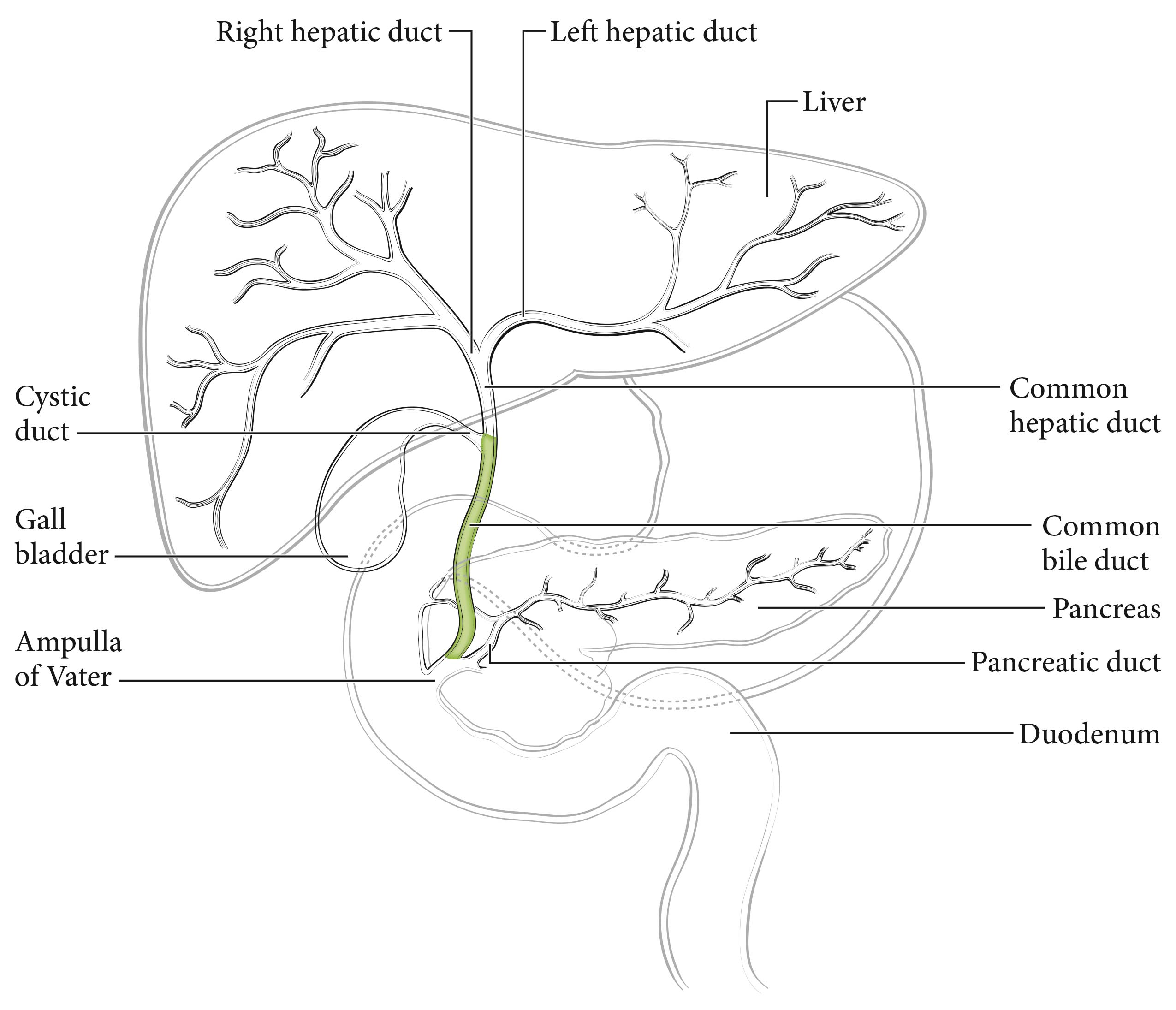
The cystic duct connects to the gallbladder and joins the common hepatic duct to form the common bile duct, which passes posterior to the first part of the duodenum, traverses the head of the pancreas, and then enters the second part of the duodenum through the ampulla of Vater. Tumors with their center located between the confluence of the cystic duct and common hepatic duct and the Ampulla of Vater (excluding ampullary carcinoma) are considered distal bile duct tumors (Figure 34.1). Histologically, the bile ducts are lined by a single layer of tall, uniform columnar calls. The mucosa usually forms irregular pleats or small longitudinal folds. The walls of the bile ducts have a layer of subepithelial connective tissue and muscle fibers. It should be noted that the muscle fibers are most prominent in the distal segment of the common bile duct. The extrahepatic ducts lack a serosa but are surrounded by varying amounts of adventitial adipose tissue. Adipose tissue surrounding the fibromuscular wall is not considered part of the bile duct mural anatomy.

Carcinomas that arise in the distal segment of the common bile duct may spread directly into the pancreas, duodenum, gallbladder, colon, stomach, or omentum.

34.1 Diagram highlighting the location of tumors to be staged as distal bile duct tumors. These tumors have an epicenter located between the confluence of the cystic duct and common hepatic duct and the ampulla of Vater (highlighted) (Modified from the College of American Pathologists).

Regional Lymph Nodes

The regional lymph nodes are the same as those resected for cancers of the head of the pancreas: nodes along the common bile duct and hepatic artery, the posterior and anterior pancreaticoduodenal nodes, and the nodes along the right lateral wall of the superior mesenteric artery.

Metastatic Sites

Distant metastases usually occur late in the course of the disease and most often are found in the liver, lungs, and peritoneum.

Clinical Classification

Most patients with distal bile duct cancer present with biliary symptoms such as painless jaundice and have abnormal liver function tests. Subsequent imaging often detects a biliary obstruction or abnormality. The ideal workup of the stricture includes direct visualization of the bile duct with targeted biopsies. Endoscopic ultrasound (EUS) may help define the lesion or bile duct wall thickening and may help direct biopsies. Delayed-contrast computed tomography (CT), magnetic resonance (MR) imaging, or MR cholangiopancreatography (MRCP) is used to further assess the lesion, adjacent vessels, and nearby lymph nodes and to detect metastatic disease. Endoscopic retrograde cholangiopancreatography (ERCP) may allow for bile duct brushings and stenting for unresectable disease. Serologic studies (carcinoembryonic antigen [CEA] and cancer antigen [CA] 19-9) may be considered. Clinical staging also may be based on findings from surgical exploration if the main tumor mass is not resected. The initial surgical assessment should rule out distant metastatic disease and determine local resectability. The presence of a dominant stricture may be a diagnostic feature of distal bile duct cancer. Positive biopsy, cytology, and/or polysomy on fluorescent in situ hybridization confirms the diagnosis.1

Most often, patients are staged following surgery and pathological examination. In one third to half of the cases, surgical resection is not attempted because of local/regional extension, and patients are treated without pathological staging. A single TNM classification applies to both clinical and pathological staging. With advances in imaging, integrated radiologic and pathological staging of patients may be achieved satisfactorily.

Imaging

Cross-sectional imaging, either contrast-enhanced, multiphasic, thin-section MR imaging or CT, typically is the preferred examination for assessing the stage of pancreatic cancers, ampullary tumors, and distal common bile duct tumors and should be performed before any interventions (e.g., biopsy, stent placement). The choice of MR imaging or CT should be based on the imaging equipment available, the expertise of the radiologists performing and interpreting the studies, and whether there are confounding issues, such as allergies to intravenous contrast or renal insufficiency (in the latter case, unenhanced MR imaging is preferred to unenhanced CT because of MR imaging's superior soft tissue contrast). As noted, imaging should be performed before interventions (e.g., stent placement, biopsy) to avoid the effects of potential postprocedure pancreatitis interfering with staging assessments.

If intravenous contrast is used, dynamic imaging (MR or CT) should be performed both during the phase of peak pancreatic enhancement (“pancreatic parenchymal” or “late arterial” phase), to enhance the conspicuity of tumor against the background pancreas (regardless of ampullary, pancreatic, or distal biliary origin), and during the portal venous phase of liver enhancement (peak liver enhancement), when veins are fully opacified, to judge extrapancreatic extent of tumor, involvement of vasculature, and the possibility of liver metastases, as liver metastases from these tumors typically are hypodense against uninvolved liver. Thin-section imaging (e.g., 2-3 mm for CT) is particularly important for judging vascular involvement and to assess for potential small sites of metastatic disease. In the setting of preoperative therapy, this technique is important, not only at baseline but also following therapy, to determine whether patients are still surgical candidates and to follow up borderline suspicious findings.

Endoscopic ultrasound may be used in conjunction with CT/MR imaging to assist in locoregional staging; however, EUS has limited utility in assessing for distant disease such as liver metastases, peritoneal implants, or adenopathy outside the surgical field. EUS and EUS/fine-needle aspiration also should be performed before ERCP, as pancreatitis may degrade the ability of EUS to visualize the tumor and stent placement makes it impossible to identify sites of duct cutoff that may be useful in guiding biopsies. ERCP subsequently may be helpful in the setting of duct abnormalities, both for treatment (stent placement) and for diagnosis (brushings). 2-19

TNM Categories of Staging by Imaging

The relationship of the tumor to relevant vessels should be reported, specifically the relation of the tumor to arteries, such as the superior mesenteric, celiac, splenic, and common hepatic arteries, as well as the aorta if the tumor extends posteriorly into the retroperitoneum. The relationship of the tumor to relevant veins, including the portal vein, splenic vein, splenoportal confluence, and superior mesenteric vein, as well as to branch vessels, such as the gastrocolic trunk, first jejunal vein, and ileocolic branches, also should be recorded.

The relationship of the tumor to the vessels should be described using terms commonly understood by the clinical community, such as degrees of circumferential involvement and the terms abutment (i.e., up to and including 180° of involvement of a given vessel by tumor) and encasement (i.e., greater than 180° of circumferential vessel involvement by tumor). Multiplanar reconstructions for CT and direct multiplanar imaging for MR imaging may be particularly helpful in visualizing the circumferential relationship of the tumor to relevant vasculature. It also is important to describe the relationship of the tumor to adjacent structures such as the stomach, spleen, colon, small bowel, and adrenal glands.

Assessment of N category (nodal) status may be a challenge for all imaging modalities, because preoperative imaging is limited and cannot detect microscopic metastatic disease. Nevertheless, it is important to fully identify the location of visibly suspicious nodes. Nodes are considered suspicious for metastatic involvement if they are greater than 1 cm in short axis or have abnormal morphology (e.g., are rounded, hypodense, or heterogeneous; have irregular margins; involve adjacent vessels or structures). Lymph nodes outside the usual surgical field, such as retroperitoneal nodes, pelvic nodes, and lymph nodes within the jejunal mesentery or ileocolic mesentery, also should be evaluated and reported if abnormal.

The most common sites of metastatic disease include the liver, peritoneum, and lung. Evaluation for potential metastatic disease is best done with contrast-enhanced CT or MR imaging.

Suggested Radiology Report Format

Tumor involvement with adjacent vasculature should be reported with terms generally understood by the oncology community, such as degrees of circumferential involvement by tumor of a given vessel, and the terms abutment and encasement, as defined earlier.

The radiology report should include detailed descriptions of the following:

  1. Primary tumor: location, size, characterization and effect on ducts (common bile duct and main pancreatic duct). Details regarding any findings suspicious for superimposed acute pancreatitis, which may distort findings relevant to staging, or chronic pancreatitis/autoimmune pancreatitis also should be reported, as these diseases may closely mimic malignancy and may be associated with duct strictures.
  2. Local extent: the relationship of the tumor, with reference to degrees of circumferential involvement using commonly understood terms, such as abutment and encasement, and occlusion with regard to adjacent arterial structures (celiac, superior mesenteric, hepatic, and splenic arteries and the aorta) and venous structures (portal, splenic, and superior mesenteric veins, and if relevant, inferior vena cava). The following observations also should be noted:
    1. How much of the vascular involvement is related to solid tumor versus stranding, and whether vessel involvement is related to direct involvement by tumor or is separate from the tumor
    2. Narrowing of vasculature, vascular thrombi, and the length of tumor involvement with the vasculature
    3. Enlarged collaterals or varices
    4. Involvement of branch vessels, such as the gastrocolic, first jejunal, and ileocolic branches of the superior mesenteric vein
  3. Relevant arterial variants: This is particularly important with regard to hepatic arterial variants, such as those arising from the superior mesenteric artery, and the nature of the variant (e.g., accessory right hepatic vs. common hepatic artery arising from the superior mesenteric artery). Confounding factors, such as narrowing of the celiac origin by arcuate ligament syndrome or atherosclerotic disease of the celiac and superior mesenteric arteries, as well as their effects on adjacent vasculature, also are important for treatment planning.
  4. Lymph node involvement: Suspicious nodes should be documented, particularly those greater than 1 cm in short axis or morphologically abnormal (e.g., rounded nodes, hypodense/heterogeneous/necrotic nodes, nodes with irregular margins); suspicious nodes outside the typical surgical field, such as retroperitoneal, pelvic, and mesenteric nodes, also should be recorded.
  5. Distant spread: Evaluation of the liver, peritoneum (including whether ascites is present or absent), bone, and lung should be recorded.
    1. Ascites should be noted, as it may indicate peritoneal metastases.

Pathological Classification

Pathological staging depends on surgical resection and pathological examination of the specimen and associated lymph nodes. The College of American Pathologists (CAP) Protocol for the Examination of Specimens from Patients with Carcinoma of the Distal Extrahepatic Bile Ducts is recommended as a guideline for the pathological evaluation of resection specimens for distal bile duct cancer (www.cap.org).

As for the T category, assessment of tumor extension may be difficult because the extrahepatic biliary tree lacks uniform smooth muscle distribution along its length, with scattered or no muscle fibers in the wall of the proximal ducts as compared with the distal bile duct.20,21 In addition to the problem created by the lack of discrete tissue boundaries, inflammatory changes in the bile ducts and desmoplastic stromal reaction to tumor may cause distortion of the bile duct wall. To overcome these difficulties, the measurement of tumor depth has been adopted in the new classification.22 This system, however, requires careful perpendicular or longitudinal sectioning of the bile duct so that the deepest tumor invasion (from the basal lamina of adjacent normal or dysplastic epithelium) can be identified and measured. If the depth of invasion is difficult to measure, a best estimate should be given. The level of invasion also should be reported separately (tumor confined to the bile duct, tumor invading beyond the bile duct wall, or tumor extending into an adjacent organ, such as the pancreas, gallbladder, duodenum, or other adjacent organ).

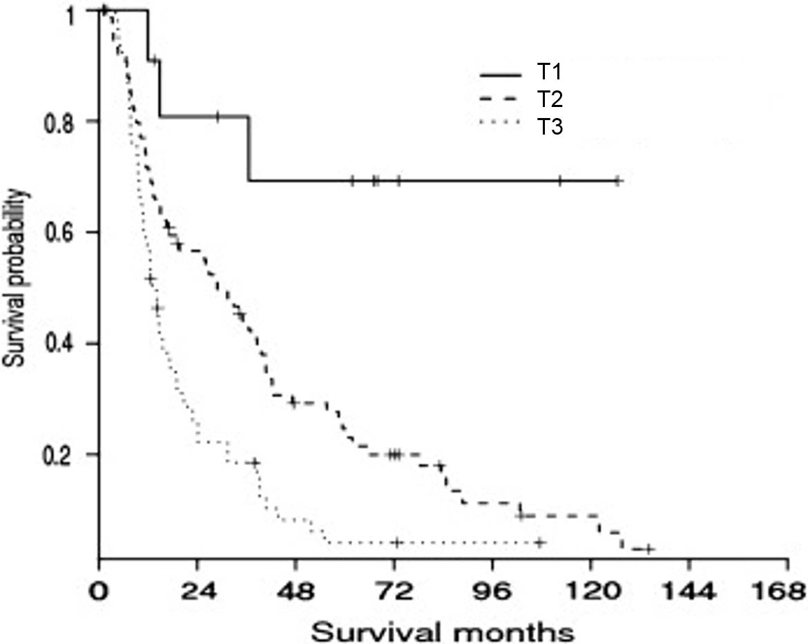
Depth of tumor invasion using the cutoff values of 0.5 cm and 1.2 cm was more powerful than the descriptive extent of tumor invasion in predicting patient outcomes (Figure 34.2) in several single-institution studies.20,22 Tumor depth should be measured from the basement membrane of adjacent normal or dysplastic epithelium to the point of deepest tumor invasion in appropriately oriented and sectioned specimens.22 AJCC Level of Evidence: II

An effort should be made to distinguish a tumor that arises in the intrapancreatic portion of the common bile duct from pancreatic cancer, given the differences in tumor biology, patient outcomes, availability of clinical trials, and staging classifications that apply to each clinical entity. Making this distinction, however, may be challenging because of the intimate association of the bile duct with the pancreas and identical immunophenotypic features.23 Tumors growing symmetrically around the common bile duct are more likely to be distal bile duct carcinomas, whereas an eccentric tumor mass with an epicenter away from the intrapancreatic bile duct more likely is a pancreatic cancer. Another helpful feature that points to distal bile duct origin is the finding of an in situ component, such as prominent biliary intraepithelial neoplasia or a biliary intraductal tubular/tubulopapillary neoplasm.23

The N category for distal bile duct cancer mirrors that of pancreatic cancer. Specifically, patients are categorized as having no regional lymph node metastasis (N0), metastasis in one to three regional lymph nodes (N1), or metastasis in four or more regional lymph nodes (N2). Tumor involvement of other nodal groups outside the region is considered distant metastasis. Although the minimal number of lymph nodes to be examined for accurate staging has not been determined, examination of at least 12 lymph nodes is recommended.

Accurate pathological staging requires that all lymph nodes that are removed be analyzed. Published studies on optimal histologic examination of a pancreaticoduodenectomy specimen for pancreatic adenocarcinoma support analysis of a minimum of 12 lymph nodes.24 If the resected lymph nodes do not contain metastatic disease but fewer than 12 are retrieved, pN0 should still be assigned. Anatomic division of regional lymph nodes is not necessary; however, separately submitted lymph nodes should be reported as submitted.

The extent of resection (R0, R1, R2) is an important stage-independent prognostic factor and should be reported.25,26 Extrahepatic bile duct carcinomas may be multifocal; thus, microscopic foci of carcinoma or intraepithelial neoplasia may be found at the margin(s) and should be reported.

Registry Data Collection Variables
  1. Tumor location (ICD code lacks specificity): cystic duct, perihilar bile ducts, or distal bile duct
  2. CEA
  3. CA 19-9

Prognostic Factors

Prognostic Factors Required for Stage Grouping

Beyond the factors used to assign T, N, or M categories, no additional prognostic factors are required for stage grouping.

Additional Factors Recommended for Clinical Care

Extent of Resection

Resection status (R0, complete resection with grossly and microscopically negative margins of resection; R1, grossly negative but microscopically positive margins of resection; R2, grossly and microscopically positive margins of resection) is not part of the TNM staging system, but complete surgical resection with microscopically negative surgical margins is an important predictor of outcome for distal bile duct cancers.25,26 It is important to confirm complete resection in intraoperative consultation, but prominent inflammation and reactive change of the surface epithelium or within the intramural mucous glands secondary to stent insertion and/or biliary obstruction may hamper the evaluation of margins on frozen section.

AJCC Level of Evidence: II

Invasion of Adjacent Organs
Invasion of adjacent organs should be described. Carcinomas that arise in the distal segment of the common bile duct may spread directly into the pancreas, duodenum, gallbladder, colon, stomach, or omentum. In particular, invasion of adjacent pancreas occurs frequently but loses prognostic significance after an adjustment is made for depth of tumor invasion.22
AJCC Level of Evidence: II

Histologic Parameters
Histologic features have a less significant impact on prognosis than stage. Nevertheless, several histologic parameters, such as high grade (poorly differentiated), perineural invasion, and lymphovascular invasion, are associated with unfavorable patient outcomes and should be noted in the pathology report.27 High-grade tumors, such as signet ring cell carcinomas, undifferentiated carcinomas, and high-grade neuroendocrine carcinomas, are associated with unfavorable patient outcomes.
AJCC Level of Evidence: II

Tumor Markers CEA and CA 19-9
CEA and CA 19-9 are not sensitive enough to be used as screening markers. In addition, these markers are not specific for bile duct cancer, as they may be elevated in other malignancies (e.g., pancreatic, gastric) and in nonneoplastic conditions (e.g., hepatolithiasis, cholangitis). An elevated CA 19-9 level, however, has been associated with unfavorable patient outcomes.25
AJCC Level of Evidence: III

Molecular Markers

Molecular markers are beginning to show promise in determining the prognosis of patients with distal bile duct tumors. Two genes, PBRM1 and BAP1 (chromatin-modulating genes), have been associated with reduced progression-free and overall survival.28 Molecular profiling may reveal potentially targetable genes among patients with advanced disease. Potentially targetable gene alterations detected in distal bile duct tumors include ERBB1 (EGFR), ERBB2 (HER2), KRAS, MET, and members of the PIK3CA/AKT/mTOR (PIK3CA and PTEN), Notch signaling, and sonic hedgehog signaling pathways.28-30

AJCC Level of Evidence: Not identified

Risk Assessment

Risk Assesment Models

The AJCC recently established guidelines that will be used to evaluate published statistical prediction models for the purpose of granting endorsement for clinical use.31 Although this is a monumental step toward the goal of precision medicine, this work was published only very recently. Therefore, the existing models that have been published or may be in clinical use have not yet been evaluated for this cancer site by the Precision Medicine Core of the AJCC. In the future, the statistical prediction models for this cancer site will be evaluated, and those that meet all AJCC criteria will be endorsed.

Recommendations

There are no recommendations for clinical trial stratification at this time.

TNM Definitions

Definition of Primary Tumor (T)

T CategoryT Criteria
TXPrimary tumor cannot be assessed
TisCarcinoma in situ/high-grade dysplasia
T1Tumor invades the bile duct wall with a depth less than 5 mm
T2Tumor invades the bile duct wall with a depth of 5-12 mm
T3Tumor invades the bile duct wall with a depth greater than 12 mm
T4Tumor involves the celiac axis, superior mesenteric artery, and/or common hepatic artery

Definition of Regional Lymph Node (N)

N CategoryN Criteria
NXRegional lymph nodes cannot be assessed
N0No regional lymph node metastasis
N1Metastasis in one to three regional lymph nodes
N2Metastasis in four or more regional lymph nodes

Definition of Distant Metastasis (M)

M CategoryM Criteria
M0No distant metastasis
M1Distant metastasis

Stage Prognostic

!!Calculator!!

When T is…and N is…and M is…Then the stage group is…
TisN0M00
T1N0M0I
T1N1M0IIA
T1N2M0IIIA
T2N0M0IIA
T2N1M0IIB
T2N2M0IIIA
T3N0M0IIB
T3N1M0IIB
T3N2M0IIIA
T4N0M0IIIB
T4N1M0IIIB
T4N2M0IIIB
Any TAny NM1IV

Histopathologic type

The staging system applies to all carcinomas that arise in the distal extrahepatic bile ducts. Sarcomas, lymphomas, and well-differentiated neuroendocrine tumors are excluded. Adenocarcinoma without specific subtype features is the most common histologic type. Carcinomas account for more than 98% of cancers of the distal extrahepatic bile ducts.

Histologic grade

HISTOLOGIC GRADE (G)

The following grading system is recommended for distal bile duct carcinomas.

GG Definition
GXGrade cannot be assessed
G1Well differentiated
G2Moderately differentiated
G3Poorly differentiated

Survival

34.2 Survival based on T category.20,22 Results from 147 US patients who underwent resection of distal bile duct carcinoma confirm earlier study results from 222 Korean patients regarding the use of depth of tumor invasion to predict prognosis and stratify T category.

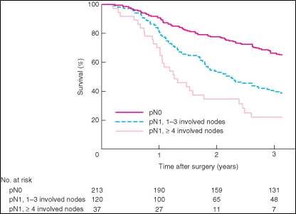
34.3 Survival based on N category.32

Bibliography

  1. Blechacz B, Komuta M, Roskams T, Gores GJ. Clinical diagnosis and staging of cholangiocarcinoma. Nature reviews. Gastroenterology & hepatology. Sep 2011;8(9):512-522.
  2. Al-Hawary MM, Francis IR, Chari ST, et al. Pancreatic ductal adenocarcinoma radiology reporting template: consensus statement of the Society of Abdominal Radiology and the American Pancreatic Association. Radiology. Jan 2014;270(1):248-260.
  3. Al-Hawary MM, Kaza RK, Wasnik AP, Francis IR. Staging of pancreatic cancer: role of imaging. Seminars in roentgenology. Jul 2013;48(3):245-252.
  4. Tamm EP, Balachandran A, Bhosale PR, et al. Imaging of pancreatic adenocarcinoma: update on staging/resectability. Radiol Clin North Am. May 2012;50(3):407-428.
  5. Brook OR, Brook A, Vollmer CM, Kent TS, Sanchez N, Pedrosa I. Structured reporting of multiphasic CT for pancreatic cancer: potential effect on staging and surgical planning. Radiology. Feb 2015;274(2):464-472.
  6. Marcal LP, Fox PS, Evans DB, et al. Analysis of free-form radiology dictations for completeness and clarity for pancreatic cancer staging. Abdom Imaging. Oct 2015;40(7):2391-2397.
  7. Gottlieb R. CT Onco Primary Pancreas Mass. RSNA Radiology Reporting Templates 2012. Accessed 8/13/2015, 2015.
  8. Tempero MA, Malafa MP, Asbun H, et al. NCCN Guidelines Version 2.2015 Pancreatic Adenocarcinoma. NCCN Guidelines [pdf]. 2015; http://www.nccn.org/professionals/physician_gls/pdf/pancreatic.pdf. Accessed 10/16/2015, 2015.
  9. Varadhachary GR, Tamm EP, Abbruzzese JL, et al. Borderline resectable pancreatic cancer: definitions, management, and role of preoperative therapy. Annals of surgical oncology. Aug 2006;13(8):1035-1046.
  10. Katz MH, Crane CH, Varadhachary G. Management of borderline resectable pancreatic cancer. Semin Radiat Oncol. Apr 2014;24(2):105-112.
  11. Valls C, andia E, Sanchez A, et al. Dual-phase helical CT of pancreatic adenocarcinoma: assessment of resectability before surgery. AJR. American journal of roentgenology. Apr 2002;178(4):821-826.
  12. Tamm EP, Loyer EM, Faria S, et al. Staging of pancreatic cancer with multidetector CT in the setting of preoperative chemoradiation therapy. Abdom Imaging. Sep-Oct 2006;31(5):568-574.
  13. Cassinotto C, Cortade J, Belleannee G, et al. An evaluation of the accuracy of CT when determining resectability of pancreatic head adenocarcinoma after neoadjuvant treatment. Eur J Radiol. Apr 2013;82(4):589-593.
  14. DeWitt J, Devereaux B, Chriswell M, et al. Comparison of endoscopic ultrasonography and multidetector computed tomography for detecting and staging pancreatic cancer.[see comment][summary for patients in Ann Intern Med. 2004 Nov 16;141(10):I46; PMID: 15545671]. Annals of internal medicine. 2004;141(10):753-763.
  15. Tamm EP, Loyer EM, Faria SC, Evans DB, Wolff RA, Charnsangavej C. Retrospective analysis of dual-phase MDCT and follow-up EUS/EUS-FNA in the diagnosis of pancreatic cancer. Abdom Imaging. Sep-Oct 2007;32(5):660-667.
  16. Nikolaidis P, Hammond NA, Day K, et al. Imaging features of benign and malignant ampullary and periampullary lesions. Radiographics : a review publication of the Radiological Society of North America, Inc. May-Jun 2014;34(3):624-641.
  17. Kim JH, Park SH, Yu ES, et al. Visually isoattenuating pancreatic adenocarcinoma at dynamic-enhanced CT: frequency, clinical and pathologic characteristics, and diagnosis at imaging examinations. Radiology. Oct 2010;257(1):87-96.
  18. Raman SP, Fishman EK. Abnormalities of the distal common bile duct and ampulla: diagnostic approach and differential diagnosis using multiplanar reformations and 3D imaging. AJR. American journal of roentgenology. Jul 2014;203(1):17-28.
  19. Motosugi U, Ichikawa T, Morisaka H, et al. Detection of pancreatic carcinoma and liver metastases with gadoxetic acid-enhanced MR imaging: comparison with contrast-enhanced multi-detector row CT. Radiology. Aug 2011;260(2):446-453.
  20. Hong SM, Cho H, Moskaluk CA, Yu E. Measurement of the invasion depth of extrahepatic bile duct carcinoma: An alternative method overcoming the current T classification problems of the AJCC staging system. The American journal of surgical pathology. Feb 2007;31(2):199-206.
  21. Hong SM, Kim MJ, Pi DY, et al. Analysis of extrahepatic bile duct carcinomas according to the New American Joint Committee on Cancer staging system focused on tumor classification problems in 222 patients. Cancer. Aug 15 2005;104(4):802-810.
  22. Hong S-M, Pawlik TM, Cho H, et al. Depth of tumor invasion better predicts prognosis than the current American Joint Committee on Cancer T classification for distal bile duct carcinoma. Surgery. 2009;146(2):250-257.
  23. Bledsoe JR, Shinagare SA, Deshpande V. Difficult Diagnostic Problems in Pancreatobiliary Neoplasia. Arch Pathol Lab Med. Jul 2015;139(7):848-857.
  24. Adsay NV, Basturk O, Altinel D, et al. The number of lymph nodes identified in a simple pancreatoduodenectomy specimen: comparison of conventional vs orange-peeling approach in pathologic assessment. Modern pathology : an official journal of the United States and Canadian Academy of Pathology, Inc. Jan 2009;22(1):107-112.
  25. Chung YJ, Choi DW, Choi SH, Heo JS, Kim DH. Prognostic factors following surgical resection of distal bile duct cancer. J Korean Surg Soc. Nov 2013;85(5):212-218.
  26. DeOliveira ML, Cunningham SC, Cameron JL, et al. Cholangiocarcinoma: thirty-one-year experience with 564 patients at a single institution. Annals of surgery. 2007;245(5):755.
  27. He P, Shi JS, Chen WK, Wang ZR, Ren H, Li H. Multivariate statistical analysis of clinicopathologic factors influencing survival of patients with bile duct carcinoma. World journal of gastroenterology : WJG. Oct 2002;8(5):943-946.
  28. Churi CR, Shroff R, Wang Y, et al. Mutation profiling in cholangiocarcinoma: prognostic and therapeutic implications. PloS one. 2014;9(12):e115383.
  29. Geynisman DM, Catenacci DV. Toward personalized treatment of advanced biliary tract cancers. Discovery medicine. Jul 2012;14(74):41-57.
  30. Oyasiji T, Zhang J, Kuvshinoff B, Iyer R, Hochwald SN. Molecular Targets in Biliary Carcinogenesis and Implications for Therapy. The oncologist. Jul 2015;20(7):742-751.
  31. Kattan MW, Hess KR, Amin MB, et al. American Joint Committee on Cancer acceptance criteria for inclusion of risk models for individualized prognosis in the practice of precision medicine. CA: a cancer journal for clinicians. Jan 19 2016.
  32. Kiriyama M, Ebata T, Aoba T, et al. Prognostic impact of lymph node metastasis in distal cholangiocarcinoma. The British journal of surgery. Mar 2015;102(4):399-406.