Cancers Staged Using This Staging System
Pancreatic ductal adenocarcinoma, acinar cell carcinoma, intraductal papillary mucinous neoplasm with associated invasive carcinoma, intraductal tubulopapillary neoplasm with associated invasive carcinoma, colloid carcinoma, mucinous cystic neoplasm with associated invasive carcinoma, solid pseudopapillary neoplasm, large cell neuroendocrine carcinoma, small cell neuroendocrine carcinoma, pancreaticoblastoma
Cancers Not Staged Using This Staging System
These histopathologic types of cancer | Are staged according to the classification for | and can be found in chapter |
---|---|---|
Well-differentiated neuroendocrine tumor | Neuroendocrine tumors of the pancreas | 34 |
Summary of Changes
Change | Details of Change | Level of Evidence |
---|---|---|
Definition of Primary Tumor (T) | T1 are subcategorized into T1a, T1b, and T1c based on size.Rationale: Size-based categorization of small invasive tumors that have been characterized as minimally invasive and have better outcome. | III |
Definition of Primary Tumor (T) | T2 and T3 categories are now based on size of invasive tumor; extrapancreatic extension is no longer part of the definition. Rationale: Size-based definitions are more objective as it is difficult to determine extrapancreatic extension. These definitions show better correlation with survival. | II |
Definition of Primary Tumor (T) | T4 categorization is now based on involvement of arteries; resectability has been removed from the definition.Rationale: Resectability is subjective, and the T category is better defined by extent of invasion. | II |
Definition of Regional Lymph Node (N) | Node-positive disease N1 has been subdivided into N1 and N2, based on number of positive lymph nodes.Rationale: Better prognostic stratification is provided based on number of positive lymph nodes. | II |
This list includes histology codes and preferred terms from the WHO Classification of Tumors and the International Classification of Diseases for Oncology (ICD-O). Most of the terms in this list represent malignant behavior. For cancer reporting purposes, behavior codes /3 (denoting malignant neoplasms), /2 (denoting in situ neoplasms), and in some cases /1 (denoting neoplasms with uncertain and unknown behavior) may be appended to the 4-digit histology codes to create a complete morphology code.
Code | Description |
---|---|
8000 | Neoplasm, malignant |
8010 | Carcinoma, NOS |
8013 | Large cell neuroendocrine carcinoma (NEC) |
8020 | Undifferentiated carcinoma |
8035 | Undifferentiated carcinoma with osteoclast-like giant cells |
8041 | Small cell neuroendocrine carcinoma (NEC) |
8140 | Adenocarcinoma, NOS |
8148 | Pancreatic intraepithelial neoplasia, grade 3 (PanIN-3) |
8154 | Mixed acinar-neuroendocrine carcinoma |
8154 | Mixed acinar-neuroendocrine-ductal carcinoma |
8154 | Mixed ductal-neuroendocrine carcinoma |
8246 | Neuroendocrine carcinoma (NEC) |
8441 | Serous cystadenocarcinoma |
8452 | Solid pseudopapillary neoplasm |
8453 | Intraductal papillary mucinous neoplasm with high-grade dysplasia |
8453 | Intraductal papillary mucinous neoplasm with associated invasive carcinoma |
8470 | Mucinous cystic neoplasm with high-grade dysplasia |
8470 | Mucinous cystic neoplasm with associated invasive carcinoma |
8480 | Colloid carcinoma (mucinous noncystic carcinoma) |
8481 | Mucin-producing adenocarcinoma |
8490 | Signet ring cell carcinoma |
8500 | Ductal adenocarcinoma |
8503 | Intraductal tubulopapillary neoplasm |
8503 | Intraductal papillary neoplasm with associated invasive carcinoma |
8510 | Medullary carcinoma |
8550 | Acinar cell carcinoma |
8551 | Acinar cell cystadenocarcinoma |
8552 | Mixed acinar-ductal carcinoma |
8560 | Adenosquamous carcinoma |
8576 | Hepatoid carcinoma |
8971 | Pancreaticoblastoma |
Histology is not ideal for clinical use in patient care, as it describes an unspecified or outdated diagnosis. Data collectors may use this code only if there is not enough information in the medical record to document a more specific diagnosis.
Bosman FT, Carneiro F, Hruban RH, Theise ND, eds. World Health Organization Classification of Tumours of the Digestive System. Lyon: IARC; 2010. Used with permission.
International Agency for Research on Cancer, World Health Organization. International Classification of Diseases for Oncology. ICD-O-3-Online.http://codes.iarc.fr/home. Accessed September 29, 2017. Used with permission.
In the United States, pancreatic cancer is the second most common malignant tumor of the gastrointestinal tract and the fourth leading cause of cancer-related death in adults. The American Cancer Society estimated nearly 49,000 new cases in the United States and approximately 41,000 deaths in 2015. Based on Surveillance, Epidemiology, and End Results (SEER) data from 2005 to 2011, the 5-year survival is 7.2%: 27.1% for localized disease, 10.7% for regional disease, and 2.4% for metastatic disease. Most pancreatic cancers are ductal adenocarcinomas. Surgical resection remains the only potentially curative approach, although multimodal therapy consisting of systemic agents, and often radiation, may improve survival. Staging of pancreatic cancers depends on the size and extent of the primary tumor and the presence and extent of metastasis.
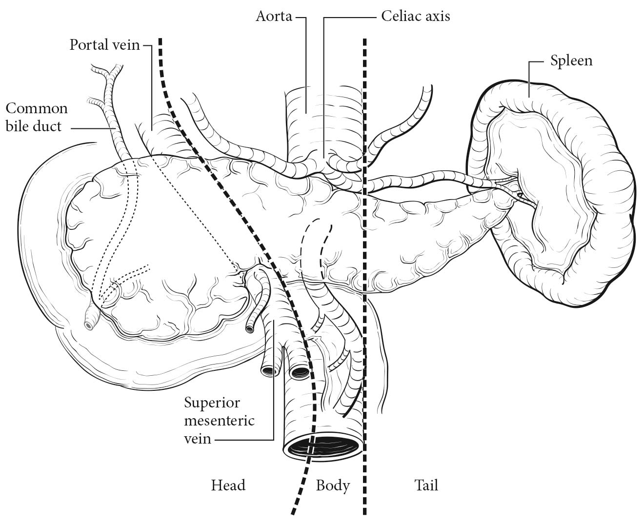
The pancreas is a coarsely lobulated gland that lies transversely across the posterior abdomen and extends from the duodenum to the splenic hilum. The organ is divided into a head with an uncinate process, a neck, a body, and a tail (Figure 36.1). The anterior aspect of the body of the pancreas is in direct contact with the posterior wall of the stomach; posteriorly, the pancreas extends to the inferior vena cava, superior mesenteric vein, splenic vein, and left kidney. The uncinate process is part of the pancreatic head and may extend posterior to the superior mesenteric vein and artery. The pancreatic head accounts for 60-70% of pancreatic adenocarcinomas, whereas 20-25% arise in the body and tail and 10-20% diffusely involve the pancreas.1
A rich lymphatic network surrounds the pancreas, and accurate tumor staging requires resection of peripancreatic lymph nodes for pathological assessment. The standard regional lymph node basins and soft tissues resected for tumors located in the head and neck of the pancreas include lymph nodes along the common bile duct, common hepatic artery, portal vein, pyloric, posterior and anterior pancreatoduodenal arcades, and along the superior mesenteric vein and right lateral wall of the superior mesenteric artery. For cancers located in the body and tail, regional lymph node basins include lymph nodes along the common hepatic artery, celiac axis, splenic artery, and splenic hilum. Tumor involvement of other nodal groups is considered distant metastasis.
Information necessary for the clinical staging of pancreatic cancer may be obtained from physical examination and three-dimensional radiographic imaging studies, which include triphasic, contrast-enhanced multislice computed tomography (CT) or magnetic resonance (MR) imaging. Endoscopic ultrasonography (if done by experienced gastroenterologists) also provides information helpful for clinical staging and is the procedure of choice for performing fine-needle aspiration (FNA) biopsy of the pancreas. The standard radiographic assessment of resectability includes evaluation for peritoneal or hepatic metastases; the patency of the superior mesenteric vein and portal vein and the relationship of these vessels and their tributaries to the tumor; and the relationship of the tumor to the superior mesenteric artery, celiac axis, and hepatic artery. If the appropriate clinical and radiographic findings are present, preoperative biopsy is not necessary before resection, but a core biopsy or an endoscopic ultrasound (EUS)-guided fine-needle aspiration biopsy specimen should be obtained to confirm the diagnosis for patients undergoing neoadjuvant therapy. Serum IgG4 levels may be helpful in identifying autoimmune pancreatitis if a tissue diagnosis has not been obtained.
Laparoscopy may be performed in patients believed to have localized, potentially resectable tumors to exclude peritoneal metastases and small metastases on the surface of the liver. Laparoscopy will reveal small (less than 1 cm) peritoneal or liver metastases and upstage (to Stage IV) approximately 10% of patients with tumors in the pancreatic head, and probably a greater percentage of patients with tumors in the body and tail. A significantly elevated preoperative cancer antigen (CA) 19-9 level (greater than 250 U/mL) increases the yield of staging laparoscopy.
Pancreatic cancers that are locally advanced have a low likelihood of resectability and a high chance of incomplete removal if resected. This scenario has given rise to the term borderline resectable tumor, which has been defined by the Americas Hepato-Pancreato-Biliary Association/Society of Surgical Oncology/Society for Surgery of the Alimentary Tract, the International Study Group of Pancreatic Surgery (ISGPS), and the National Comprehensive Cancer Network (NCCN).2,3 The superior mesenteric artery may have tumor abutment up to but not more than 180o of the circumference of the vessel and may have short segment involvement of the hepatic artery that does not extend onto the celiac axis. Venous involvement of the superior mesenteric vein/portal vein is defined as tumor abutment with or without impingement and narrowing of the lumen or short-segment venous occlusion resulting from either tumor thrombus or encasement but with suitable vessel proximal and distal to the area of vessel involvement. In general, these locally advanced cancers should be treated as part of a clinical trial or receive neoadjuvant therapy.
Imaging
Cross-sectional imaging, either contrast-enhanced, multiphasic, thin-section MR imaging or CT, typically is the preferred examination for assessing the stage of pancreatic cancers, ampullary tumors, and distal common bile duct tumors and should be performed before any interventions (e.g., biopsy, stent placement). The choice of MR imaging or CT should be based on the imaging equipment available, the expertise of the radiologists performing and interpreting the studies, and whether there are confounding issues, such as allergies to intravenous contrast or renal insufficiency (in the latter case, unenhanced MR imaging is preferred to unenhanced CT because of MR imaging's superior soft tissue contrast). Imaging should be performed before interventions (e.g. stent placement, biopsy) to avoid the effects of potential postprocedure pancreatitis interfering with staging assessments.
If intravenous contrast is used, dynamic imaging (MR imaging or CT) should be performed both during the phase of peak pancreatic enhancement (pancreatic parenchymal or late arterial phase), to enhance the conspicuity of tumor against the background pancreas (regardless of ampullary, pancreatic, or distal biliary origin), and during the portal venous phase of liver enhancement (peak liver enhancement) and when veins are fully opacified, to judge extrapancreatic extent of tumor, involvement of vasculature, and the possibility of liver metastases, as liver metastases from these tumors typically are hypodense against uninvolved liver. Thin-section imaging (2-3 mm for CT, thicker but as thin as reasonably possible for MR imaging) is particularly important for judging vascular involvement and to assess for potential small sites of metastatic disease. In the setting of preoperative therapy, such technique is important not only at baseline but also following therapy to determine whether patients are still surgical candidates and to follow up borderline suspicious findings.
Endoscopic ultrasound may be used next after CT/MR imaging for problem-solving, given that it is a relatively invasive technique and has limited utility for assessing for distant disease such as liver metastases, peritoneal implants, or adenopathy outside the surgical field. However, the ability of EUS to detect small tumors and guide biopsy is particularly helpful for tumors that may appear isodense to, and therefore indistinguishable from, background pancreas on CT and/or MR imaging. EUS and EUS-FNA also should be performed before ERCP, as pancreatitis may degrade the ability of EUS to visualize tumor and the placement of stents eliminates the ability to identify sites of duct cutoff that may be useful in guiding biopsies. ERCP subsequently may be helpful in the setting of duct abnormalities, both for treatment (stent placement) and for diagnosis (brushings).4-21
TNM Categories of Staging by Imaging
The T category is assessed by measuring the largest diameter of the tumor in the axial plane, whether by CT or by MR imaging. With regard to MR imaging, the measurement should be made on the sequence that best delineates the tumor. Note should be made if there is suspicion that pancreatitis may be present, which may alter the apparent size of the tumor.
The relationship of tumor to relevant vessels should be reported, including its relationship to arteries such as the superior mesenteric, celiac, splenic, and common hepatic arteries, as well as the aorta if the tumor extends sufficiently posteriorly into the retroperitoneum. The tumor's relationship to relevant veins includes the portal vein, splenic vein, splenoportal confluence, and superior mesenteric vein, as well as branch vessels such as the gastrocolic trunk, first jejunal vein, and ileocolic branches. The goal is to delineate tumor extent sufficiently to provide useful information for potential en bloc resection with vascular graft placement.
The relationship of tumor to vessels should be described using terms commonly understood by the clinical community, such as degrees of circumferential involvement and the terms abutment (i.e., less than or equal to 180° of involvement of a given vessel by tumor) and encasement (i.e., greater than 180° of circumferential vessel involvement by tumor). Multiplanar reconstructions for CT and direct multiplanar imaging for MR imaging may be particularly helpful in visualizing the circumferential relationship of tumor to relevant vasculature. It also is important to describe the relationship of tumor to adjacent structures, such as the stomach, spleen, colon, small bowel, and adrenal glands.
Assessment of N category (nodal) status is a challenge for all imaging modalities, because all are limited with regard to detection of microscopic metastatic disease to nodes. Nevertheless, it is important to fully identify the location of visibly suspicious nodes. Nodes are considered suspicious for metastatic involvement if they are greater than 1 cm in short axis or have abnormal morphology (e.g., are rounded, are hypodense or heterogeneous, have irregular margins, involve adjacent vessels or structures).
The most common sites of metastatic disease include the liver, peritoneum, lung, and bone, with metastases to the latter two sites usually occurring late in the disease. Evaluations for potential metastases are best done with contrast-enhanced CT and MR imaging; MR imaging likely provides superior capability for assessing potential liver metastases and involvement of bone, whereas CT is better for evaluating potential lung metastases.
Suggested Radiology Report Format
With the development of neoadjuvant therapy and the category of borderline resectable disease, it is particularly important that radiology reports use commonly understood terminology and that borderline suspicious findings, whether of tumor involvement of vasculature or of potential metastatic disease, such as to the liver, be noted so that they can be monitored on follow-up.
Details of the radiology report should include descriptions of:
Partial resection (pancreaticoduodenectomy or distal pancreatectomy) or complete resection of the pancreas, including the tumor and associated regional lymph nodes, provides the information necessary for pathological staging. In pancreaticoduodenectomy specimens, the bile duct, pancreatic parenchymal, uncinate, proximal (duodenal or gastric), and distal duodenal margins should be evaluated. The pancreatic parenchymal margin is also referred to as the pancreatic duct margin, pancreatic neck margin, and distal pancreatic resection margin. The uncinate margin has also been termed the superior mesenteric artery margin, retroperitoneal margin, mesopancreatic margin, posterior-inferior margin, deep margin, and radial margin. All margins except the pancreatic parenchymal margin should be assessed in total pancreatectomy specimens. The College of American Pathologists (CAP) Checklist for Exocrine Pancreatic Tumors is recommended as a guide for the pathological evaluation of pancreatic resection specimens (www.cap.org).
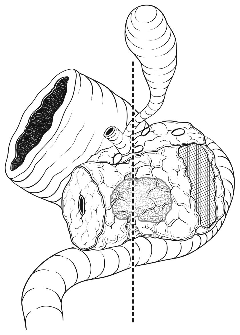
Most local recurrences arise in the pancreatic bed in the region of the uncinate margin. The soft tissue in this area is richly innervated and is adjacent to the right lateral aspect of the superior mesenteric artery (Figure 36.2). The uncinate margin should be inked as part of the gross evaluation of the specimen; the specimen is then cut perpendicular to the inked margin for histologic analysis. The closest microscopic approach of the tumor to the margin should be recorded. The smooth area adjacent to the uncinate process corresponds to the superior mesenteric and portal veins, and is referred to as the vascular groove or vascular bed. This area, as well as the posterior surface (including the nonuncinate posterior surface of the pancreatic head) and anterior surface (corresponding to the peritoneum), is not considered a true surgical margin in the CAP protocol, although this practice is not universally accepted. Histologic assessment of these areas for tumor is recommended but not mandated by the CAP protocol.
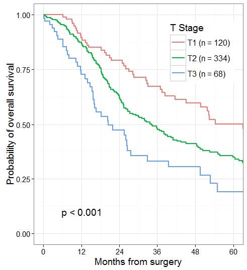
The T categories are based on tumor size. For invasive carcinomas in association with intraductal mucinous neoplasm, intraductal tubulopapillary neoplasm, and mucinous cystic neoplasm, the T category should be determined by the size of the invasive component. The invasive carcinomas in this setting often are small and have a favorable outcome. These tumors have been referred to as minimally invasive carcinomas, with various criteria being used to define this term. T1 subcategories (T1a, T1b, and T1c) provide objective criteria for describing small invasive tumors, justifying the incorporation of these strata into the current system. The current cut-off points of less than or equal to 2 cm, greater than 2 to 4 cm, and greater than 4 cm for definitions of T1 to T3 are based on recent reviews of large databases.1,22-26 T4 for pancreatic cancer is defined as involvement of the superior mesenteric artery, celiac axis, and/or common hepatic artery, which in most cases renders the tumor unresectable. This situation usually is determined by radiographic and endoscopic findings. Hence, T4 category is not determined by pathological examination of surgical resection specimens. Adenocarcinomas of the head of the pancreas often show direct extension into the ampulla of Vater, intrapancreatic portion of the common bile duct, duodenum, peritoneum, and peripancreatic soft tissue. Adenocarcinomas of the body and tail of the pancreas may directly invade the stomach, spleen, left adrenal gland, and peritoneum. In the absence of arterial involvement (celiac axis, superior mesenteric artery, common hepatic artery), the T category is based on size, regardless of invasion of adjacent organs or veins. Extrapancreatic extension may be difficult to determine because the pancreas does not have a capsule, and the distinction between pancreas and extrapancreatic soft tissue often is obscured by fibrosis as part of the tumor or chronic pancreatitis. This parameter is no longer included in the definition of T categories.
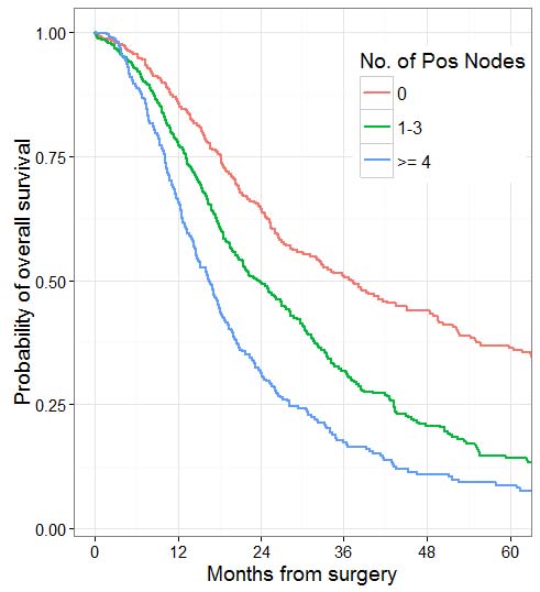
Nodal involvement, regardless of direct extension or metastases to peripancreatic nodes, has been associated with unfavorable outcomes in multiple studies.27 Thus, it is important to identify and properly assess as many regional lymph nodes in the specimen as possible. Based on survival data and a review of the number of lymph nodes that can be practically obtained from resection specimens, evaluation of a minimum of 12 lymph nodes is recommended to accurately stage N0 tumors.28,29 Recent studies show that the total number of positive lymph nodes and/or lymph node ratio (LNR) are also strong prognostic predictors.30-33 The total number of positive lymph nodes outperformed LNR in studies with sufficient numbers of lymph nodes obtained and evaluated.32,34 Thus, lymph node-positive categories based on the number of positive lymph nodes have been added to the N category classification of the pancreas, similar to other gastrointestinal sites. Although different cutoffs have been used in different studies,31,32,34 the cutoffs of zero versus one to three versus four or more have been adopted in the current staging scheme based on available data.34,35Anatomic division of regional lymph nodes is not necessary. However, separately submitted lymph nodes should be reported as labeled by the surgeon. Seeding of the peritoneum (even if limited to the lesser sac region), as well as peritoneal fluid with microscopic evidence of carcinoma, is considered M1.
Patients who undergo surgical resection for localized nonmetastatic adenocarcinoma of the pancreas have a long-term survival rate of approximately 27% and a median survival of 12-20 months. Patients with resectable tumors showing regional lymph node involvement and without distant metastasis have a 5-year survival of approximately 11% and a median survival of 6-10 months. Patients with metastatic disease have a short survival (3-6 months), the length of which depends on the extent of disease, performance status, and response to systemic therapy.
Prognostic Factors Required for Stage Grouping
Beyond the factors used to assign T, N, or M categories, no additional prognostic factors are required for stage grouping.
Additional Factors Recommended for Clinical Care
Recent improvements in vascular surgery have led to a profusion of small reports on arterial resection and reconstruction for T4 pancreatic cancers.36,37 These reports demonstrate two main points. First, these operations are still associated with a much higher rate of morbidity and mortality compared with resections without vascular involvement or resections associated with venous resection and reconstruction. Additionally, in patients surviving the perioperative period, the long-term (1-, 3-, and 5-year) survival is not as good as that of patients who have undergone resection without arterial involvement or those who have had venous resection and reconstruction. However, their long-term survival is better than that of patients with similar arterial involvement who do not undergo resection. Therefore, involvement of the celiac and/or superior mesenteric artery remains, at a minimum, a relative contraindication to resection. If contemplated, resection with arterial resection and reconstruction should be performed at expert centers.
The only serum biomarker approved by the US Food and Drug Administration for pancreatic ductal adenocarcinoma is CA 19-9. This marker, however, has limitations in its specificity and sensitivity, and an estimated 15% of the population cannot produce the CA 19-9 (sialyl Lewis a) antigen. Despite these limitations, CA 19-9 may be a useful prognostic marker in the settings of both localized and metastatic disease.38,39 Several reports have noted that an elevated preoperative CA 19-9 level is associated with an increased likelihood of radiographically occult metastatic disease being found at staging laparoscopy in patients about to undergo resection.40 Preoperative CA 19-9 level also is a strong predictor of resectability in the absence of metastatic disease. Many studies also have shown that an increased CA 19-9 level is associated with higher pathological stage and decreased survival.41 In addition, postresection CA 19-9 levels have been associated with postresection survival and have been used as a stratification variable in randomized trials of adjuvant therapy.
The uncinate margin represents the plane of abutment of the uncinate process with the superior mesenteric artery. Because only a scant buffer of connective tissue separates the uncinate process from the superior mesenteric artery and the neural and lymphatic plexus around the celiac trunk, this margin is at highest risk for residual disease in tumors involving the pancreatic head.42 The margin is considered positive if the tumor is at or within 1 mm of the margin. Several studies showed that the recurrence rates are similar in both these situations.43-46 Incomplete resection resulting in a grossly positive uncinate margin provides no survival advantage with surgical resection (compared with chemoradiation and no surgery). The resection status is not part of the TNM staging system, but because of its prognostic significance, it should be recorded in the pathology report as follows: complete resection with grossly and microscopically negative margins of resection (R0), grossly negative but microscopically positive margin(s) of resection (R1), or grossly and microscopically positive margin(s) of resection (R2). The nonuncinate posterior surface, anterior surface, and the vascular groove (corresponding to the superior mesenteric and portal veins) are regarded as resection margins by some groups but are not considered true resection margins by CAP or AJCC. Because involvement of these surfaces may have prognostic significance, it is recommended that this information be included in the pathology report.
Given the increasing number of studies investigating the use of neoadjuvant treatment for ductal adenocarcinoma of the pancreas, it is important to assess the response of tumor to preoperative chemotherapy and/or radiation therapy. Several grading schemes for the extent of residual tumor in posttreatment pancreatectomy specimens have been proposed. The CAP protocol recommends a four-tiered grading system (modified Ryan scheme) similar to that for the rectum.47
Histologic features have less impact on outcome than stage. Several histologic parameters, such as high grade (poorly differentiated), perineural invasion, lymphovascular invasion, and involvement of muscular vessels, have been shown to adversely affect survival and should be noted in pathology reports.48-50 Perineural and lymphovascular invasion also are important prognostic factors after neoadjuvant therapy.51
Human equilibrative nucleoside transporter 1 (hENT1) plays an important role in transport of gemcitabine into tumor cells, and its expression in tumor cells is associated with increased survival in patients treated with gemcitabine but not with other drugs, such as 5-fluorouracil.52 Expression of SPARC (secreted protein acidic and rich in cysteine) in the tumor and stroma also is a favorable prognostic factor in patients receiving gemcitabine as adjuvant therapy53 and may be responsible for higher efficacy of nanoparticle albumin-bound (nab)-paclitaxel.51 Targeting the epidermal growth factor receptor (EGFR) pathway by cetuximab and the angiogenesis pathway by the vascular endothelial growth factor (VEGF) inhibitor bevacizumab and anti-HER2 antagonist trastuzumab so far has not yielded positive results.54,55 Clinical trials also have focused on other signaling pathways using insulin-like growth factor inhibitors, Hedgehog signaling pathway inhibitors, and Akt/PTEN/mTOR inhibitors, but success has been limited and better patient selection is necessary for future trials.55 Immunotherapies such as macrophage-directed therapy and cytotoxic T-cell-directed therapy have shown some promising results in preclinical studies, whereas others, such as tumor vaccines and interferon, have not shown favorable outcomes.55
The AJCC recently established guidelines that will be used to evaluate published statistical prediction models for the purpose of granting endorsement for clinical use.56 Although this is a monumental step toward the goal of precision medicine, this work was published only very recently. Therefore, the existing models that have been published or may be in clinical use have not yet been evaluated for this cancer site by the Precision Medicine Core of the AJCC. In the future, the statistical prediction models for this cancer site will be evaluated, and those that meet all AJCC criteria will be endorsed.
Clinical T (cT)
cT Category | cT Criteria |
---|---|
cTX | Primary tumor cannot be assessed |
cT0 | No evidence of primary tumor |
cTis | Carcinoma in situ. This includes high-grade pancreatic intraepithelial neoplasia (PanIn-3), intraductal papillary mucinous neoplasm with high-grade dysplasia, intraductal tubulopapillary neoplasm with high-grade dysplasia, and mucinous cystic neoplasm with high-grade dysplasia. |
cT1 | Tumor less than or equal to 2 cm in greatest dimension |
cT1a | Tumor less than or equal to 0.5 cm in greatest dimension |
cT1b | Tumor greater than 0.5 cm and less than 1 cm in greatest dimension |
cT1c | Tumor 1-2 cm in greatest dimension |
cT2 | Tumor greater than 2 cm and less than or equal to 4 cm in greatest dimension |
cT3 | Tumor greater than 4 cm in greatest dimension |
cT4 | Tumor involves celiac axis, superior mesenteric artery, and/or common hepatic artery, regardless of size |
Pathological T (pT)
pT Category | pT Criteria |
---|---|
pTX | Primary tumor cannot be assessed |
pT0 | No evidence of primary tumor |
pTis | Carcinoma in situ. This includes high-grade pancreatic intraepithelial neoplasia (PanIn-3), intraductal papillary mucinous neoplasm with high-grade dysplasia, intraductal tubulopapillary neoplasm with high-grade dysplasia, and mucinous cystic neoplasm with high-grade dysplasia. |
pT1 | Tumor less than or equal to 2 cm in greatest dimension |
pT1a | Tumor less than or equal to 0.5 cm in greatest dimension |
pT1b | Tumor greater than 0.5 cm and less than 1 cm in greatest dimension |
pT1c | Tumor 1-2 cm in greatest dimension |
pT2 | Tumor greater than 2 cm and less than or equal to 4 cm in greatest dimension |
pT3 | Tumor greater than 4 cm in greatest dimension |
pT4 | Tumor involves celiac axis, superior mesenteric artery, and/or common hepatic artery, regardless of size |
cTX | Primary tumor cannot be assessed |
cT0 | No evidence of primary tumor |
cTis | Carcinoma in situ. This includes high-grade pancreatic intraepithelial neoplasia (PanIn-3), intraductal papillary mucinous neoplasm with high-grade dysplasia, intraductal tubulopapillary neoplasm with high-grade dysplasia, and mucinous cystic neoplasm with high-grade dysplasia. |
cT1 | Tumor less than or equal to 2 cm in greatest dimension |
cT1a | Tumor less than or equal to 0.5 cm in greatest dimension |
cT1b | Tumor greater than 0.5 cm and less than 1 cm in greatest dimension |
cT1c | Tumor 1-2 cm in greatest dimension |
cT2 | Tumor greater than 2 cm and less than or equal to 4 cm in greatest dimension |
cT3 | Tumor greater than 4 cm in greatest dimension |
cT4 | Tumor involves celiac axis, superior mesenteric artery, and/or common hepatic artery, regardless of size |
Neoadjuvant Clinical T (yT)
ycT Category | ycT Criteria |
---|---|
ycTX | Primary tumor cannot be assessed |
ycT0 | No evidence of primary tumor |
ycTis | Carcinoma in situ. This includes high-grade pancreatic intraepithelial neoplasia (PanIn-3), intraductal papillary mucinous neoplasm with high-grade dysplasia, intraductal tubulopapillary neoplasm with high-grade dysplasia, and mucinous cystic neoplasm with high-grade dysplasia. |
ycT1 | Tumor less than or equal to 2 cm in greatest dimension |
ycT1a | Tumor less than or equal to 0.5 cm in greatest dimension |
ycT1b | Tumor greater than 0.5 cm and less than 1 cm in greatest dimension |
ycT1c | Tumor 1-2 cm in greatest dimension |
ycT2 | Tumor greater than 2 cm and less than or equal to 4 cm in greatest dimension |
ycT3 | Tumor greater than 4 cm in greatest dimension |
ycT4 | Tumor involves celiac axis, superior mesenteric artery, and/or common hepatic artery, regardless of size |
Neoadjuvant Pathological T (yT)
ypT Category | ypT Criteria |
---|---|
ypTX | Primary tumor cannot be assessed |
ypT0 | No evidence of primary tumor |
ypTis | Carcinoma in situ. This includes high-grade pancreatic intraepithelial neoplasia (PanIn-3), intraductal papillary mucinous neoplasm with high-grade dysplasia, intraductal tubulopapillary neoplasm with high-grade dysplasia, and mucinous cystic neoplasm with high-grade dysplasia. |
ypT1 | Tumor less than or equal to 2 cm in greatest dimension |
ypT1a | Tumor less than or equal to 0.5 cm in greatest dimension |
ypT1b | Tumor greater than 0.5 cm and less than 1 cm in greatest dimension |
ypT1c | Tumor 1-2 cm in greatest dimension |
ypT2 | Tumor greater than 2 cm and less than or equal to 4 cm in greatest dimension |
ypT3 | Tumor greater than 4 cm in greatest dimension |
ypT4 | Tumor involves celiac axis, superior mesenteric artery, and/or common hepatic artery, regardless of size |
Definition of Regional Lymph Node (N)
Clinical N (cN)cN Category | cN Criteria |
---|---|
cNX | Regional lymph nodes cannot be assessed |
cN0 | No regional lymph node metastases |
cN1 | Metastasis in one to three regional lymph nodes |
cN2 | Metastasis in four or more regional lymph nodes |
pN Category | pN Criteria |
---|---|
pNX | Regional lymph nodes cannot be assessed |
pN0 | No regional lymph node metastases |
pN1 | Metastasis in one to three regional lymph nodes |
pN2 | Metastasis in four or more regional lymph nodes |
cNX | Regional lymph nodes cannot be assessed |
cN0 | No regional lymph node metastases |
cN1 | Metastasis in one to three regional lymph nodes |
cN2 | Metastasis in four or more regional lymph nodes |
ycN Category | ycN Criteria |
---|---|
ycNX | Regional lymph nodes cannot be assessed |
ycN0 | No regional lymph node metastases |
ycN1 | Metastasis in one to three regional lymph nodes |
ycN2 | Metastasis in four or more regional lymph nodes |
ypN Category | ypN Criteria |
---|---|
ypNX | Regional lymph nodes cannot be assessed |
ypN0 | No regional lymph node metastases |
ypN1 | Metastasis in one to three regional lymph nodes |
ypN2 | Metastasis in four or more regional lymph nodes |
Definition of Distant Metastasis (M)- Clinical M (cN)
cM Category | cM Criteria |
---|---|
cM0 | No distant metastasis |
cM1 | Distant metastasis |
pM1 | Microscopic evidence of distant metastasis |
Definition of Distant Metastasis (M)- Pathological M (pN)
pM Category | pM Criteria |
---|---|
cM0 | No distant metastasis |
cM1 | Distant metastasis |
pM1 | Microscopic evidence of distant metastasis |
Definition of Distant Metastasis (M)- Neoadjuvant Clinical M (pY)
ycM Category | ycM Criteria |
---|---|
cM0 | No distant metastasis |
cM1 | Distant metastasis |
pM1 | Microscopic evidence of distant metastasis |
Definition of Distant Metastasis (M)- Neoadjuvant Pathological M (pY)
ypM Category | ypM Criteria |
---|---|
cM0 | No distant metastasis |
cM1 | Distant metastasis |
pM1 | Microscopic evidence of distant metastasis |
Clinical
When T is | and N is | and M is | Then the Clinical Prognostic Stage Group is |
---|---|---|---|
cTis | cN0 | cM0 | 0 |
cT1 | cN0 | cM0 | IA |
cT1a | cN0 | cM0 | IA |
cT1b | cN0 | cM0 | IA |
cT1c | cN0 | cM0 | IA |
cT1 | cN1 | cM0 | IIB |
cT1a | cN1 | cM0 | IIB |
cT1b | cN1 | cM0 | IIB |
cT1c | cN1 | cM0 | IIB |
cT1 | cN2 | cM0 | III |
cT1a | cN2 | cM0 | III |
cT1b | cN2 | cM0 | III |
cT1c | cN2 | cM0 | III |
cT2 | cN0 | cM0 | IB |
cT2 | cN1 | cM0 | IIB |
cT2 | cN2 | cM0 | III |
cT3 | cN0 | cM0 | IIA |
cT3 | cN1 | cM0 | IIB |
cT3 | cN2 | cM0 | III |
cT4 | cNX | cM0 | III |
cT4 | cN0 | cM0 | III |
cT4 | cN1 | cM0 | III |
cT4 | cN2 | cM0 | III |
cTX | cNX | cM1 | IV |
cTX | cN0 | cM1 | IV |
cTX | cN1 | cM1 | IV |
cTX | cN2 | cM1 | IV |
cT1 | cNX | cM1 | IV |
cT1 | cN0 | cM1 | IV |
cT1 | cN1 | cM1 | IV |
cT1 | cN2 | cM1 | IV |
cT1a | cNX | cM1 | IV |
cT1a | cN0 | cM1 | IV |
cT1a | cN1 | cM1 | IV |
cT1a | cN2 | cM1 | IV |
cT1b | cNX | cM1 | IV |
cT1b | cN0 | cM1 | IV |
cT1b | cN1 | cM1 | IV |
cT1b | cN2 | cM1 | IV |
cT1c | cNX | cM1 | IV |
cT1c | cN0 | cM1 | IV |
cT1c | cN1 | cM1 | IV |
cT1c | cN2 | cM1 | IV |
cT2 | cNX | cM1 | IV |
cT2 | cN0 | cM1 | IV |
cT2 | cN1 | cM1 | IV |
cT2 | cN2 | cM1 | IV |
cT3 | cNX | cM1 | IV |
cT3 | cN0 | cM1 | IV |
cT3 | cN1 | cM1 | IV |
cT3 | cN2 | cM1 | IV |
cT4 | cNX | cM1 | IV |
cT4 | cN0 | cM1 | IV |
cT4 | cN1 | cM1 | IV |
cT4 | cN2 | cM1 | IV |
cTX | cNX | pM1 | IV |
cTX | cN0 | pM1 | IV |
cTX | cN1 | pM1 | IV |
cTX | cN2 | pM1 | IV |
cT1 | cNX | pM1 | IV |
cT1 | cN0 | pM1 | IV |
cT1 | cN1 | pM1 | IV |
cT1 | cN2 | pM1 | IV |
cT1a | cNX | pM1 | IV |
cT1a | cN0 | pM1 | IV |
cT1a | cN1 | pM1 | IV |
cT1a | cN2 | pM1 | IV |
cT1b | cNX | pM1 | IV |
cT1b | cN0 | pM1 | IV |
cT1b | cN1 | pM1 | IV |
cT1b | cN2 | pM1 | IV |
cT1c | cNX | pM1 | IV |
cT1c | cN0 | pM1 | IV |
cT1c | cN1 | pM1 | IV |
cT1c | cN2 | pM1 | IV |
cT2 | cNX | pM1 | IV |
cT2 | cN0 | pM1 | IV |
cT2 | cN1 | pM1 | IV |
cT2 | cN2 | pM1 | IV |
cT3 | cNX | pM1 | IV |
cT3 | cN0 | pM1 | IV |
cT3 | cN1 | pM1 | IV |
cT3 | cN2 | pM1 | IV |
cT4 | cNX | pM1 | IV |
cT4 | cN0 | pM1 | IV |
cT4 | cN1 | pM1 | IV |
cT4 | cN2 | pM1 | IV |
Pathological
When T is | and N is | and M is | Then the Pathological Prognostic Stage Group is |
---|---|---|---|
pTis | pN0 | cM0 | 0 |
pTis | cN0 | cM0 | 0 |
pT1 | pN0 | cM0 | IA |
pT1a | pN0 | cM0 | IA |
pT1b | pN0 | cM0 | IA |
pT1c | pN0 | cM0 | IA |
pT1 | pN1 | cM0 | IIB |
pT1a | pN1 | cM0 | IIB |
pT1b | pN1 | cM0 | IIB |
pT1c | pN1 | cM0 | IIB |
pT1 | pN2 | cM0 | III |
pT1a | pN2 | cM0 | III |
pT1b | pN2 | cM0 | III |
pT1c | pN2 | cM0 | III |
pT2 | pN0 | cM0 | IB |
pT2 | pN1 | cM0 | IIB |
pT2 | pN2 | cM0 | III |
pT3 | pN0 | cM0 | IIA |
pT3 | pN1 | cM0 | IIB |
pT3 | pN2 | cM0 | III |
pT4 | pNX | cM0 | III |
pT4 | pN0 | cM0 | III |
pT4 | pN1 | cM0 | III |
pT4 | pN2 | cM0 | III |
pTX | pNX | cM1 | IV |
pTX | pN0 | cM1 | IV |
pTX | pN1 | cM1 | IV |
pTX | pN2 | cM1 | IV |
pT1 | pNX | cM1 | IV |
pT1 | pN0 | cM1 | IV |
pT1 | pN1 | cM1 | IV |
pT1 | pN2 | cM1 | IV |
pT1a | pNX | cM1 | IV |
pT1a | pN0 | cM1 | IV |
pT1a | pN1 | cM1 | IV |
pT1a | pN2 | cM1 | IV |
pT1b | pNX | cM1 | IV |
pT1b | pN0 | cM1 | IV |
pT1b | pN1 | cM1 | IV |
pT1b | pN2 | cM1 | IV |
pT1c | pNX | cM1 | IV |
pT1c | pN0 | cM1 | IV |
pT1c | pN1 | cM1 | IV |
pT1c | pN2 | cM1 | IV |
pT2 | pNX | cM1 | IV |
pT2 | pN0 | cM1 | IV |
pT2 | pN1 | cM1 | IV |
pT2 | pN2 | cM1 | IV |
pT3 | pNX | cM1 | IV |
pT3 | pN0 | cM1 | IV |
pT3 | pN1 | cM1 | IV |
pT3 | pN2 | cM1 | IV |
pT4 | pNX | cM1 | IV |
pT4 | pN0 | cM1 | IV |
pT4 | pN1 | cM1 | IV |
pT4 | pN2 | cM1 | IV |
pTX | pNX | pM1 | IV |
pTX | pN0 | pM1 | IV |
pTX | pN1 | pM1 | IV |
pTX | pN2 | pM1 | IV |
pT1 | pNX | pM1 | IV |
pT1 | pN0 | pM1 | IV |
pT1 | pN1 | pM1 | IV |
pT1 | pN2 | pM1 | IV |
pT1a | pNX | pM1 | IV |
pT1a | pN0 | pM1 | IV |
pT1a | pN1 | pM1 | IV |
pT1a | pN2 | pM1 | IV |
pT1b | pNX | pM1 | IV |
pT1b | pN0 | pM1 | IV |
pT1b | pN1 | pM1 | IV |
pT1b | pN2 | pM1 | IV |
pT1c | pNX | pM1 | IV |
pT1c | pN0 | pM1 | IV |
pT1c | pN1 | pM1 | IV |
pT1c | pN2 | pM1 | IV |
pT2 | pNX | pM1 | IV |
pT2 | pN0 | pM1 | IV |
pT2 | pN1 | pM1 | IV |
pT2 | pN2 | pM1 | IV |
pT3 | pNX | pM1 | IV |
pT3 | pN0 | pM1 | IV |
pT3 | pN1 | pM1 | IV |
pT3 | pN2 | pM1 | IV |
pT4 | pNX | pM1 | IV |
pT4 | pN0 | pM1 | IV |
pT4 | pN1 | pM1 | IV |
pT4 | pN2 | pM1 | IV |
cTX | cNX | pM1 | IV |
cTX | cN0 | pM1 | IV |
cTX | cN1 | pM1 | IV |
cTX | cN2 | pM1 | IV |
cT1 | cNX | pM1 | IV |
cT1 | cN0 | pM1 | IV |
cT1 | cN1 | pM1 | IV |
cT1 | cN2 | pM1 | IV |
cT1a | cNX | pM1 | IV |
cT1a | cN0 | pM1 | IV |
cT1a | cN1 | pM1 | IV |
cT1a | cN2 | pM1 | IV |
cT1b | cNX | pM1 | IV |
cT1b | cN0 | pM1 | IV |
cT1b | cN1 | pM1 | IV |
cT1b | cN2 | pM1 | IV |
cT1c | cNX | pM1 | IV |
cT1c | cN0 | pM1 | IV |
cT1c | cN1 | pM1 | IV |
cT1c | cN2 | pM1 | IV |
cT2 | cNX | pM1 | IV |
cT2 | cN0 | pM1 | IV |
cT2 | cN1 | pM1 | IV |
cT2 | cN2 | pM1 | IV |
cT3 | cNX | pM1 | IV |
cT3 | cN0 | pM1 | IV |
cT3 | cN1 | pM1 | IV |
cT3 | cN2 | pM1 | IV |
cT4 | cNX | pM1 | IV |
cT4 | cN0 | pM1 | IV |
cT4 | cN1 | pM1 | IV |
cT4 | cN2 | pM1 | IV |
Neoadjuvant Clinical
There is no Postneoadjuvant Clinical Therapy (ycTNM) stage group available at this time.
Neoadjuvant Pathological
When T is | and N is | and M is | Then the Postneoadjuvant Pathological Stage Group is |
---|---|---|---|
ypTis | ypN0 | cM0 | 0 |
ypT1 | ypN0 | cM0 | IA |
ypT1a | ypN0 | cM0 | IA |
ypT1b | ypN0 | cM0 | IA |
ypT1c | ypN0 | cM0 | IA |
ypT1 | ypN1 | cM0 | IIB |
ypT1a | ypN1 | cM0 | IIB |
ypT1b | ypN1 | cM0 | IIB |
ypT1c | ypN1 | cM0 | IIB |
ypT1 | ypN2 | cM0 | III |
ypT1a | ypN2 | cM0 | III |
ypT1b | ypN2 | cM0 | III |
ypT1c | ypN2 | cM0 | III |
ypT2 | ypN0 | cM0 | IB |
ypT2 | ypN1 | cM0 | IIB |
ypT2 | ypN2 | cM0 | III |
ypT3 | ypN0 | cM0 | IIA |
ypT3 | ypN1 | cM0 | IIB |
ypT3 | ypN2 | cM0 | III |
ypT4 | ypNX | cM0 | III |
ypT4 | ypN0 | cM0 | III |
ypT4 | ypN1 | cM0 | III |
ypT4 | ypN2 | cM0 | III |
ypTX | ypNX | cM1 | IV |
ypTX | ypN0 | cM1 | IV |
ypTX | ypN1 | cM1 | IV |
ypTX | ypN2 | cM1 | IV |
ypT0 | ypNX | cM1 | IV |
ypT0 | ypN0 | cM1 | IV |
ypT0 | ypN1 | cM1 | IV |
ypT0 | ypN2 | cM1 | IV |
ypT1 | ypNX | cM1 | IV |
ypT1 | ypN0 | cM1 | IV |
ypT1 | ypN1 | cM1 | IV |
ypT1 | ypN2 | cM1 | IV |
ypT1a | ypNX | cM1 | IV |
ypT1a | ypN0 | cM1 | IV |
ypT1a | ypN1 | cM1 | IV |
ypT1a | ypN2 | cM1 | IV |
ypT1b | ypNX | cM1 | IV |
ypT1b | ypN0 | cM1 | IV |
ypT1b | ypN1 | cM1 | IV |
ypT1b | ypN2 | cM1 | IV |
ypT1c | ypNX | cM1 | IV |
ypT1c | ypN0 | cM1 | IV |
ypT1c | ypN1 | cM1 | IV |
ypT1c | ypN2 | cM1 | IV |
ypT2 | ypNX | cM1 | IV |
ypT2 | ypN0 | cM1 | IV |
ypT2 | ypN1 | cM1 | IV |
ypT2 | ypN2 | cM1 | IV |
ypT3 | ypNX | cM1 | IV |
ypT3 | ypN0 | cM1 | IV |
ypT3 | ypN1 | cM1 | IV |
ypT3 | ypN2 | cM1 | IV |
ypT4 | ypNX | cM1 | IV |
ypT4 | ypN0 | cM1 | IV |
ypT4 | ypN1 | cM1 | IV |
ypT4 | ypN2 | cM1 | IV |
ypTX | ypNX | pM1 | IV |
ypTX | ypN0 | pM1 | IV |
ypTX | ypN1 | pM1 | IV |
ypTX | ypN2 | pM1 | IV |
ypT0 | ypNX | pM1 | IV |
ypT0 | ypN0 | pM1 | IV |
ypT0 | ypN1 | pM1 | IV |
ypT0 | ypN2 | pM1 | IV |
ypT1 | ypNX | pM1 | IV |
ypT1 | ypN0 | pM1 | IV |
ypT1 | ypN1 | pM1 | IV |
ypT1 | ypN2 | pM1 | IV |
ypT1a | ypNX | pM1 | IV |
ypT1a | ypN0 | pM1 | IV |
ypT1a | ypN1 | pM1 | IV |
ypT1a | ypN2 | pM1 | IV |
ypT1b | ypNX | pM1 | IV |
ypT1b | ypN0 | pM1 | IV |
ypT1b | ypN1 | pM1 | IV |
ypT1b | ypN2 | pM1 | IV |
ypT1c | ypNX | pM1 | IV |
ypT1c | ypN0 | pM1 | IV |
ypT1c | ypN1 | pM1 | IV |
ypT1c | ypN2 | pM1 | IV |
ypT2 | ypNX | pM1 | IV |
ypT2 | ypN0 | pM1 | IV |
ypT2 | ypN1 | pM1 | IV |
ypT2 | ypN2 | pM1 | IV |
ypT3 | ypNX | pM1 | IV |
ypT3 | ypN0 | pM1 | IV |
ypT3 | ypN1 | pM1 | IV |
ypT3 | ypN2 | pM1 | IV |
ypT4 | ypNX | pM1 | IV |
ypT4 | ypN0 | pM1 | IV |
ypT4 | ypN1 | pM1 | IV |
ypT4 | ypN2 | pM1 | IV |
Infiltrating ductal adenocarcinoma of the pancreas is characterized by invasive neoplastic glands, desmoplastic reaction, and frequent perineural and vascular invasion. Acinar cell carcinomas account for less than 2% of all pancreatic cancers and are composed of cells with acinar differentiation. Invasive carcinomas associated with intraductal papillary mucinous neoplasms range from ductal adenocarcinomas to colloid carcinomas.
For ductal adenocarcinomas, the grading scheme recommended by the World Health Organization (Kloeppel grading scheme)59 is based on glandular differentiation, mucin production, mitoses, and nuclear pleomorphism. Variation in these features is common within the same tumor, and the highest grade is reported. Histologic grade has been shown to have prognostic significance, with grade 3 being an unfavorable prognostic factor.57-59 Other grading schemes have been proposed but have not been adopted widely.
G | G Definition |
---|---|
GX | Grade cannot be assessed |
G1 | Well differentiated |
G2 | Moderately differentiated |
G3 | Poorly differentiated |