section name header

Authors

Chapter Summary

Cancers Staged Using This Staging System

Malignant tumors arising in the ovary, fallopian tube, and primary peritoneum, with the exception of gastrointestinal stromal tumors (GIST) and specific sarcomas of the peritoneum, are staged in this chapter.

Cancers Not Staged Using This Staging System

These histopathologic types of cancerAre staged according to the classification for…and can be found in chapter…
Extra-gastrointestinal stromal tumor (8936) arising in peritoneumGastrointestinal stromal tumor43
Specific sarcomas of peritoneum, including fibrosarcoma (8810), solitary fibrous tumor (8815), inflammatory myofibroblastic tumor (8825), and Leiomyomatosis peritonealis disseminata (8890)Soft tissue sarcoma of the retroperitoneum44

Summary of Changes

ChangeDetails of ChangeLevel of Evidence
Cancers staged using this staging systemFallopian tube carcinoma now shares the same staging system as ovary and primary peritoneal carcinoma.N/A
AJCC Prognostic Stage GroupsStage I: intraoperative rupture (“surgical spill”; Stage IC1) is separated from capsule ruptured before surgery (Stage IC2). Positive washings in the presence or absence of capsule rupture are considered indicative of Stage IC3.II
AJCC Prognostic Stage GroupsStage II: Tumors confined to the pelvis are substaged as Stage IIA (extension to and/or implants on the uterus and/or fallopian tubes and/or ovaries) or IIB (extension to other pelvic intraperitoneal tissues). Former substage IIC (i.e., IIA or IIB but with tumor on surface, capsule ruptured, or ascites or positive peritoneal washing) was thought to be redundant and therefore was eliminated.III
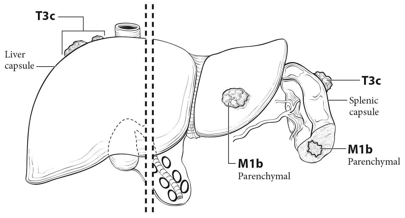
AJCC Prognostic Stage GroupsStage III: Spread to the retroperitoneal (pelvic and/or para-aortic) lymph nodes without extrapelvic peritoneal dissemination is Stage IIIA1, whereas microscopic extrapelvic (above the pelvic brim) peritoneal involvement with or without positive retroperitoneal lymph nodes is Stage IIIA2. In Stage IIIC, there is macroscopic peritoneal metastasis beyond the pelvis, >2 cm in size, with or without metastasis to the retroperitoneal lymph nodes. Tumor extension or metastasis to the liver and/or splenic capsule without parenchymal involvement is still Stage IIIC.I
AJCC Prognostic Stage GroupsStage III: IIIA1 is subdivided into IIIA1(i)—metastasis up to and including 10 mm in greatest dimension—and IIIA1(ii)—metastasis more than 10 mm in greatest dimensionIII
AJCC Prognostic Stage GroupsStage IV: Parenchymal liver or splenic involvement by tumor extension or isolated metastasis is now Stage IVB and should be identified and distinguished from splenic or liver capsular involvement only. Status of splenic involvement as defined here is new with this edition. Transmural intestinal involvement is now Stage IVB.I

ICD-O-3 Topography Codes

CodeDescription
C48.1Specified parts of peritoneum
C48.2Peritoneum (female only)
C48.8Overlapping lesion of retroperitoneum and peritoneum (female only)
C56.9Ovary
C57.0Fallopian tube

Excludes topography 48.1, 48.2, 48.8

WHO Classification of Tumors

This list includes histology codes and preferred terms from the WHO Classification of Tumors and the International Classification of Diseases for Oncology (ICD-O). Most of the terms in this list represent malignant behavior. For cancer reporting purposes, behavior codes /3 (denoting malignant neoplasms), /2 (denoting in situ neoplasms), and in some cases /1 (denoting neoplasms with uncertain and unknown behavior) may be appended to the 4-digit histology codes to create a complete morphology code.

CodeDescription
8000Neoplasm, malignant
8010Carcinoma, NOS
8020Undifferentiated carcinoma
8041Small cell carcinoma, pulmonary type
8044Small cell carcinoma, hypercalcemic type
8070Squamous cell carcinoma
8120Transitional cell carcinoma
8140Adenocarcinoma
8240Carcinoid
8243Mucinous carcinoid
8310Clear cell carcinoma
8313Clear cell borderline tumor / atypical proliferative clear cell tumor
8323Mixed cell adenocarcinoma
8380Endometrioid borderline tumor / atypical proliferative endometrioid tumor
8380Endometrioid carcinoma
8410Sebaceous carcinoma
8441Serous tubal intraepithelial carcinoma
8441Serous cystadenocarcinoma, NOS
8442Serous borderline tumor / atypical proliferative serous tumor
8452Solid pseudopapillary neoplasm
8460Low-grade serous carcinoma
8461High-grade serous carcinoma
8470Mucinous cystadenocarcinoma, NOS
8472Mucinous borderline tumor / atypical proliferative mucinous tumor
8474Seromucinous borderline tumor / atypical proliferative seromucinous tumor
8474Seromucinous carcinoma
8480Mucinous carcinoma
8590Sex cord-stromal tumor, NOS
8594Mixed germ cell sex cord-stromal tumor, unclassified
8620Adult granulosa cell tumor
8622Juvenile granulosa cell tumor
8623Sex cord tumor with annular tubules
8631Sertoli-Leydig cell tumor, moderately differentiated
8631Sertoli-Leydig cell tumor, poorly differentiated
8631Sertoli-Leydig cell tumor, well differentiated
8633Sertoli-Leydig cell tumor, retiform
8634Sertoli-Leydig cell tumor, moderately differentiated with heterologous elements
8634Sertoli-Leydig cell tumor, poorly differentiated with heterologous elements
8634Sertoli-Leydig cell tumor, retiform with heterologous elements
8640Sertoli cell tumor
8670Steroid cell tumor, malignant
8806Desmoplastic small round cell tumor
8810Fibrosarcoma
8815Solitary fibrous tumor, malignant
8822Pelvic fibromatosis
8825Inflammatory myofibroblastic tumor
8890Leiomyomatosis peritonealis disseminata
8930High-grade endometrioid stromal sarcoma
8931Low-grade endometrioid stromal sarcoma
8933Adenosarcoma
8936Extra-gastrointestinal stromal tumor
8950Müllerian mixed tumor
8960Wilms tumor
8980Carcinosarcoma
9000Borderline Brenner tumor
9000Malignant Brenner tumor
9050Mesothelioma
9052Well-differentiated papillary mesothelioma
9060Dysgerminoma
9070Embryonal carcinoma
9071Yolk sac tumor
9073Gonadoblastoma, including gonadoblastoma with malignant germ cell tumor
9080Immature teratoma
9085Mixed germ cell tumor
9090Struma ovarii, malignant
9091Strumal carcinoid
9100Non-gestational choriocarcinoma
9110Adenocarcinoma of rete ovarii
9110Wolffian tumor

Histology is not ideal for clinical use in patient care, as it describes an unspecified or outdated diagnosis. Data collectors may use this code only if there is not enough information in the medical record to document a more specific diagnosis.

Excludes histology 8810, 8815, 8825, 8890, and 8936.

Kurman RJ, Carcangiu ML, Herrington CS, Young RH, eds. World Health Organization Classification of Tumours of the Female Reproductive System. Lyon: IARC; 2014.

International Agency for Research on Cancer, World Health Organization. International Classification of Diseases for Oncology. ICD-O-3-Online.http://codes.iarc.fr/home. Accessed August 16, 2017. Used with permission.

Introduction

Ovarian cancer represents a heterogeneous group of distinct diseases. Approximately 90% of ovarian cancers are carcinomas (malignant epithelial tumors). Much less common are malignant germ cell tumors (3%) and potentially malignant sex cord-stromal tumors (1-2%). The most common type of epithelial ovarian cancer, high-grade serous carcinoma (HGSC), rarely may present as primary fallopian tube cancer or primary peritoneal cancer. Clinically, however, these three cancers are treated similarly, and a single staging system recently was proposed by the Federation Internationale de Gynecologie et d'Obstetrique (FIGO). Whereas some HGSCs, mainly (breast cancer susceptibility gene-positive [BRCA+] cases, seem to originate in the fimbriated end of the fallopian tube, other cases most likely arise from embryonic progenitors in the peritoneum or the ovarian surface epithelium. High-grade serous tubal intraepithelial carcinoma (STIC) can metastasize and, therefore, cannot be considered carcinoma in situ. The staging of cancer of the ovary, fallopian tube, and peritoneum in the AJCC Cancer Staging Manual, 8th Edition mirrors that of FIGO: for Stage I tumors, intraoperative rupture (“surgical spill”) is Stage IC1, capsule ruptured before surgery or tumor on ovarian or fallopian tube surface is Stage IC2, and positive peritoneal cytology with or without rupture is Stage IC3. The new staging preserves the separation between pelvic (Stage II) and extrapelvic (Stage III) spread but includes a revision of Stage III tumors; assignment to Stage IIIA1 is based on spread to the retroperitoneal (pelvic and/or para-aortic) lymph nodes without extrapelvic peritoneal dissemination (formerly classified as Stage IIIC). Extension and/or metastasis of tumor to the liver or splenic parenchyma qualifies as Stage IVB and should be distinguished from splenic or liver capsular involvement only (Stage IIIC).

Ovarian cancer is the fifth most common cancer diagnosis among women in higher-resource regions.1 Primary peritoneal cancer and primary fallopian tube cancer are rare malignancies, usually HGSCs—that is, identical to the most common histotype of ovarian cancer and the prototype tumor occurring in women with BRCA1 or BRCA2 germline mutations. Clinically, these three cancers are treated similarly,2 and a single staging system has been adopted by FIGO.3

During the past 30 years, it has been recognized that ovarian cancer is not a homogeneous disease but rather a group of diseases—each with different morphology and biological behavior. Approximately 90% of ovarian cancers are carcinomas (malignant epithelial tumors), and based on histopathology, immunohistochemistry, and molecular genetic analysis, at least five main types are distinguished: HGSC (70%), endometrioid carcinoma (EC; 10%), clear cell carcinoma (CCC; 10%), mucinous carcinoma (MC; 3%), and low-grade serous carcinoma (LGSC; less than 5%). These tumor types, which account for 98% of ovarian carcinomas, can be reproducibly diagnosed by light microscopy and are inherently different diseases, as indicated by differences in epidemiologic and genetic risk factors, precursor lesions, patterns of spread, molecular genetic abnormalities, response to chemotherapy, and prognosis.4-6 The vast majority of borderline tumors (formerly referred to as “tumors of low malignant potential”) rarely recur or metastasize; however, up to 10% may recur. At the time of recurrence, some have the features of carcinoma, particularly of the serous and mucinous types. Much less common are malignant germ cell tumors (dysgerminomas, yolk sac tumors, and immature teratomas [3% of ovarian cancers] and potentially malignant sex cord-stromal tumors [1-2%, mainly granulosa cell tumors]). Ovarian cancers differ primarily based on histologic type.

Reproducible histopathologic diagnosis of tumor cell type is required for successful treatment. Different tumor histotypes respond differently to chemotherapy. Even if different patterns of dissemination would justify the use of separate staging systems for each type of ovarian carcinoma, such a complex classification would not be practical. For the sake of simplicity, a flexible staging system that takes into account the most relevant prognostic parameters shared by all tumor types should be used. Histologic type should be designated at staging (i.e., HGSC, EC, CCC, MC, LGSC, and borderline tumors; other or cannot be classified; and malignant germ cell tumors and potentially malignant sex cord-stromal tumors).

Patients with BRCA mutation (breast-ovarian cancer syndrome) undergoing risk-reducing salpingo-oophorectomy (RRSO) are found to have high-grade STIC, particularly in the fimbria.7 Although STIC can metastasize and therefore cannot be considered carcinoma in situ, compelling evidence for a tubal origin of BRCA+ HGSC has accumulated during the past decade.8,9 High-grade STIC also has been found in an undetermined number of advanced-stage sporadic HGSCs associated with ovarian tumor masses and in rare cases of primary tubal or peritoneal HGSCs without obvious ovarian involvement. The relative proportion of HGSCs of ovarian and tubal derivation is unknown, probably because in advanced-stage cancers, tumor growth conceals the primary site. Even in cases involving BRCA mutation, evidence of a tubal origin of HGSCs is incomplete and a multicentric origin of these tumors cannot be excluded.

Whereas asymptomatic BRCA+ women undergoing RRSO are found to have STIC in 6% of cases, symptomatic, rapidly progressive BRCA+ tumors discovered at advanced stage in younger patients are less likely to be associated with STIC. This paradox questions the effectiveness of salpingectomy alone in preventing HGSC in BRCA+ women.10

The aforementioned findings suggest that the fallopian tube is linked to only some HGSCs and that the remaining cases originate from the nearby peritoneum/ovarian surface epithelium. Recently, it was hypothesized that cytokeratin 7-positive embryonic/stem cells may be capable of Müllerian differentiation in cortical epithelial inclusion cysts resulting from ovarian surface epithelium (mesothelium) invaginations. Thus, embryonic progenitors may give rise to immunophenotypically distinct neoplastic progeny,11 which would support the old concept of “Müllerian neometaplasia.”

HGSCs and LGSCs are fundamentally different tumor types and, consequently, different diseases. HGSCs are the most common ovarian carcinomas, and most patients (approximately 80%) present with advanced-stage disease; tumors confined to the ovary at diagnosis are distinctly uncommon (less than 10%). By contrast, LGSCs are much less common, usually contain a serous borderline component, and carry KRAS and BRAF mutations.12,13 HGSCs are not associated with serous borderline tumors and typically exhibit TP53 mutations and BRCA abnormalities, resulting in chromosomal instability and widespread DNA copy number changes. This highly aberrant genome is the hallmark of HGSC and allows further evolution into different molecular subtypes associated with clinical outcome.

A putative tubal or peritoneal origin applies exclusively to HGSCs and not to the vast majority of ECs and CCCs, which are thought to arise in the ovary from endometriosis. Although a significant number of HGSCs might not arise from the ovary, and the term ovarian cancer may not be pathogenically precise in every case, ovarian involvement is the rule in almost all cases. In view of the rarity of HGSCs associated with tubal tumor masses, it is unlikely that all HGSCs originate in the fallopian tube. The term HGSC of ovary should be kept until the different origins of ovarian tumors are better understood. Terms such as Müllerian or pelvic serous carcinoma are not recommended because they create confusion for patients, physicians, and medical investigators.14

Classification Rules

Primary Site(s)

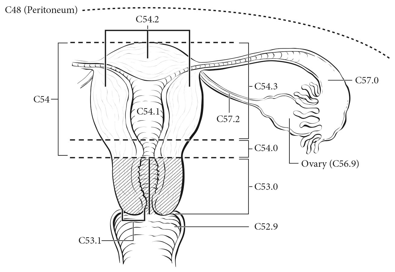
The ovaries are a pair of solid, flattened ovoids 2 to 4 cm in diameter that are connected by a peritoneal fold to the broad ligament and by the infundibulopelvic ligament to the lateral wall of the pelvis. They are attached medially to the uterus by the utero-ovarian ligament (Figure 68.1).

The fallopian tube extends from the posterior superior aspect of the uterine fundus laterally and anterior to the ovary. Its length is approximately 10 cm. The medial end arises in the cornual portion of the uterine cavity, and the lateral(fimbrial) end opens to the peritoneal cavity.

The peritoneum is the serous membrane of the abdominal cavity that lines the walls of the abdomen (parietal peritoneum) and covers the abdominal organs (visceral peritoneum). The pelvic peritoneum covers the fundus of the urinary bladder and the front of the rectum. In females, it lines the anterior and posterior surface of the uterus and the upper posterior vagina. There are two potential spaces posterior to the bladder (the uterovesical pouch) and posterior to the uterus (the rectouterine pouch of Douglas).On the anterior and posterior surfaces of the uterus, the peritoneum is reflected laterally to the pelvic sidewalls as the broad ligaments, containing the fallopian tubes.

68.1 Anatomic sites of the ovary (C56.9), fallopian tube (C57.0) and primary peritoneum (C48).

Regional Lymph Nodes

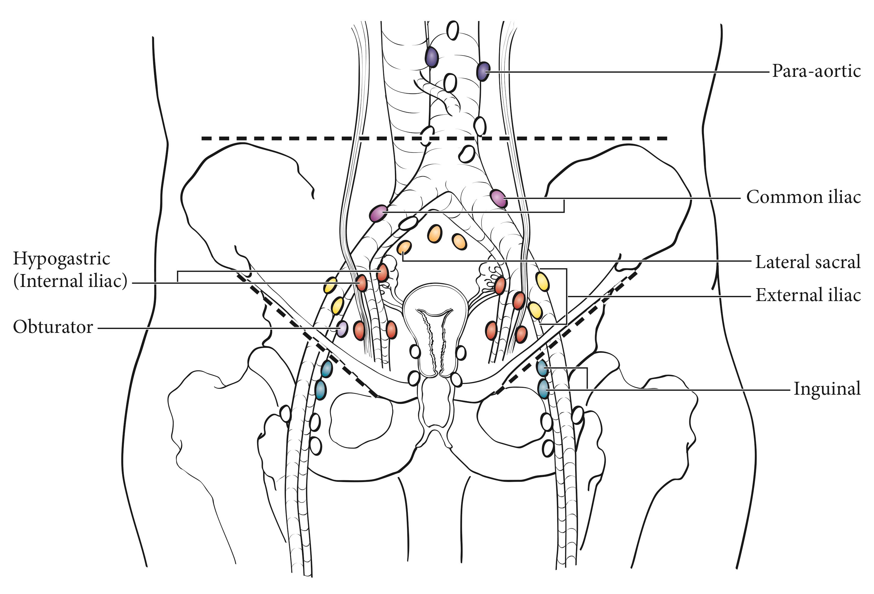
The lymphatic drainage occurs by the infundibulopelvic and round ligament trunks and an external iliac accessory route into the following regional nodes (Figure 68.2):

  • External iliac
  • Internal iliac (hypogastric)
  • Obturator
  • Common iliac
  • Para-aortic
  • Pelvic, NOS
  • Retroperitoneal, NOS

68.2 Regional lymph nodes of ovary, fallopian tube and primary peritoneal carcinomas.

Metastatic Sites

The peritoneum, including the omentum and the pelvic and abdominal visceral and parietal peritoneum, comprises common sites for seeding. Diaphragmatic and liver surface involvement also is common. However, to be consistent with FIGO staging, these implants within the abdominal cavity (T3) are not considered distant metastases. Extraperitoneal sites, including parenchymal liver, lung, spleen, and skeletal metastases, and inguinal, supraclavicular, and axillary nodes, are M1.

Clinical Classification

Ovarian, fallopian tube, and peritoneal cancer is surgically and pathologically staged. There should be histologic confirmation of the ovarian, fallopian tube, and peritoneal disease. Laparotomy or operative laparoscopy with resection of the ovarian mass, as well as hysterectomy, forms the basis for staging. Biopsies of all frequently involved sites, such as the omentum, mesentery, diaphragm, peritoneal surfaces, pelvic nodes, and para-aortic nodes, are required for ideal staging of early disease. For example, to stage a patient confidently as Stage IA (T1 N0 M0), negative biopsies of all of the aforementioned sites should be obtained to exclude microscopic metastases. Assignment to Stage IIIA1 is based on spread to the retroperitoneal lymph nodes without intraperitoneal dissemination, because an analysis of these patients indicates that their survival is significantly better than that of patients with intraperitoneal dissemination.3,15-19 On the other hand, a single biopsy from an omental mass 2 cm or greater showing metastatic carcinoma is adequate to classify a patient as Stage IIIC, thus making other biopsies unnecessary from a staging standpoint. The final histologic and cytologic findings after surgery are to be considered in the staging. Operative findings before tumor debulking determine stage, which may be modified by histopathologic as well as clinical or radiologic evaluation (e.g., palpable supraclavicular node or pulmonary metastases on chest X-ray).

Although clinical assessments similar to those for other sites may be performed, surgical-pathological evaluation of the abdomen and pelvis is necessary to establish a definitive diagnosis of ovarian/fallopian tube/peritoneal cancer and to rule out other primary malignancies that may present with similar preoperative findings (e.g., bowel, uterine, and pancreatic cancers or, occasionally, lymphoma). Although laparotomy is the most widely accepted procedure for surgical-pathological staging, occasionally laparoscopy may be used. Occasionally, patients with advanced disease and/or women who are medically unsuitable candidates for surgery may be presumed to have ovarian cancer on the basis of cytology of ascites or pleural effusion showing typical carcinoma, combined with imaging studies demonstrating enlarged ovaries/fallopian tubes, and/or peritoneal involvement. Such patients usually are considered unstaged (TX), although positive cytology of a pleural effusion or supraclavicular lymph node occasionally allows designation of M1 or FIGO Stage IV disease. The presence of ascites does not affect staging unless malignant cells are present.

Imaging studies often are done in conjunction with definitive abdominal-pelvic surgery, and chest X-ray, bone scans, computed tomography (CT), or positron emission tomography (PET) may identify lung, bone, or brain metastases that should be considered in the final stage. Pleural effusions should be evaluated with cytology. In the future, pretreatment imaging will be more relevant to staging because of the increasing use of neoadjuvant chemotherapy in many women diagnosed with ovarian cancer.20

As with all gynecologic cancers, the final stage should be established at the time of initial treatment. It should not be modified or changed on the basis of subsequent findings.

Findings related to procedures such as laparoscopy or laparotomy after initial chemotherapy do not change the patient's original stage.

Imaging

CT currently is the preferred modality for the staging of ovarian cancer. Magnetic resonance (MR) imaging is excellent for characterizing adnexal masses. PET/CT is useful in evaluating distant disease but not for diagnosing primary ovarian cancer.

TNM Components of Tumor Staging

In clinical category T1, disease is limited to the ovaries; either MR imaging or ultrasound may be helpful in diagnosing the malignant adnexal mass. Contrast-enhanced CT is useful in assessing peritoneal disease. If the disease is confined to the pelvis, it is clinical category T2. Category T3a/b includes retroperitoneal lymph node metastases. PET/CT has been advocated to assess for lymph node metastases, as it has better specificity than contrast-enhanced CT or MR imaging. Category T3c includes surface involvement of the liver and spleen without any parenchymal metastases, which can be assessed with contrast-enhanced CT. PET/CT, if available, may be used as a single modality to assess both peritoneal disease and lung parenchymal disease in patients with advanced cancer.

The imaging criteria used to assess lymph node metastases are based on node size, with abnormal being greater than 1 cm in the short axial dimension on cross-sectional scans. CT and MR imaging are shown to perform equally well in assessing adenopathy. However, because there may be false positive causes of enlarged nodes from benign disease, PET/CT is considered superior for assessing lymph node metastases. Metabolically active lymph nodes of any size on PET/CT are considered metastatic.

Suggested Imaging Report Format

  1. Primary tumor
    1. One or both ovaries
  2. Local extent
    1. Ascites
    2. Indicate whether localized to the pelvis or whether extrapelvic disease is present
    3. Retroperitoneal adenopathy
    4. Liver of splenic surface disease or lung parenchymal disease
    5. Pleural effusion

Pathological Classification

Surgery and biopsy of all suspected sites of involvement provide the basis for staging. Histologic and cytologic data are required. This is the preferred method of staging for ovarian cancer. The operative notes and/or the pathology report should describe the location and size of metastatic lesions and the primary tumors for optimal staging. In addition, the size of the tumor outside the pelvis must be determined, and noted and documented in the operative report. Size is reported in centimeters and represents the largest implant, regardless of whether it was resected during surgical exploration.

Carcinoma of the fallopian tube almost always is HGSC, which may be accompanied by STIC. The tumor invades locally into the muscular wall of the tube and then into the peritubal soft tissue or adjacent organs, such as the uterus or ovary, or through the serosa of the tube into the peritoneal cavity. Metastatic tumor implants may be found throughout the peritoneal cavity. The tumor may obstruct the tubal lumen and present as a ruptured or unruptured hydrosalpinx or hematosalpinx. It has been suggested that carcinomas in the fimbriated end without invasion have a worse prognosis than those invading the wall of the tube because of direct access to the peritoneal cavity.15

Examination of prophylactic salpingo-oophorectomy specimens from BRCA+ patients has shown thatmost early carcinomas detected in these samples occur in the tubal fimbria, and some of them arestill confined to the mucosa in the form of STIC.16,17 To detect these early carcinomas, serial longitudinal sections of the fallopian tube fimbria at 2- to 3-mm intervals should be obtained to examine most of the plicae surface.

Advanced invasive HGSC associated with STIC may be ovarian or tubal in origin without clinical relevance. For tumors limited to one ovary associated with STIC, there are three possibilities: a) STIC extending to one ovary, b) ovarian HGSC extending to the fallopian tube, and c) synchronous or metachronous tumor involving the ovary and fallopian tube. With regard to staging, these tumors are considered Stage IA ovarian carcinoma with STIC unless there is evidence of direct extension from STIC to the ovary, in which case they would be stage IIA fallopian tube carcinoma.

In some cases, an adenocarcinoma is primary in the peritoneum. The ovaries are not involved or are involved only with minimal surface implants. The clinical presentation, surgical therapy, chemotherapy, and prognosis of these peritoneal tumors mirror those of HGSC of the ovary. Patients who undergo prophylactic salpingo-oophorectomy for a familial history of ovarian cancer appear to retain a 1-2% chance of developing peritoneal adenocarcinoma, which is histopathologically and clinically similar to primary ovarian cancer. It is not possible to have Stage I peritoneal cancer.

Intranodal single tumor cells or small clusters of cells not more than 0.2 mm in greatest diameter are classified as isolated tumor cells. These may be detected by routine histology or by immunohistochemical methods. They are designated N0(i+).

For pN0, histologic examination should include both pelvic and para-aortic lymph nodes.

For patients receiving neoadjuvant therapy, it is important to record the clinical stage before treatment. Surgical staging after neoadjuvant therapy should be classified as “yp.”

Registry Data Collection Variables
  1. FIGO stage
  2. Preoperative CA-125 level
  3. Gross residual tumor after primary cytoreductive surgery
  4. Residual tumor volume after primary cytoreductive surgery
  5. Residual tumor location after primary cytoreductive surgery
Prognostic Factors

Prognostic Factors Required for Stage Grouping

Beyond the factors used to assign T, N, or M categories, no additional prognostic factors are required for stage grouping.

Additional Factors Recommended for Clinical Care

FIGO Stage

FIGO stage3 is the strongest predictor of outcome in ovarian cancer. Whereas complete staging would be sufficient surgical treatment for tumors in Stages I and II, patients with advanced disease require cytoreductive surgery. Stage I ovarian cancer is confined to the ovaries, and fewer than 5% of HGSCs are Stage I tumors. Tumor rupture, ovarian surface involvement by tumor cells, or the presence of malignant cells in peritoneal washings or ascitic fluid warrants a Stage IC classification.

Fewer than 10% of HGSCs are found in Stage II, that is, extending or metastasizing to extraovarian/extratubal pelvic organs or tissues. Stage II includes examples of direct extension to the tubes/ovaries and pelvic sidewall, as well as pelvic peritoneal metastases. Thus, it includes resectable and curable tumors that have extended to adjacent organs, as well as tumors that have seeded the pelvic peritoneum and are associated with a poor prognosis.

HGSC most commonly presents in Stage III, and the vast majority of these cases are Stage IIIC. These tumors typically spread along the abdominopelvic peritoneum, involving the omentum, serosa of the small and large bowel, mesentery, paracolic gutters, diaphragm, and peritoneal surfaces of the liver and spleen. Ascites is found in almost all cases, and positive lymph nodes are found in many patients who undergo node sampling or lymphadenectomy and in almost 80% of those with advanced-stage tumors.

The new FIGO staging system includes a revision of Stage III criteria. The designation of Stage IIIA1 is based on spread to the retroperitoneal lymph nodes without intraperitoneal dissemination, because an analysis of these patients indicates that their survival is significantly better than that of patients with intraperitoneal dissemination.17-20,22 Nodal metastasis without peritoneal metastasis is relatively uncommon (about 9% of cases).23 Most of these patients have positive para-aortic nodes.

AJCC Level of Evidence: I
Histology and Grade

Histology and grade are important prognostic factors. Women with borderline tumors have an excellent prognosis, even when noninvasive extraovarian disease (noninvasive implants) is found. In patients with invasive ovarian cancer, low-grade tumors have a better prognosis than high-grade tumors, stage for stage. Histologic type, which includes the histologic grade, also is extremely important. Some stromal tumors (granulosa and Sertoli-Leydig cell tumors) have an excellent prognosis, whereas malignant epithelial tumors in general have a less favorable outcome. For this reason, epithelial cell types generally are reported together and sex cord-stromal tumors and germ cell tumors are reported separately. Tumor cell type also helps guide the type of chemotherapy that is recommended.

AJCC Level of Evidence: I
Residual Disease

In advanced disease, the most important prognostic factor is residual disease after initial surgical management. Even among patients with advanced-stage cancer, those with no gross residual disease after surgical debulking have a considerably better prognosis than those with minimal or extensive residual disease. Besides the size of the residual tumor, the number of sites of residual disease also appears to be important (tumor volume).

AJCC Level of Evidence: I
Preoperative Cancer Antigen 125

The tumor marker cancer antigen 125 (CA-125) is useful for following the response to therapy in patients with epithelial ovarian cancer, who have elevated levels of this marker. The rate of regression during chemotherapy may have prognostic significance. Women with germ cell tumors also may have elevated serum tumor markers, namely α-fetoprotein or human chorionic gonadotropin. Other factors, such as growth factors and oncogene amplification, currently are under investigation.

AJCC Level of Evidence: I
Gross Residual Tumor after Primary Cytoreductive Surgery

Gross residual tumor after primary cytoreductive surgery is a prognostic factor that has been demonstrated in several large studies. Whether patients undergo neoadjuvant chemotherapy or primary cytoreduction, the best prognostic category after surgery includes those who are left with no gross residual disease. Physicians should record the presence or absence of residual disease; if residual disease is observed, the size of the largest visible lesion should be documented.
AJCC Level of Evidence: I
Residual Tumor Volume after Primary Cytoreductive Surgery

Although volume of residual disease is an important prognostic factor in most studies, it applies only to Stages IIIC and IV. The parameter that defines optimal cytoreduction is residual disease less than 1 cm. Physicians should record no gross tumor, tumor less than or equal to 1 cm, or tumor greater than 1 cm.

AJCC Level of Evidence: I
Residual Tumor Location following Primary Cytoreductive Surgery
Residual tumor location should be recorded in the operative notes.
AJCC Level of Evidence: I

The authors have not provided recommendations for clinical trial stratification.

Risk Assessment

Risk Assesment Models

The AJCC recently established guidelines that will be used to evaluate published statistical prediction models for the purpose of granting endorsement for clinical use.24 Although this is a monumental step toward the goal of precision medicine, this work was published only very recently. For this reason, the existing models that have been published or may be in clinical use have not yet been evaluated for this cancer site by the Precision Medicine Core of the AJCC. In the future, the statistical prediction models for this cancer site will be evaluated, and those that meet all AJCC criteria will be endorsed.

Recommendations

The authors have not provided recommendations for clinical trial stratification.

TNM Definitions

The definitions of the T categories correspond to the stages accepted by the Federation Internationale de Gynecologie et d'Obstetrique (FIGO).3 Both systems are included for comparison.

Clinical T (cT)

cT CategoryFIGO StagecT Criteria
cTXN/APrimary tumor cannot be assessed
cT0N/ANo evidence of primary tumor
cT1ITumor limited to ovaries (one or both) or fallopian tube(s)
cT1aIATumor limited to one ovary (capsule intact) or fallopian tube, no tumor on ovarian or fallopian tube surface; no malignant cells in ascites or peritoneal washings
cT1bIBTumor limited to both ovaries (capsules intact) or fallopian tubes; no tumor on ovarian or fallopian tube surface; no malignant cells in ascites or peritoneal washings
cT1cICTumor limited to one or both ovaries or fallopian tubes, with any of the following:
cT1c1IC1Surgical spill
cT1c2IC2Capsule ruptured before surgery or tumor on ovarian or fallopian tube surface
cT1c3IC3Malignant cells in ascites or peritoneal washings
cT2IITumor involves one or both ovaries or fallopian tubes with pelvic extension below pelvic brim or primary peritoneal cancer
cT2aIIAExtension and/or implants on the uterus and/or fallopian tube(s) and/or ovaries
cT2bIIBExtension to and/or implants on other pelvic tissues
cT3IIITumor involves one or both ovaries or fallopian tubes, or primary peritoneal cancer, with microscopically confirmed peritoneal metastasis outside the pelvis and/or metastasis to the retroperitoneal (pelvic and/or para-aortic) lymph nodes
cT3aIIIA2Microscopic extrapelvic (above the pelvic brim) peritoneal involvement with or without positive retroperitoneal lymph nodes
cT3bIIIBMacroscopic peritoneal metastasis beyond pelvis 2 cm or less in greatest dimension with or without metastasis to the retroperitoneal lymph nodes
cT3cIIICMacroscopic peritoneal metastasis beyond the pelvis more than 2 cm in greatest dimension with or without metastasis to the retroperitoneal lymph nodes (includes extension of tumor to capsule of liver and spleen without parenchymal involvement of either organ)

Pathological T (pT)

pT CategoryFIGO StagepT Criteria
pTXN/APrimary tumor cannot be assessed
pT0N/ANo evidence of primary tumor
pT1ITumor limited to ovaries (one or both) or fallopian tube(s)
pT1aIATumor limited to one ovary (capsule intact) or fallopian tube, no tumor on ovarian or fallopian tube surface; no malignant cells in ascites or peritoneal washings
pT1bIBTumor limited to both ovaries (capsules intact) or fallopian tubes; no tumor on ovarian or fallopian tube surface; no malignant cells in ascites or peritoneal washings
pT1cICTumor limited to one or both ovaries or fallopian tubes, with any of the following:
pT1c1IC1Surgical spill
pT1c2IC2Capsule ruptured before surgery or tumor on ovarian or fallopian tube surface
pT1c3IC3Malignant cells in ascites or peritoneal washings
pT2IITumor involves one or both ovaries or fallopian tubes with pelvic extension below pelvic brim or primary peritoneal cancer
pT2aIIAExtension and/or implants on the uterus and/or fallopian tube(s) and/or ovaries
pT2bIIBExtension to and/or implants on other pelvic tissues
pT3IIITumor involves one or both ovaries or fallopian tubes, or primary peritoneal cancer, with microscopically confirmed peritoneal metastasis outside the pelvis and/or metastasis to the retroperitoneal (pelvic and/or para-aortic) lymph nodes
pT3aIIIA2Microscopic extrapelvic (above the pelvic brim) peritoneal involvement with or without positive retroperitoneal lymph nodes
pT3bIIIBMacroscopic peritoneal metastasis beyond pelvis 2 cm or less in greatest dimension with or without metastasis to the retroperitoneal lymph nodes
pT3cIIICMacroscopic peritoneal metastasis beyond the pelvis more than 2 cm in greatest dimension with or without metastasis to the retroperitoneal lymph nodes (includes extension of tumor to capsule of liver and spleen without parenchymal involvement of either organ)
cTXN/APrimary tumor cannot be assessed
cT0N/ANo evidence of primary tumor
cT1ITumor limited to ovaries (one or both) or fallopian tube(s)
cT1aIATumor limited to one ovary (capsule intact) or fallopian tube, no tumor on ovarian or fallopian tube surface; no malignant cells in ascites or peritoneal washings
cT1bIBTumor limited to both ovaries (capsules intact) or fallopian tubes; no tumor on ovarian or fallopian tube surface; no malignant cells in ascites or peritoneal washings
cT1cICTumor limited to one or both ovaries or fallopian tubes, with any of the following:
cT1c1IC1Surgical spill
cT1c2IC2Capsule ruptured before surgery or tumor on ovarian or fallopian tube surface
cT1c3IC3Malignant cells in ascites or peritoneal washings
cT2IITumor involves one or both ovaries or fallopian tubes with pelvic extension below pelvic brim or primary peritoneal cancer
cT2aIIAExtension and/or implants on the uterus and/or fallopian tube(s) and/or ovaries
cT2bIIBExtension to and/or implants on other pelvic tissues
cT3IIITumor involves one or both ovaries or fallopian tubes, or primary peritoneal cancer, with microscopically confirmed peritoneal metastasis outside the pelvis and/or metastasis to the retroperitoneal (pelvic and/or para-aortic) lymph nodes
cT3aIIIA2Microscopic extrapelvic (above the pelvic brim) peritoneal involvement with or without positive retroperitoneal lymph nodes
cT3bIIIBMacroscopic peritoneal metastasis beyond pelvis 2 cm or less in greatest dimension with or without metastasis to the retroperitoneal lymph nodes
cT3cIIICMacroscopic peritoneal metastasis beyond the pelvis more than 2 cm in greatest dimension with or without metastasis to the retroperitoneal lymph nodes (includes extension of tumor to capsule of liver and spleen without parenchymal involvement of either organ)

Neoadjuvant Clinical T (yT)

ycT CategoryFIGO StageycT Criteria
ycTXN/APrimary tumor cannot be assessed
ycT0N/ANo evidence of primary tumor
ycT1ITumor limited to ovaries (one or both) or fallopian tube(s)
ycT1aIATumor limited to one ovary (capsule intact) or fallopian tube, no tumor on ovarian or fallopian tube surface; no malignant cells in ascites or peritoneal washings
ycT1bIBTumor limited to both ovaries (capsules intact) or fallopian tubes; no tumor on ovarian or fallopian tube surface; no malignant cells in ascites or peritoneal washings
ycT1cICTumor limited to one or both ovaries or fallopian tubes, with any of the following:
ycT1c1IC1Surgical spill
ycT1c2IC2Capsule ruptured before surgery or tumor on ovarian or fallopian tube surface
ycT1c3IC3Malignant cells in ascites or peritoneal washings
ycT2IITumor involves one or both ovaries or fallopian tubes with pelvic extension below pelvic brim or primary peritoneal cancer
ycT2aIIAExtension and/or implants on the uterus and/or fallopian tube(s) and/or ovaries
ycT2bIIBExtension to and/or implants on other pelvic tissues
ycT3IIITumor involves one or both ovaries or fallopian tubes, or primary peritoneal cancer, with microscopically confirmed peritoneal metastasis outside the pelvis and/or metastasis to the retroperitoneal (pelvic and/or para-aortic) lymph nodes
ycT3aIIIA2Microscopic extrapelvic (above the pelvic brim) peritoneal involvement with or without positive retroperitoneal lymph nodes
ycT3bIIIBMacroscopic peritoneal metastasis beyond pelvis 2 cm or less in greatest dimension with or without metastasis to the retroperitoneal lymph nodes
ycT3cIIICMacroscopic peritoneal metastasis beyond the pelvis more than 2 cm in greatest dimension with or without metastasis to the retroperitoneal lymph nodes (includes extension of tumor to capsule of liver and spleen without parenchymal involvement of either organ)

Neoadjuvant Pathological T (yT)

ypT CategoryFIGO StageypT Criteria
ypTXN/APrimary tumor cannot be assessed
ypT0N/ANo evidence of primary tumor
ypT1ITumor limited to ovaries (one or both) or fallopian tube(s)
ypT1aIATumor limited to one ovary (capsule intact) or fallopian tube, no tumor on ovarian or fallopian tube surface; no malignant cells in ascites or peritoneal washings
ypT1bIBTumor limited to both ovaries (capsules intact) or fallopian tubes; no tumor on ovarian or fallopian tube surface; no malignant cells in ascites or peritoneal washings
ypT1cICTumor limited to one or both ovaries or fallopian tubes, with any of the following:
ypT1c1IC1Surgical spill
ypT1c2IC2Capsule ruptured before surgery or tumor on ovarian or fallopian tube surface
ypT1c3IC3Malignant cells in ascites or peritoneal washings
ypT2IITumor involves one or both ovaries or fallopian tubes with pelvic extension below pelvic brim or primary peritoneal cancer
ypT2aIIAExtension and/or implants on the uterus and/or fallopian tube(s) and/or ovaries
ypT2bIIBExtension to and/or implants on other pelvic tissues
ypT3IIITumor involves one or both ovaries or fallopian tubes, or primary peritoneal cancer, with microscopically confirmed peritoneal metastasis outside the pelvis and/or metastasis to the retroperitoneal (pelvic and/or para-aortic) lymph nodes
ypT3aIIIA2Microscopic extrapelvic (above the pelvic brim) peritoneal involvement with or without positive retroperitoneal lymph nodes
ypT3bIIIBMacroscopic peritoneal metastasis beyond pelvis 2 cm or less in greatest dimension with or without metastasis to the retroperitoneal lymph nodes
ypT3cIIICMacroscopic peritoneal metastasis beyond the pelvis more than 2 cm in greatest dimension with or without metastasis to the retroperitoneal lymph nodes (includes extension of tumor to capsule of liver and spleen without parenchymal involvement of either organ)

Definition of Regional Lymph Node (N)

Clinical N (cN)
cN CategoryFIGO StagecN Criteria
cNXN/ARegional lymph nodes cannot be assessed
cN0N/ANo regional lymph node metastasis
cN0(i+)N/AIsolated tumor cells in regional lymph node(s) no greater than 0.2 mm
cN1IIIA1Positive retroperitoneal lymph nodes only (histologically confirmed)
cN1aIIIA1iMetastasis up to and including 10 mm in greatest dimension
cN1bIIIA1iiMetastasis more than 10 mm in greatest dimension
Pathological N (pN)
pN CategoryFIGO StagepN Criteria
pNXN/ARegional lymph nodes cannot be assessed
pN0N/ANo regional lymph node metastasis
pN0(i+)N/AIsolated tumor cells in regional lymph node(s) no greater than 0.2 mm
pN1IIIA1Positive retroperitoneal lymph nodes only (histologically confirmed)
pN1aIIIA1iMetastasis up to and including 10 mm in greatest dimension
pN1bIIIA1iiMetastasis more than 10 mm in greatest dimension
cNXN/ARegional lymph nodes cannot be assessed
cN0N/ANo regional lymph node metastasis
cN0(i+)N/AIsolated tumor cells in regional lymph node(s) no greater than 0.2 mm
cN1IIIA1Positive retroperitoneal lymph nodes only (histologically confirmed)
cN1aIIIA1iMetastasis up to and including 10 mm in greatest dimension
cN1bIIIA1iiMetastasis more than 10 mm in greatest dimension
Neoadjuvant Clinical N (pY)
ycN CategoryFIGO StageycN Criteria
ycNXN/ARegional lymph nodes cannot be assessed
ycN0N/ANo regional lymph node metastasis
ycN0(i+)N/AIsolated tumor cells in regional lymph node(s) no greater than 0.2 mm
ycN1IIIA1Positive retroperitoneal lymph nodes only (histologically confirmed)
ycN1aIIIA1iMetastasis up to and including 10 mm in greatest dimension
ycN1bIIIA1iiMetastasis more than 10 mm in greatest dimension
Neoadjuvant Pathological N (pY)
ypN CategoryFIGO StageypN Criteria
ypNXN/ARegional lymph nodes cannot be assessed
ypN0N/ANo regional lymph node metastasis
ypN0(i+)N/AIsolated tumor cells in regional lymph node(s) no greater than 0.2 mm
ypN1IIIA1Positive retroperitoneal lymph nodes only (histologically confirmed)
ypN1aIIIA1iMetastasis up to and including 10 mm in greatest dimension
ypN1bIIIA1iiMetastasis more than 10 mm in greatest dimension

Definition of Distant Metastasis (M)- Clinical M (cN)

cM CategoryFIGO StagecM Criteria
cM0N/ANo distant metastasis
cM1IVDistant metastasis, including pleural effusion with positive cytology; liver or splenic parenchymal metastasis; metastasis to extra-abdominal organs (including inguinal lymph nodes and lymph nodes outside the abdominal cavity); and transmural involvement of intestine
cM1bIVBLiver or splenic parenchymal metastases; metastases to extra-abdominal organs (including inguinal lymph nodes and lymph nodes outside the abdominal cavity); transmural involvement of intestine
pM1IVMicroscopic evidence of distant metastasis, including pleural effusion with positive cytology; liver or splenic parenchymal metastasis; metastasis to extra-abdominal organs (including inguinal lymph nodes and lymph nodes outside the abdominal cavity); and transmural involvement of intestine
pM1aIVAMicroscopic evidence of pleural effusion with positive cytology
pM1bIVBMicroscopic evidence of liver or splenic parenchymal metastases; metastases to extra-abdominal organs (including inguinal lymph nodes and lymph nodes outside the abdominal cavity); transmural involvement of intestine

Definition of Distant Metastasis (M)- Pathological M (pN)

pM CategoryFIGO StagepM Criteria
cM0N/ANo distant metastasis
cM1IVDistant metastasis, including pleural effusion with positive cytology; liver or splenic parenchymal metastasis; metastasis to extra-abdominal organs (including inguinal lymph nodes and lymph nodes outside the abdominal cavity); and transmural involvement of intestine
cM1bIVBLiver or splenic parenchymal metastases; metastases to extra-abdominal organs (including inguinal lymph nodes and lymph nodes outside the abdominal cavity); transmural involvement of intestine
pM1IVMicroscopic evidence of distant metastasis, including pleural effusion with positive cytology; liver or splenic parenchymal metastasis; metastasis to extra-abdominal organs (including inguinal lymph nodes and lymph nodes outside the abdominal cavity); and transmural involvement of intestine
pM1aIVAMicroscopic evidence of pleural effusion with positive cytology
pM1bIVBMicroscopic evidence of liver or splenic parenchymal metastases; metastases to extra-abdominal organs (including inguinal lymph nodes and lymph nodes outside the abdominal cavity); transmural involvement of intestine

Definition of Distant Metastasis (M)- Neoadjuvant Clinical M (pY)

ycM CategoryFIGO StageycM Criteria
cM0N/ANo distant metastasis
cM1IVDistant metastasis, including pleural effusion with positive cytology; liver or splenic parenchymal metastasis; metastasis to extra-abdominal organs (including inguinal lymph nodes and lymph nodes outside the abdominal cavity); and transmural involvement of intestine
cM1bIVBLiver or splenic parenchymal metastases; metastases to extra-abdominal organs (including inguinal lymph nodes and lymph nodes outside the abdominal cavity); transmural involvement of intestine
pM1IVMicroscopic evidence of distant metastasis, including pleural effusion with positive cytology; liver or splenic parenchymal metastasis; metastasis to extra-abdominal organs (including inguinal lymph nodes and lymph nodes outside the abdominal cavity); and transmural involvement of intestine
pM1aIVAMicroscopic evidence of pleural effusion with positive cytology
pM1bIVBMicroscopic evidence of liver or splenic parenchymal metastases; metastases to extra-abdominal organs (including inguinal lymph nodes and lymph nodes outside the abdominal cavity); transmural involvement of intestine

Definition of Distant Metastasis (M)- Neoadjuvant Pathological M (pY)

ypM CategoryFIGO StageypM Criteria
cM0N/ANo distant metastasis
cM1IVDistant metastasis, including pleural effusion with positive cytology; liver or splenic parenchymal metastasis; metastasis to extra-abdominal organs (including inguinal lymph nodes and lymph nodes outside the abdominal cavity); and transmural involvement of intestine
cM1bIVBLiver or splenic parenchymal metastases; metastases to extra-abdominal organs (including inguinal lymph nodes and lymph nodes outside the abdominal cavity); transmural involvement of intestine
pM1IVMicroscopic evidence of distant metastasis, including pleural effusion with positive cytology; liver or splenic parenchymal metastasis; metastasis to extra-abdominal organs (including inguinal lymph nodes and lymph nodes outside the abdominal cavity); and transmural involvement of intestine
pM1aIVAMicroscopic evidence of pleural effusion with positive cytology
pM1bIVBMicroscopic evidence of liver or splenic parenchymal metastases; metastases to extra-abdominal organs (including inguinal lymph nodes and lymph nodes outside the abdominal cavity); transmural involvement of intestine

Stage Prognostic

Clinical

When T is…and N is…and M is…Then the Clinical Prognostic Stage Group is…
cT1cN0cM0I
cT1cN0(i+)cM0I
cT1acN0cM0IA
cT1acN0(i+)cM0IA
cT1bcN0cM0IB
cT1bcN0(i+)cM0IB
cT1ccN0cM0IC
cT1ccN0(i+)cM0IC
cT1c1cN0cM0IC
cT1c1cN0(i+)cM0IC
cT1c2cN0cM0IC
cT1c2cN0(i+)cM0IC
cT1c3cN0cM0IC
cT1c3cN0(i+)cM0IC
cT2cN0cM0II
cT2cN0(i+)cM0II
cT2acN0cM0IIA
cT2acN0(i+)cM0IIA
cT2bcN0cM0IIB
cT2bcN0(i+)cM0IIB
cT1cN1cM0IIIA1
cT1cN1acM0IIIA1
cT1cN1bcM0IIIA1
cT1acN1cM0IIIA1
cT1acN1acM0IIIA1
cT1acN1bcM0IIIA1
cT1bcN1cM0IIIA1
cT1bcN1acM0IIIA1
cT1bcN1bcM0IIIA1
cT1ccN1cM0IIIA1
cT1ccN1acM0IIIA1
cT1ccN1bcM0IIIA1
cT1c1cN1cM0IIIA1
cT1c1cN1acM0IIIA1
cT1c1cN1bcM0IIIA1
cT1c2cN1cM0IIIA1
cT1c2cN1acM0IIIA1
cT1c2cN1bcM0IIIA1
cT1c3cN1cM0IIIA1
cT1c3cN1acM0IIIA1
cT1c3cN1bcM0IIIA1
cT2cN1cM0IIIA1
cT2cN1acM0IIIA1
cT2cN1bcM0IIIA1
cT2acN1cM0IIIA1
cT2acN1acM0IIIA1
cT2acN1bcM0IIIA1
cT2bcN1cM0IIIA1
cT2bcN1acM0IIIA1
cT2bcN1bcM0IIIA1
cT3acNXcM0IIIA2
cT3acN0cM0IIIA2
cT3acN0(i+)cM0IIIA2
cT3acN1cM0IIIA2
cT3acN1acM0IIIA2
cT3acN1bcM0IIIA2
cT3bcNXcM0IIIB
cT3bcN0cM0IIIB
cT3bcN0(i+)cM0IIIB
cT3bcN1cM0IIIB
cT3bcN1acM0IIIB
cT3bcN1bcM0IIIB
cT3ccNXcM0IIIC
cT3ccN0cM0IIIC
cT3ccN0(i+)cM0IIIC
cT3ccN1cM0IIIC
cT3ccN1acM0IIIC
cT3ccN1bcM0IIIC
cTXcNXcM1IV
cTXcN0cM1IV
cTXcN0(i+)cM1IV
cTXcN1cM1IV
cTXcN1acM1IV
cTXcN1bcM1IV
cT0cNXcM1IV
cT0cN0cM1IV
cT0cN0(i+)cM1IV
cT0cN1cM1IV
cT0cN1acM1IV
cT0cN1bcM1IV
cT1cNXcM1IV
cT1cN0cM1IV
cT1cN0(i+)cM1IV
cT1cN1cM1IV
cT1cN1acM1IV
cT1cN1bcM1IV
cT1acNXcM1IV
cT1acN0cM1IV
cT1acN0(i+)cM1IV
cT1acN1cM1IV
cT1acN1acM1IV
cT1acN1bcM1IV
cT1bcNXcM1IV
cT1bcN0cM1IV
cT1bcN0(i+)cM1IV
cT1bcN1cM1IV
cT1bcN1acM1IV
cT1bcN1bcM1IV
cT1ccNXcM1IV
cT1ccN0cM1IV
cT1ccN0(i+)cM1IV
cT1ccN1cM1IV
cT1ccN1acM1IV
cT1ccN1bcM1IV
cT1c1cNXcM1IV
cT1c1cN0cM1IV
cT1c1cN0(i+)cM1IV
cT1c1cN1cM1IV
cT1c1cN1acM1IV
cT1c1cN1bcM1IV
cT1c2cNXcM1IV
cT1c2cN0cM1IV
cT1c2cN0(i+)cM1IV
cT1c2cN1cM1IV
cT1c2cN1acM1IV
cT1c2cN1bcM1IV
cT1c3cNXcM1IV
cT1c3cN0cM1IV
cT1c3cN0(i+)cM1IV
cT1c3cN1cM1IV
cT1c3cN1acM1IV
cT1c3cN1bcM1IV
cT2cNXcM1IV
cT2cN0cM1IV
cT2cN0(i+)cM1IV
cT2cN1cM1IV
cT2cN1acM1IV
cT2cN1bcM1IV
cT2acNXcM1IV
cT2acN0cM1IV
cT2acN0(i+)cM1IV
cT2acN1cM1IV
cT2acN1acM1IV
cT2acN1bcM1IV
cT2bcNXcM1IV
cT2bcN0cM1IV
cT2bcN0(i+)cM1IV
cT2bcN1cM1IV
cT2bcN1acM1IV
cT2bcN1bcM1IV
cT3cNXcM1IV
cT3cN0cM1IV
cT3cN0(i+)cM1IV
cT3cN1cM1IV
cT3cN1acM1IV
cT3cN1bcM1IV
cT3acNXcM1IV
cT3acN0cM1IV
cT3acN0(i+)cM1IV
cT3acN1cM1IV
cT3acN1acM1IV
cT3acN1bcM1IV
cT3bcNXcM1IV
cT3bcN0cM1IV
cT3bcN0(i+)cM1IV
cT3bcN1cM1IV
cT3bcN1acM1IV
cT3bcN1bcM1IV
cT3ccNXcM1IV
cT3ccN0cM1IV
cT3ccN0(i+)cM1IV
cT3ccN1cM1IV
cT3ccN1acM1IV
cT3ccN1bcM1IV
cTXcNXcM1bIVB
cTXcN0cM1bIVB
cTXcN0(i+)cM1bIVB
cTXcN1cM1bIVB
cTXcN1acM1bIVB
cTXcN1bcM1bIVB
cT0cNXcM1bIVB
cT0cN0cM1bIVB
cT0cN0(i+)cM1bIVB
cT0cN1cM1bIVB
cT0cN1acM1bIVB
cT0cN1bcM1bIVB
cT1cNXcM1bIVB
cT1cN0cM1bIVB
cT1cN0(i+)cM1bIVB
cT1cN1cM1bIVB
cT1cN1acM1bIVB
cT1cN1bcM1bIVB
cT1acNXcM1bIVB
cT1acN0cM1bIVB
cT1acN0(i+)cM1bIVB
cT1acN1cM1bIVB
cT1acN1acM1bIVB
cT1acN1bcM1bIVB
cT1bcNXcM1bIVB
cT1bcN0cM1bIVB
cT1bcN0(i+)cM1bIVB
cT1bcN1cM1bIVB
cT1bcN1acM1bIVB
cT1bcN1bcM1bIVB
cT1ccNXcM1bIVB
cT1ccN0cM1bIVB
cT1ccN0(i+)cM1bIVB
cT1ccN1cM1bIVB
cT1ccN1acM1bIVB
cT1ccN1bcM1bIVB
cT1c1cNXcM1bIVB
cT1c1cN0cM1bIVB
cT1c1cN0(i+)cM1bIVB
cT1c1cN1cM1bIVB
cT1c1cN1acM1bIVB
cT1c1cN1bcM1bIVB
cT1c2cNXcM1bIVB
cT1c2cN0cM1bIVB
cT1c2cN0(i+)cM1bIVB
cT1c2cN1cM1bIVB
cT1c2cN1acM1bIVB
cT1c2cN1bcM1bIVB
cT1c3cNXcM1bIVB
cT1c3cN0cM1bIVB
cT1c3cN0(i+)cM1bIVB
cT1c3cN1cM1bIVB
cT1c3cN1acM1bIVB
cT1c3cN1bcM1bIVB
cT2cNXcM1bIVB
cT2cN0cM1bIVB
cT2cN0(i+)cM1bIVB
cT2cN1cM1bIVB
cT2cN1acM1bIVB
cT2cN1bcM1bIVB
cT2acNXcM1bIVB
cT2acN0cM1bIVB
cT2acN0(i+)cM1bIVB
cT2acN1cM1bIVB
cT2acN1acM1bIVB
cT2acN1bcM1bIVB
cT2bcNXcM1bIVB
cT2bcN0cM1bIVB
cT2bcN0(i+)cM1bIVB
cT2bcN1cM1bIVB
cT2bcN1acM1bIVB
cT2bcN1bcM1bIVB
cT3cNXcM1bIVB
cT3cN0cM1bIVB
cT3cN0(i+)cM1bIVB
cT3cN1cM1bIVB
cT3cN1acM1bIVB
cT3cN1bcM1bIVB
cT3acNXcM1bIVB
cT3acN0cM1bIVB
cT3acN0(i+)cM1bIVB
cT3acN1cM1bIVB
cT3acN1acM1bIVB
cT3acN1bcM1bIVB
cT3bcNXcM1bIVB
cT3bcN0cM1bIVB
cT3bcN0(i+)cM1bIVB
cT3bcN1cM1bIVB
cT3bcN1acM1bIVB
cT3bcN1bcM1bIVB
cT3ccNXcM1bIVB
cT3ccN0cM1bIVB
cT3ccN0(i+)cM1bIVB
cT3ccN1cM1bIVB
cT3ccN1acM1bIVB
cT3ccN1bcM1bIVB
cTXcNXpM1IV
cTXcN0pM1IV
cTXcN0(i+)pM1IV
cTXcN1pM1IV
cTXcN1apM1IV
cTXcN1bpM1IV
cT0cNXpM1IV
cT0cN0pM1IV
cT0cN0(i+)pM1IV
cT0cN1pM1IV
cT0cN1apM1IV
cT0cN1bpM1IV
cT1cNXpM1IV
cT1cN0pM1IV
cT1cN0(i+)pM1IV
cT1cN1pM1IV
cT1cN1apM1IV
cT1cN1bpM1IV
cT1acNXpM1IV
cT1acN0pM1IV
cT1acN0(i+)pM1IV
cT1acN1pM1IV
cT1acN1apM1IV
cT1acN1bpM1IV
cT1bcNXpM1IV
cT1bcN0pM1IV
cT1bcN0(i+)pM1IV
cT1bcN1pM1IV
cT1bcN1apM1IV
cT1bcN1bpM1IV
cT1ccNXpM1IV
cT1ccN0pM1IV
cT1ccN0(i+)pM1IV
cT1ccN1pM1IV
cT1ccN1apM1IV
cT1ccN1bpM1IV
cT1c1cNXpM1IV
cT1c1cN0pM1IV
cT1c1cN0(i+)pM1IV
cT1c1cN1pM1IV
cT1c1cN1apM1IV
cT1c1cN1bpM1IV
cT1c2cNXpM1IV
cT1c2cN0pM1IV
cT1c2cN0(i+)pM1IV
cT1c2cN1pM1IV
cT1c2cN1apM1IV
cT1c2cN1bpM1IV
cT1c3cNXpM1IV
cT1c3cN0pM1IV
cT1c3cN0(i+)pM1IV
cT1c3cN1pM1IV
cT1c3cN1apM1IV
cT1c3cN1bpM1IV
cT2cNXpM1IV
cT2cN0pM1IV
cT2cN0(i+)pM1IV
cT2cN1pM1IV
cT2cN1apM1IV
cT2cN1bpM1IV
cT2acNXpM1IV
cT2acN0pM1IV
cT2acN0(i+)pM1IV
cT2acN1pM1IV
cT2acN1apM1IV
cT2acN1bpM1IV
cT2bcNXpM1IV
cT2bcN0pM1IV
cT2bcN0(i+)pM1IV
cT2bcN1pM1IV
cT2bcN1apM1IV
cT2bcN1bpM1IV
cT3cNXpM1IV
cT3cN0pM1IV
cT3cN0(i+)pM1IV
cT3cN1pM1IV
cT3cN1apM1IV
cT3cN1bpM1IV
cT3acNXpM1IV
cT3acN0pM1IV
cT3acN0(i+)pM1IV
cT3acN1pM1IV
cT3acN1apM1IV
cT3acN1bpM1IV
cT3bcNXpM1IV
cT3bcN0pM1IV
cT3bcN0(i+)pM1IV
cT3bcN1pM1IV
cT3bcN1apM1IV
cT3bcN1bpM1IV
cT3ccNXpM1IV
cT3ccN0pM1IV
cT3ccN0(i+)pM1IV
cT3ccN1pM1IV
cT3ccN1apM1IV
cT3ccN1bpM1IV
cTXcNXpM1aIVA
cTXcN0pM1aIVA
cTXcN0(i+)pM1aIVA
cTXcN1pM1aIVA
cTXcN1apM1aIVA
cTXcN1bpM1aIVA
cT0cNXpM1aIVA
cT0cN0pM1aIVA
cT0cN0(i+)pM1aIVA
cT0cN1pM1aIVA
cT0cN1apM1aIVA
cT0cN1bpM1aIVA
cT1cNXpM1aIVA
cT1cN0pM1aIVA
cT1cN0(i+)pM1aIVA
cT1cN1pM1aIVA
cT1cN1apM1aIVA
cT1cN1bpM1aIVA
cT1acNXpM1aIVA
cT1acN0pM1aIVA
cT1acN0(i+)pM1aIVA
cT1acN1pM1aIVA
cT1acN1apM1aIVA
cT1acN1bpM1aIVA
cT1bcNXpM1aIVA
cT1bcN0pM1aIVA
cT1bcN0(i+)pM1aIVA
cT1bcN1pM1aIVA
cT1bcN1apM1aIVA
cT1bcN1bpM1aIVA
cT1ccNXpM1aIVA
cT1ccN0pM1aIVA
cT1ccN0(i+)pM1aIVA
cT1ccN1pM1aIVA
cT1ccN1apM1aIVA
cT1ccN1bpM1aIVA
cT1c1cNXpM1aIVA
cT1c1cN0pM1aIVA
cT1c1cN0(i+)pM1aIVA
cT1c1cN1pM1aIVA
cT1c1cN1apM1aIVA
cT1c1cN1bpM1aIVA
cT1c2cNXpM1aIVA
cT1c2cN0pM1aIVA
cT1c2cN0(i+)pM1aIVA
cT1c2cN1pM1aIVA
cT1c2cN1apM1aIVA
cT1c2cN1bpM1aIVA
cT1c3cNXpM1aIVA
cT1c3cN0pM1aIVA
cT1c3cN0(i+)pM1aIVA
cT1c3cN1pM1aIVA
cT1c3cN1apM1aIVA
cT1c3cN1bpM1aIVA
cT2cNXpM1aIVA
cT2cN0pM1aIVA
cT2cN0(i+)pM1aIVA
cT2cN1pM1aIVA
cT2cN1apM1aIVA
cT2cN1bpM1aIVA
cT2acNXpM1aIVA
cT2acN0pM1aIVA
cT2acN0(i+)pM1aIVA
cT2acN1pM1aIVA
cT2acN1apM1aIVA
cT2acN1bpM1aIVA
cT2bcNXpM1aIVA
cT2bcN0pM1aIVA
cT2bcN0(i+)pM1aIVA
cT2bcN1pM1aIVA
cT2bcN1apM1aIVA
cT2bcN1bpM1aIVA
cT3cNXpM1aIVA
cT3cN0pM1aIVA
cT3cN0(i+)pM1aIVA
cT3cN1pM1aIVA
cT3cN1apM1aIVA
cT3cN1bpM1aIVA
cT3acNXpM1aIVA
cT3acN0pM1aIVA
cT3acN0(i+)pM1aIVA
cT3acN1pM1aIVA
cT3acN1apM1aIVA
cT3acN1bpM1aIVA
cT3bcNXpM1aIVA
cT3bcN0pM1aIVA
cT3bcN0(i+)pM1aIVA
cT3bcN1pM1aIVA
cT3bcN1apM1aIVA
cT3bcN1bpM1aIVA
cT3ccNXpM1aIVA
cT3ccN0pM1aIVA
cT3ccN0(i+)pM1aIVA
cT3ccN1pM1aIVA
cT3ccN1apM1aIVA
cT3ccN1bpM1aIVA
cTXcNXpM1bIVB
cTXcN0pM1bIVB
cTXcN0(i+)pM1bIVB
cTXcN1pM1bIVB
cTXcN1apM1bIVB
cTXcN1bpM1bIVB
cT0cNXpM1bIVB
cT0cN0pM1bIVB
cT0cN0(i+)pM1bIVB
cT0cN1pM1bIVB
cT0cN1apM1bIVB
cT0cN1bpM1bIVB
cT1cNXpM1bIVB
cT1cN0pM1bIVB
cT1cN0(i+)pM1bIVB
cT1cN1pM1bIVB
cT1cN1apM1bIVB
cT1cN1bpM1bIVB
cT1acNXpM1bIVB
cT1acN0pM1bIVB
cT1acN0(i+)pM1bIVB
cT1acN1pM1bIVB
cT1acN1apM1bIVB
cT1acN1bpM1bIVB
cT1bcNXpM1bIVB
cT1bcN0pM1bIVB
cT1bcN0(i+)pM1bIVB
cT1bcN1pM1bIVB
cT1bcN1apM1bIVB
cT1bcN1bpM1bIVB
cT1ccNXpM1bIVB
cT1ccN0pM1bIVB
cT1ccN0(i+)pM1bIVB
cT1ccN1pM1bIVB
cT1ccN1apM1bIVB
cT1ccN1bpM1bIVB
cT1c1cNXpM1bIVB
cT1c1cN0pM1bIVB
cT1c1cN0(i+)pM1bIVB
cT1c1cN1pM1bIVB
cT1c1cN1apM1bIVB
cT1c1cN1bpM1bIVB
cT1c2cNXpM1bIVB
cT1c2cN0pM1bIVB
cT1c2cN0(i+)pM1bIVB
cT1c2cN1pM1bIVB
cT1c2cN1apM1bIVB
cT1c2cN1bpM1bIVB
cT1c3cNXpM1bIVB
cT1c3cN0pM1bIVB
cT1c3cN0(i+)pM1bIVB
cT1c3cN1pM1bIVB
cT1c3cN1apM1bIVB
cT1c3cN1bpM1bIVB
cT2cNXpM1bIVB
cT2cN0pM1bIVB
cT2cN0(i+)pM1bIVB
cT2cN1pM1bIVB
cT2cN1apM1bIVB
cT2cN1bpM1bIVB
cT2acNXpM1bIVB
cT2acN0pM1bIVB
cT2acN0(i+)pM1bIVB
cT2acN1pM1bIVB
cT2acN1apM1bIVB
cT2acN1bpM1bIVB
cT2bcNXpM1bIVB
cT2bcN0pM1bIVB
cT2bcN0(i+)pM1bIVB
cT2bcN1pM1bIVB
cT2bcN1apM1bIVB
cT2bcN1bpM1bIVB
cT3cNXpM1bIVB
cT3cN0pM1bIVB
cT3cN0(i+)pM1bIVB
cT3cN1pM1bIVB
cT3cN1apM1bIVB
cT3cN1bpM1bIVB
cT3acNXpM1bIVB
cT3acN0pM1bIVB
cT3acN0(i+)pM1bIVB
cT3acN1pM1bIVB
cT3acN1apM1bIVB
cT3acN1bpM1bIVB
cT3bcNXpM1bIVB
cT3bcN0pM1bIVB
cT3bcN0(i+)pM1bIVB
cT3bcN1pM1bIVB
cT3bcN1apM1bIVB
cT3bcN1bpM1bIVB
cT3ccNXpM1bIVB
cT3ccN0pM1bIVB
cT3ccN0(i+)pM1bIVB
cT3ccN1pM1bIVB
cT3ccN1apM1bIVB
cT3ccN1bpM1bIVB

Pathological

When T is…and N is…and M is…Then the Pathological Prognostic Stage Group is…
pT1pN0cM0I
pT1pN0(i+)cM0I
pT1apN0cM0IA
pT1apN0(i+)cM0IA
pT1bpN0cM0IB
pT1bpN0(i+)cM0IB
pT1cpN0cM0IC
pT1cpN0(i+)cM0IC
pT1c1pN0cM0IC
pT1c1pN0(i+)cM0IC
pT1c2pN0cM0IC
pT1c2pN0(i+)cM0IC
pT1c3pN0cM0IC
pT1c3pN0(i+)cM0IC
pT2pN0cM0II
pT2pN0(i+)cM0II
pT2apN0cM0IIA
pT2apN0(i+)cM0IIA
pT2bpN0cM0IIB
pT2bpN0(i+)cM0IIB
pT1pN1cM0IIIA1
pT1pN1acM0IIIA1
pT1pN1bcM0IIIA1
pT1apN1cM0IIIA1
pT1apN1acM0IIIA1
pT1apN1bcM0IIIA1
pT1bpN1cM0IIIA1
pT1bpN1acM0IIIA1
pT1bpN1bcM0IIIA1
pT1cpN1cM0IIIA1
pT1cpN1acM0IIIA1
pT1cpN1bcM0IIIA1
pT1c1pN1cM0IIIA1
pT1c1pN1acM0IIIA1
pT1c1pN1bcM0IIIA1
pT1c2pN1cM0IIIA1
pT1c2pN1acM0IIIA1
pT1c2pN1bcM0IIIA1
pT1c3pN1cM0IIIA1
pT1c3pN1acM0IIIA1
pT1c3pN1bcM0IIIA1
pT2pN1cM0IIIA1
pT2pN1acM0IIIA1
pT2pN1bcM0IIIA1
pT2apN1cM0IIIA1
pT2apN1acM0IIIA1
pT2apN1bcM0IIIA1
pT2bpN1cM0IIIA1
pT2bpN1acM0IIIA1
pT2bpN1bcM0IIIA1
pT3apNXcM0IIIA2
pT3apN0cM0IIIA2
pT3apN0(i+)cM0IIIA2
pT3apN1cM0IIIA2
pT3apN1acM0IIIA2
pT3apN1bcM0IIIA2
pT3bpNXcM0IIIB
pT3bpN0cM0IIIB
pT3bpN0(i+)cM0IIIB
pT3bpN1cM0IIIB
pT3bpN1acM0IIIB
pT3bpN1bcM0IIIB
pT3cpNXcM0IIIC
pT3cpN0cM0IIIC
pT3cpN0(i+)cM0IIIC
pT3cpN1cM0IIIC
pT3cpN1acM0IIIC
pT3cpN1bcM0IIIC
pTXpNXpM1IV
pTXpN0pM1IV
pTXpN0(i+)pM1IV
pTXpN1pM1IV
pTXpN1apM1IV
pTXpN1bpM1IV
pT0pNXpM1IV
pT0pN0pM1IV
pT0pN0(i+)pM1IV
pT0pN1pM1IV
pT0pN1apM1IV
pT0pN1bpM1IV
pT1pNXpM1IV
pT1pN0pM1IV
pT1pN0(i+)pM1IV
pT1pN1pM1IV
pT1pN1apM1IV
pT1pN1bpM1IV
pT1apNXpM1IV
pT1apN0pM1IV
pT1apN0(i+)pM1IV
pT1apN1pM1IV
pT1apN1apM1IV
pT1apN1bpM1IV
pT1bpNXpM1IV
pT1bpN0pM1IV
pT1bpN0(i+)pM1IV
pT1bpN1pM1IV
pT1bpN1apM1IV
pT1bpN1bpM1IV
pT1cpNXpM1IV
pT1cpN0pM1IV
pT1cpN0(i+)pM1IV
pT1cpN1pM1IV
pT1cpN1apM1IV
pT1cpN1bpM1IV
pT1c1pNXpM1IV
pT1c1pN0pM1IV
pT1c1pN0(i+)pM1IV
pT1c1pN1pM1IV
pT1c1pN1apM1IV
pT1c1pN1bpM1IV
pT1c2pNXpM1IV
pT1c2pN0pM1IV
pT1c2pN0(i+)pM1IV
pT1c2pN1pM1IV
pT1c2pN1apM1IV
pT1c2pN1bpM1IV
pT1c3pNXpM1IV
pT1c3pN0pM1IV
pT1c3pN0(i+)pM1IV
pT1c3pN1pM1IV
pT1c3pN1apM1IV
pT1c3pN1bpM1IV
pT2pNXpM1IV
pT2pN0pM1IV
pT2pN0(i+)pM1IV
pT2pN1pM1IV
pT2pN1apM1IV
pT2pN1bpM1IV
pT2apNXpM1IV
pT2apN0pM1IV
pT2apN0(i+)pM1IV
pT2apN1pM1IV
pT2apN1apM1IV
pT2apN1bpM1IV
pT2bpNXpM1IV
pT2bpN0pM1IV
pT2bpN0(i+)pM1IV
pT2bpN1pM1IV
pT2bpN1apM1IV
pT2bpN1bpM1IV
pT3pNXpM1IV
pT3pN0pM1IV
pT3pN0(i+)pM1IV
pT3pN1pM1IV
pT3pN1apM1IV
pT3pN1bpM1IV
pT3apNXpM1IV
pT3apN0pM1IV
pT3apN0(i+)pM1IV
pT3apN1pM1IV
pT3apN1apM1IV
pT3apN1bpM1IV
pT3bpNXpM1IV
pT3bpN0pM1IV
pT3bpN0(i+)pM1IV
pT3bpN1pM1IV
pT3bpN1apM1IV
pT3bpN1bpM1IV
pT3cpNXpM1IV
pT3cpN0pM1IV
pT3cpN0(i+)pM1IV
pT3cpN1pM1IV
pT3cpN1apM1IV
pT3cpN1bpM1IV
pTXpNXpM1aIVA
pTXpN0pM1aIVA
pTXpN0(i+)pM1aIVA
pTXpN1pM1aIVA
pTXpN1apM1aIVA
pTXpN1bpM1aIVA
pT0pNXpM1aIVA
pT0pN0pM1aIVA
pT0pN0(i+)pM1aIVA
pT0pN1pM1aIVA
pT0pN1apM1aIVA
pT0pN1bpM1aIVA
pT1pNXpM1aIVA
pT1pN0pM1aIVA
pT1pN0(i+)pM1aIVA
pT1pN1pM1aIVA
pT1pN1apM1aIVA
pT1pN1bpM1aIVA
pT1apNXpM1aIVA
pT1apN0pM1aIVA
pT1apN0(i+)pM1aIVA
pT1apN1pM1aIVA
pT1apN1apM1aIVA
pT1apN1bpM1aIVA
pT1bpNXpM1aIVA
pT1bpN0pM1aIVA
pT1bpN0(i+)pM1aIVA
pT1bpN1pM1aIVA
pT1bpN1apM1aIVA
pT1bpN1bpM1aIVA
pT1cpNXpM1aIVA
pT1cpN0pM1aIVA
pT1cpN0(i+)pM1aIVA
pT1cpN1pM1aIVA
pT1cpN1apM1aIVA
pT1cpN1bpM1aIVA
pT1c1pNXpM1aIVA
pT1c1pN0pM1aIVA
pT1c1pN0(i+)pM1aIVA
pT1c1pN1pM1aIVA
pT1c1pN1apM1aIVA
pT1c1pN1bpM1aIVA
pT1c2pNXpM1aIVA
pT1c2pN0pM1aIVA
pT1c2pN0(i+)pM1aIVA
pT1c2pN1pM1aIVA
pT1c2pN1apM1aIVA
pT1c2pN1bpM1aIVA
pT1c3pNXpM1aIVA
pT1c3pN0pM1aIVA
pT1c3pN0(i+)pM1aIVA
pT1c3pN1pM1aIVA
pT1c3pN1apM1aIVA
pT1c3pN1bpM1aIVA
pT2pNXpM1aIVA
pT2pN0pM1aIVA
pT2pN0(i+)pM1aIVA
pT2pN1pM1aIVA
pT2pN1apM1aIVA
pT2pN1bpM1aIVA
pT2apNXpM1aIVA
pT2apN0pM1aIVA
pT2apN0(i+)pM1aIVA
pT2apN1pM1aIVA
pT2apN1apM1aIVA
pT2apN1bpM1aIVA
pT2bpNXpM1aIVA
pT2bpN0pM1aIVA
pT2bpN0(i+)pM1aIVA
pT2bpN1pM1aIVA
pT2bpN1apM1aIVA
pT2bpN1bpM1aIVA
pT3pNXpM1aIVA
pT3pN0pM1aIVA
pT3pN0(i+)pM1aIVA
pT3pN1pM1aIVA
pT3pN1apM1aIVA
pT3pN1bpM1aIVA
pT3apNXpM1aIVA
pT3apN0pM1aIVA
pT3apN0(i+)pM1aIVA
pT3apN1pM1aIVA
pT3apN1apM1aIVA
pT3apN1bpM1aIVA
pT3bpNXpM1aIVA
pT3bpN0pM1aIVA
pT3bpN0(i+)pM1aIVA
pT3bpN1pM1aIVA
pT3bpN1apM1aIVA
pT3bpN1bpM1aIVA
pT3cpNXpM1aIVA
pT3cpN0pM1aIVA
pT3cpN0(i+)pM1aIVA
pT3cpN1pM1aIVA
pT3cpN1apM1aIVA
pT3cpN1bpM1aIVA
pTXpNXpM1bIVB
pTXpN0pM1bIVB
pTXpN0(i+)pM1bIVB
pTXpN1pM1bIVB
pTXpN1apM1bIVB
pTXpN1bpM1bIVB
pT0pNXpM1bIVB
pT0pN0pM1bIVB
pT0pN0(i+)pM1bIVB
pT0pN1pM1bIVB
pT0pN1apM1bIVB
pT0pN1bpM1bIVB
pT1pNXpM1bIVB
pT1pN0pM1bIVB
pT1pN0(i+)pM1bIVB
pT1pN1pM1bIVB
pT1pN1apM1bIVB
pT1pN1bpM1bIVB
pT1apNXpM1bIVB
pT1apN0pM1bIVB
pT1apN0(i+)pM1bIVB
pT1apN1pM1bIVB
pT1apN1apM1bIVB
pT1apN1bpM1bIVB
pT1bpNXpM1bIVB
pT1bpN0pM1bIVB
pT1bpN0(i+)pM1bIVB
pT1bpN1pM1bIVB
pT1bpN1apM1bIVB
pT1bpN1bpM1bIVB
pT1cpNXpM1bIVB
pT1cpN0pM1bIVB
pT1cpN0(i+)pM1bIVB
pT1cpN1pM1bIVB
pT1cpN1apM1bIVB
pT1cpN1bpM1bIVB
pT1c1pNXpM1bIVB
pT1c1pN0pM1bIVB
pT1c1pN0(i+)pM1bIVB
pT1c1pN1pM1bIVB
pT1c1pN1apM1bIVB
pT1c1pN1bpM1bIVB
pT1c2pNXpM1bIVB
pT1c2pN0pM1bIVB
pT1c2pN0(i+)pM1bIVB
pT1c2pN1pM1bIVB
pT1c2pN1apM1bIVB
pT1c2pN1bpM1bIVB
pT1c3pNXpM1bIVB
pT1c3pN0pM1bIVB
pT1c3pN0(i+)pM1bIVB
pT1c3pN1pM1bIVB
pT1c3pN1apM1bIVB
pT1c3pN1bpM1bIVB
pT2pNXpM1bIVB
pT2pN0pM1bIVB
pT2pN0(i+)pM1bIVB
pT2pN1pM1bIVB
pT2pN1apM1bIVB
pT2pN1bpM1bIVB
pT2apNXpM1bIVB
pT2apN0pM1bIVB
pT2apN0(i+)pM1bIVB
pT2apN1pM1bIVB
pT2apN1apM1bIVB
pT2apN1bpM1bIVB
pT2bpNXpM1bIVB
pT2bpN0pM1bIVB
pT2bpN0(i+)pM1bIVB
pT2bpN1pM1bIVB
pT2bpN1apM1bIVB
pT2bpN1bpM1bIVB
pT3pNXpM1bIVB
pT3pN0pM1bIVB
pT3pN0(i+)pM1bIVB
pT3pN1pM1bIVB
pT3pN1apM1bIVB
pT3pN1bpM1bIVB
pT3apNXpM1bIVB
pT3apN0pM1bIVB
pT3apN0(i+)pM1bIVB
pT3apN1pM1bIVB
pT3apN1apM1bIVB
pT3apN1bpM1bIVB
pT3bpNXpM1bIVB
pT3bpN0pM1bIVB
pT3bpN0(i+)pM1bIVB
pT3bpN1pM1bIVB
pT3bpN1apM1bIVB
pT3bpN1bpM1bIVB
pT3cpNXpM1bIVB
pT3cpN0pM1bIVB
pT3cpN0(i+)pM1bIVB
pT3cpN1pM1bIVB
pT3cpN1apM1bIVB
pT3cpN1bpM1bIVB
pTXpNXcM1IV
pTXpN0cM1IV
pTXpN0(i+)cM1IV
pTXpN1cM1IV
pTXpN1acM1IV
pTXpN1bcM1IV
pT0pNXcM1IV
pT0pN0cM1IV
pT0pN0(i+)cM1IV
pT0pN1cM1IV
pT0pN1acM1IV
pT0pN1bcM1IV
pT1pNXcM1IV
pT1pN0cM1IV
pT1pN0(i+)cM1IV
pT1pN1cM1IV
pT1pN1acM1IV
pT1pN1bcM1IV
pT1apNXcM1IV
pT1apN0cM1IV
pT1apN0(i+)cM1IV
pT1apN1cM1IV
pT1apN1acM1IV
pT1apN1bcM1IV
pT1bpNXcM1IV
pT1bpN0cM1IV
pT1bpN0(i+)cM1IV
pT1bpN1cM1IV
pT1bpN1acM1IV
pT1bpN1bcM1IV
pT1cpNXcM1IV
pT1cpN0cM1IV
pT1cpN0(i+)cM1IV
pT1cpN1cM1IV
pT1cpN1acM1IV
pT1cpN1bcM1IV
pT1c1pNXcM1IV
pT1c1pN0cM1IV
pT1c1pN0(i+)cM1IV
pT1c1pN1cM1IV
pT1c1pN1acM1IV
pT1c1pN1bcM1IV
pT1c2pNXcM1IV
pT1c2pN0cM1IV
pT1c2pN0(i+)cM1IV
pT1c2pN1cM1IV
pT1c2pN1acM1IV
pT1c2pN1bcM1IV
pT1c3pNXcM1IV
pT1c3pN0cM1IV
pT1c3pN0(i+)cM1IV
pT1c3pN1cM1IV
pT1c3pN1acM1IV
pT1c3pN1bcM1IV
pT2pNXcM1IV
pT2pN0cM1IV
pT2pN0(i+)cM1IV
pT2pN1cM1IV
pT2pN1acM1IV
pT2pN1bcM1IV
pT2apNXcM1IV
pT2apN0cM1IV
pT2apN0(i+)cM1IV
pT2apN1cM1IV
pT2apN1acM1IV
pT2apN1bcM1IV
pT2bpNXcM1IV
pT2bpN0cM1IV
pT2bpN0(i+)cM1IV
pT2bpN1cM1IV
pT2bpN1acM1IV
pT2bpN1bcM1IV
pT3pNXcM1IV
pT3pN0cM1IV
pT3pN0(i+)cM1IV
pT3pN1cM1IV
pT3pN1acM1IV
pT3pN1bcM1IV
pT3apNXcM1IV
pT3apN0cM1IV
pT3apN0(i+)cM1IV
pT3apN1cM1IV
pT3apN1acM1IV
pT3apN1bcM1IV
pT3bpNXcM1IV
pT3bpN0cM1IV
pT3bpN0(i+)cM1IV
pT3bpN1cM1IV
pT3bpN1acM1IV
pT3bpN1bcM1IV
pT3cpNXcM1IV
pT3cpN0cM1IV
pT3cpN0(i+)cM1IV
pT3cpN1cM1IV
pT3cpN1acM1IV
pT3cpN1bcM1IV
pTXpNXcM1bIVB
pTXpN0cM1bIVB
pTXpN0(i+)cM1bIVB
pTXpN1cM1bIVB
pTXpN1acM1bIVB
pTXpN1bcM1bIVB
pT0pNXcM1bIVB
pT0pN0cM1bIVB
pT0pN0(i+)cM1bIVB
pT0pN1cM1bIVB
pT0pN1acM1bIVB
pT0pN1bcM1bIVB
pT1pNXcM1bIVB
pT1pN0cM1bIVB
pT1pN0(i+)cM1bIVB
pT1pN1cM1bIVB
pT1pN1acM1bIVB
pT1pN1bcM1bIVB
pT1apNXcM1bIVB
pT1apN0cM1bIVB
pT1apN0(i+)cM1bIVB
pT1apN1cM1bIVB
pT1apN1acM1bIVB
pT1apN1bcM1bIVB
pT1bpNXcM1bIVB
pT1bpN0cM1bIVB
pT1bpN0(i+)cM1bIVB
pT1bpN1cM1bIVB
pT1bpN1acM1bIVB
pT1bpN1bcM1bIVB
pT1cpNXcM1bIVB
pT1cpN0cM1bIVB
pT1cpN0(i+)cM1bIVB
pT1cpN1cM1bIVB
pT1cpN1acM1bIVB
pT1cpN1bcM1bIVB
pT1c1pNXcM1bIVB
pT1c1pN0cM1bIVB
pT1c1pN0(i+)cM1bIVB
pT1c1pN1cM1bIVB
pT1c1pN1acM1bIVB
pT1c1pN1bcM1bIVB
pT1c2pNXcM1bIVB
pT1c2pN0cM1bIVB
pT1c2pN0(i+)cM1bIVB
pT1c2pN1cM1bIVB
pT1c2pN1acM1bIVB
pT1c2pN1bcM1bIVB
pT1c3pNXcM1bIVB
pT1c3pN0cM1bIVB
pT1c3pN0(i+)cM1bIVB
pT1c3pN1cM1bIVB
pT1c3pN1acM1bIVB
pT1c3pN1bcM1bIVB
pT2pNXcM1bIVB
pT2pN0cM1bIVB
pT2pN0(i+)cM1bIVB
pT2pN1cM1bIVB
pT2pN1acM1bIVB
pT2pN1bcM1bIVB
pT2apNXcM1bIVB
pT2apN0cM1bIVB
pT2apN0(i+)cM1bIVB
pT2apN1cM1bIVB
pT2apN1acM1bIVB
pT2apN1bcM1bIVB
pT2bpNXcM1bIVB
pT2bpN0cM1bIVB
pT2bpN0(i+)cM1bIVB
pT2bpN1cM1bIVB
pT2bpN1acM1bIVB
pT2bpN1bcM1bIVB
pT3pNXcM1bIVB
pT3pN0cM1bIVB
pT3pN0(i+)cM1bIVB
pT3pN1cM1bIVB
pT3pN1acM1bIVB
pT3pN1bcM1bIVB
pT3apNXcM1bIVB
pT3apN0cM1bIVB
pT3apN0(i+)cM1bIVB
pT3apN1cM1bIVB
pT3apN1acM1bIVB
pT3apN1bcM1bIVB
pT3bpNXcM1bIVB
pT3bpN0cM1bIVB
pT3bpN0(i+)cM1bIVB
pT3bpN1cM1bIVB
pT3bpN1acM1bIVB
pT3bpN1bcM1bIVB
pT3cpNXcM1bIVB
pT3cpN0cM1bIVB
pT3cpN0(i+)cM1bIVB
pT3cpN1cM1bIVB
pT3cpN1acM1bIVB
pT3cpN1bcM1bIVB
cTXcNXpM1IV
cTXcN0pM1IV
cTXcN0(i+)pM1IV
cTXcN1pM1IV
cTXcN1apM1IV
cTXcN1bpM1IV
cT0cNXpM1IV
cT0cN0pM1IV
cT0cN0(i+)pM1IV
cT0cN1pM1IV
cT0cN1apM1IV
cT0cN1bpM1IV
cT1cNXpM1IV
cT1cN0pM1IV
cT1cN0(i+)pM1IV
cT1cN1pM1IV
cT1cN1apM1IV
cT1cN1bpM1IV
cT1acNXpM1IV
cT1acN0pM1IV
cT1acN0(i+)pM1IV
cT1acN1pM1IV
cT1acN1apM1IV
cT1acN1bpM1IV
cT1bcNXpM1IV
cT1bcN0pM1IV
cT1bcN0(i+)pM1IV
cT1bcN1pM1IV
cT1bcN1apM1IV
cT1bcN1bpM1IV
cT1ccNXpM1IV
cT1ccN0pM1IV
cT1ccN0(i+)pM1IV
cT1ccN1pM1IV
cT1ccN1apM1IV
cT1ccN1bpM1IV
cT1c1cNXpM1IV
cT1c1cN0pM1IV
cT1c1cN0(i+)pM1IV
cT1c1cN1pM1IV
cT1c1cN1apM1IV
cT1c1cN1bpM1IV
cT1c2cNXpM1IV
cT1c2cN0pM1IV
cT1c2cN0(i+)pM1IV
cT1c2cN1pM1IV
cT1c2cN1apM1IV
cT1c2cN1bpM1IV
cT1c3cNXpM1IV
cT1c3cN0pM1IV
cT1c3cN0(i+)pM1IV
cT1c3cN1pM1IV
cT1c3cN1apM1IV
cT1c3cN1bpM1IV
cT2cNXpM1IV
cT2cN0pM1IV
cT2cN0(i+)pM1IV
cT2cN1pM1IV
cT2cN1apM1IV
cT2cN1bpM1IV
cT2acNXpM1IV
cT2acN0pM1IV
cT2acN0(i+)pM1IV
cT2acN1pM1IV
cT2acN1apM1IV
cT2acN1bpM1IV
cT2bcNXpM1IV
cT2bcN0pM1IV
cT2bcN0(i+)pM1IV
cT2bcN1pM1IV
cT2bcN1apM1IV
cT2bcN1bpM1IV
cT3cNXpM1IV
cT3cN0pM1IV
cT3cN0(i+)pM1IV
cT3cN1pM1IV
cT3cN1apM1IV
cT3cN1bpM1IV
cT3acNXpM1IV
cT3acN0pM1IV
cT3acN0(i+)pM1IV
cT3acN1pM1IV
cT3acN1apM1IV
cT3acN1bpM1IV
cT3bcNXpM1IV
cT3bcN0pM1IV
cT3bcN0(i+)pM1IV
cT3bcN1pM1IV
cT3bcN1apM1IV
cT3bcN1bpM1IV
cT3ccNXpM1IV
cT3ccN0pM1IV
cT3ccN0(i+)pM1IV
cT3ccN1pM1IV
cT3ccN1apM1IV
cT3ccN1bpM1IV
cTXcNXpM1aIVA
cTXcN0pM1aIVA
cTXcN0(i+)pM1aIVA
cTXcN1pM1aIVA
cTXcN1apM1aIVA
cTXcN1bpM1aIVA
cT0cNXpM1aIVA
cT0cN0pM1aIVA
cT0cN0(i+)pM1aIVA
cT0cN1pM1aIVA
cT0cN1apM1aIVA
cT0cN1bpM1aIVA
cT1cNXpM1aIVA
cT1cN0pM1aIVA
cT1cN0(i+)pM1aIVA
cT1cN1pM1aIVA
cT1cN1apM1aIVA
cT1cN1bpM1aIVA
cT1acNXpM1aIVA
cT1acN0pM1aIVA
cT1acN0(i+)pM1aIVA
cT1acN1pM1aIVA
cT1acN1apM1aIVA
cT1acN1bpM1aIVA
cT1bcNXpM1aIVA
cT1bcN0pM1aIVA
cT1bcN0(i+)pM1aIVA
cT1bcN1pM1aIVA
cT1bcN1apM1aIVA
cT1bcN1bpM1aIVA
cT1ccNXpM1aIVA
cT1ccN0pM1aIVA
cT1ccN0(i+)pM1aIVA
cT1ccN1pM1aIVA
cT1ccN1apM1aIVA
cT1ccN1bpM1aIVA
cT1c1cNXpM1aIVA
cT1c1cN0pM1aIVA
cT1c1cN0(i+)pM1aIVA
cT1c1cN1pM1aIVA
cT1c1cN1apM1aIVA
cT1c1cN1bpM1aIVA
cT1c2cNXpM1aIVA
cT1c2cN0pM1aIVA
cT1c2cN0(i+)pM1aIVA
cT1c2cN1pM1aIVA
cT1c2cN1apM1aIVA
cT1c2cN1bpM1aIVA
cT1c3cNXpM1aIVA
cT1c3cN0pM1aIVA
cT1c3cN0(i+)pM1aIVA
cT1c3cN1pM1aIVA
cT1c3cN1apM1aIVA
cT1c3cN1bpM1aIVA
cT2cNXpM1aIVA
cT2cN0pM1aIVA
cT2cN0(i+)pM1aIVA
cT2cN1pM1aIVA
cT2cN1apM1aIVA
cT2cN1bpM1aIVA
cT2acNXpM1aIVA
cT2acN0pM1aIVA
cT2acN0(i+)pM1aIVA
cT2acN1pM1aIVA
cT2acN1apM1aIVA
cT2acN1bpM1aIVA
cT2bcNXpM1aIVA
cT2bcN0pM1aIVA
cT2bcN0(i+)pM1aIVA
cT2bcN1pM1aIVA
cT2bcN1apM1aIVA
cT2bcN1bpM1aIVA
cT3cNXpM1aIVA
cT3cN0pM1aIVA
cT3cN0(i+)pM1aIVA
cT3cN1pM1aIVA
cT3cN1apM1aIVA
cT3cN1bpM1aIVA
cT3acNXpM1aIVA
cT3acN0pM1aIVA
cT3acN0(i+)pM1aIVA
cT3acN1pM1aIVA
cT3acN1apM1aIVA
cT3acN1bpM1aIVA
cT3bcNXpM1aIVA
cT3bcN0pM1aIVA
cT3bcN0(i+)pM1aIVA
cT3bcN1pM1aIVA
cT3bcN1apM1aIVA
cT3bcN1bpM1aIVA
cT3ccNXpM1aIVA
cT3ccN0pM1aIVA
cT3ccN0(i+)pM1aIVA
cT3ccN1pM1aIVA
cT3ccN1apM1aIVA
cT3ccN1bpM1aIVA
cTXcNXpM1bIVB
cTXcN0pM1bIVB
cTXcN0(i+)pM1bIVB
cTXcN1pM1bIVB
cTXcN1apM1bIVB
cTXcN1bpM1bIVB
cT0cNXpM1bIVB
cT0cN0pM1bIVB
cT0cN0(i+)pM1bIVB
cT0cN1pM1bIVB
cT0cN1apM1bIVB
cT0cN1bpM1bIVB
cT1cNXpM1bIVB
cT1cN0pM1bIVB
cT1cN0(i+)pM1bIVB
cT1cN1pM1bIVB
cT1cN1apM1bIVB
cT1cN1bpM1bIVB
cT1acNXpM1bIVB
cT1acN0pM1bIVB
cT1acN0(i+)pM1bIVB
cT1acN1pM1bIVB
cT1acN1apM1bIVB
cT1acN1bpM1bIVB
cT1bcNXpM1bIVB
cT1bcN0pM1bIVB
cT1bcN0(i+)pM1bIVB
cT1bcN1pM1bIVB
cT1bcN1apM1bIVB
cT1bcN1bpM1bIVB
cT1ccNXpM1bIVB
cT1ccN0pM1bIVB
cT1ccN0(i+)pM1bIVB
cT1ccN1pM1bIVB
cT1ccN1apM1bIVB
cT1ccN1bpM1bIVB
cT1c1cNXpM1bIVB
cT1c1cN0pM1bIVB
cT1c1cN0(i+)pM1bIVB
cT1c1cN1pM1bIVB
cT1c1cN1apM1bIVB
cT1c1cN1bpM1bIVB
cT1c2cNXpM1bIVB
cT1c2cN0pM1bIVB
cT1c2cN0(i+)pM1bIVB
cT1c2cN1pM1bIVB
cT1c2cN1apM1bIVB
cT1c2cN1bpM1bIVB
cT1c3cNXpM1bIVB
cT1c3cN0pM1bIVB
cT1c3cN0(i+)pM1bIVB
cT1c3cN1pM1bIVB
cT1c3cN1apM1bIVB
cT1c3cN1bpM1bIVB
cT2cNXpM1bIVB
cT2cN0pM1bIVB
cT2cN0(i+)pM1bIVB
cT2cN1pM1bIVB
cT2cN1apM1bIVB
cT2cN1bpM1bIVB
cT2acNXpM1bIVB
cT2acN0pM1bIVB
cT2acN0(i+)pM1bIVB
cT2acN1pM1bIVB
cT2acN1apM1bIVB
cT2acN1bpM1bIVB
cT2bcNXpM1bIVB
cT2bcN0pM1bIVB
cT2bcN0(i+)pM1bIVB
cT2bcN1pM1bIVB
cT2bcN1apM1bIVB
cT2bcN1bpM1bIVB
cT3cNXpM1bIVB
cT3cN0pM1bIVB
cT3cN0(i+)pM1bIVB
cT3cN1pM1bIVB
cT3cN1apM1bIVB
cT3cN1bpM1bIVB
cT3acNXpM1bIVB
cT3acN0pM1bIVB
cT3acN0(i+)pM1bIVB
cT3acN1pM1bIVB
cT3acN1apM1bIVB
cT3acN1bpM1bIVB
cT3bcNXpM1bIVB
cT3bcN0pM1bIVB
cT3bcN0(i+)pM1bIVB
cT3bcN1pM1bIVB
cT3bcN1apM1bIVB
cT3bcN1bpM1bIVB
cT3ccNXpM1bIVB
cT3ccN0pM1bIVB
cT3ccN0(i+)pM1bIVB
cT3ccN1pM1bIVB
cT3ccN1apM1bIVB
cT3ccN1bpM1bIVB

Neoadjuvant Clinical

There is no Postneoadjuvant Clinical Therapy (ycTNM) stage group available at this time.

Neoadjuvant Pathological

When T is…and N is…and M is…Then the Postneoadjuvant Pathological Stage Group is…
ypT1ypN0cM0I
ypT1ypN0(i+)cM0I
ypT1aypN0cM0IA
ypT1aypN0(i+)cM0IA
ypT1bypN0cM0IB
ypT1bypN0(i+)cM0IB
ypT1cypN0cM0IC
ypT1cypN0(i+)cM0IC
ypT1c1ypN0cM0IC
ypT1c1ypN0(i+)cM0IC
ypT1c2ypN0cM0IC
ypT1c2ypN0(i+)cM0IC
ypT1c3ypN0cM0IC
ypT1c3ypN0(i+)cM0IC
ypT2ypN0cM0II
ypT2ypN0(i+)cM0II
ypT2aypN0cM0IIA
ypT2aypN0(i+)cM0IIA
ypT2bypN0cM0IIB
ypT2bypN0(i+)cM0IIB
ypT1ypN1cM0IIIA1
ypT1ypN1acM0IIIA1
ypT1ypN1bcM0IIIA1
ypT1aypN1cM0IIIA1
ypT1aypN1acM0IIIA1
ypT1aypN1bcM0IIIA1
ypT1bypN1cM0IIIA1
ypT1bypN1acM0IIIA1
ypT1bypN1bcM0IIIA1
ypT1cypN1cM0IIIA1
ypT1cypN1acM0IIIA1
ypT1cypN1bcM0IIIA1
ypT1c1ypN1cM0IIIA1
ypT1c1ypN1acM0IIIA1
ypT1c1ypN1bcM0IIIA1
ypT1c2ypN1cM0IIIA1
ypT1c2ypN1acM0IIIA1
ypT1c2ypN1bcM0IIIA1
ypT1c3ypN1cM0IIIA1
ypT1c3ypN1acM0IIIA1
ypT1c3ypN1bcM0IIIA1
ypT2ypN1cM0IIIA1
ypT2ypN1acM0IIIA1
ypT2ypN1bcM0IIIA1
ypT2aypN1cM0IIIA1
ypT2aypN1acM0IIIA1
ypT2aypN1bcM0IIIA1
ypT2bypN1cM0IIIA1
ypT2bypN1acM0IIIA1
ypT2bypN1bcM0IIIA1
ypT3aypNXcM0IIIA2
ypT3aypN0cM0IIIA2
ypT3aypN0(i+)cM0IIIA2
ypT3aypN1cM0IIIA2
ypT3aypN1acM0IIIA2
ypT3aypN1bcM0IIIA2
ypT3bypNXcM0IIIB
ypT3bypN0cM0IIIB
ypT3bypN0(i+)cM0IIIB
ypT3bypN1cM0IIIB
ypT3bypN1acM0IIIB
ypT3bypN1bcM0IIIB
ypT3cypNXcM0IIIC
ypT3cypN0cM0IIIC
ypT3cypN0(i+)cM0IIIC
ypT3cypN1cM0IIIC
ypT3cypN1acM0IIIC
ypT3cypN1bcM0IIIC
ypTXypNXpM1IV
ypTXypN0pM1IV
ypTXypN0(i+)pM1IV
ypTXypN1pM1IV
ypTXypN1apM1IV
ypTXypN1bpM1IV
ypT0ypNXpM1IV
ypT0ypN0pM1IV
ypT0ypN0(i+)pM1IV
ypT0ypN1pM1IV
ypT0ypN1apM1IV
ypT0ypN1bpM1IV
ypT1ypNXpM1IV
ypT1ypN0pM1IV
ypT1ypN0(i+)pM1IV
ypT1ypN1pM1IV
ypT1ypN1apM1IV
ypT1ypN1bpM1IV
ypT1aypNXpM1IV
ypT1aypN0pM1IV
ypT1aypN0(i+)pM1IV
ypT1aypN1pM1IV
ypT1aypN1apM1IV
ypT1aypN1bpM1IV
ypT1bypNXpM1IV
ypT1bypN0pM1IV
ypT1bypN0(i+)pM1IV
ypT1bypN1pM1IV
ypT1bypN1apM1IV
ypT1bypN1bpM1IV
ypT1cypNXpM1IV
ypT1cypN0pM1IV
ypT1cypN0(i+)pM1IV
ypT1cypN1pM1IV
ypT1cypN1apM1IV
ypT1cypN1bpM1IV
ypT1c1ypNXpM1IV
ypT1c1ypN0pM1IV
ypT1c1ypN0(i+)pM1IV
ypT1c1ypN1pM1IV
ypT1c1ypN1apM1IV
ypT1c1ypN1bpM1IV
ypT1c2ypNXpM1IV
ypT1c2ypN0pM1IV
ypT1c2ypN0(i+)pM1IV
ypT1c2ypN1pM1IV
ypT1c2ypN1apM1IV
ypT1c2ypN1bpM1IV
ypT1c3ypNXpM1IV
ypT1c3ypN0pM1IV
ypT1c3ypN0(i+)pM1IV
ypT1c3ypN1pM1IV
ypT1c3ypN1apM1IV
ypT1c3ypN1bpM1IV
ypT2ypNXpM1IV
ypT2ypN0pM1IV
ypT2ypN0(i+)pM1IV
ypT2ypN1pM1IV
ypT2ypN1apM1IV
ypT2ypN1bpM1IV
ypT2aypNXpM1IV
ypT2aypN0pM1IV
ypT2aypN0(i+)pM1IV
ypT2aypN1pM1IV
ypT2aypN1apM1IV
ypT2aypN1bpM1IV
ypT2bypNXpM1IV
ypT2bypN0pM1IV
ypT2bypN0(i+)pM1IV
ypT2bypN1pM1IV
ypT2bypN1apM1IV
ypT2bypN1bpM1IV
ypT3ypNXpM1IV
ypT3ypN0pM1IV
ypT3ypN0(i+)pM1IV
ypT3ypN1pM1IV
ypT3ypN1apM1IV
ypT3ypN1bpM1IV
ypT3aypNXpM1IV
ypT3aypN0pM1IV
ypT3aypN0(i+)pM1IV
ypT3aypN1pM1IV
ypT3aypN1apM1IV
ypT3aypN1bpM1IV
ypT3bypNXpM1IV
ypT3bypN0pM1IV
ypT3bypN0(i+)pM1IV
ypT3bypN1pM1IV
ypT3bypN1apM1IV
ypT3bypN1bpM1IV
ypT3cypNXpM1IV
ypT3cypN0pM1IV
ypT3cypN0(i+)pM1IV
ypT3cypN1pM1IV
ypT3cypN1apM1IV
ypT3cypN1bpM1IV
ypTXypNXpM1aIVA
ypTXypN0pM1aIVA
ypTXypN0(i+)pM1aIVA
ypTXypN1pM1aIVA
ypTXypN1apM1aIVA
ypTXypN1bpM1aIVA
ypT0ypNXpM1aIVA
ypT0ypN0pM1aIVA
ypT0ypN0(i+)pM1aIVA
ypT0ypN1pM1aIVA
ypT0ypN1apM1aIVA
ypT0ypN1bpM1aIVA
ypT1ypNXpM1aIVA
ypT1ypN0pM1aIVA
ypT1ypN0(i+)pM1aIVA
ypT1ypN1pM1aIVA
ypT1ypN1apM1aIVA
ypT1ypN1bpM1aIVA
ypT1aypNXpM1aIVA
ypT1aypN0pM1aIVA
ypT1aypN0(i+)pM1aIVA
ypT1aypN1pM1aIVA
ypT1aypN1apM1aIVA
ypT1aypN1bpM1aIVA
ypT1bypNXpM1aIVA
ypT1bypN0pM1aIVA
ypT1bypN0(i+)pM1aIVA
ypT1bypN1pM1aIVA
ypT1bypN1apM1aIVA
ypT1bypN1bpM1aIVA
ypT1cypNXpM1aIVA
ypT1cypN0pM1aIVA
ypT1cypN0(i+)pM1aIVA
ypT1cypN1pM1aIVA
ypT1cypN1apM1aIVA
ypT1cypN1bpM1aIVA
ypT1c1ypNXpM1aIVA
ypT1c1ypN0pM1aIVA
ypT1c1ypN0(i+)pM1aIVA
ypT1c1ypN1pM1aIVA
ypT1c1ypN1apM1aIVA
ypT1c1ypN1bpM1aIVA
ypT1c2ypNXpM1aIVA
ypT1c2ypN0pM1aIVA
ypT1c2ypN0(i+)pM1aIVA
ypT1c2ypN1pM1aIVA
ypT1c2ypN1apM1aIVA
ypT1c2ypN1bpM1aIVA
ypT1c3ypNXpM1aIVA
ypT1c3ypN0pM1aIVA
ypT1c3ypN0(i+)pM1aIVA
ypT1c3ypN1pM1aIVA
ypT1c3ypN1apM1aIVA
ypT1c3ypN1bpM1aIVA
ypT2ypNXpM1aIVA
ypT2ypN0pM1aIVA
ypT2ypN0(i+)pM1aIVA
ypT2ypN1pM1aIVA
ypT2ypN1apM1aIVA
ypT2ypN1bpM1aIVA
ypT2aypNXpM1aIVA
ypT2aypN0pM1aIVA
ypT2aypN0(i+)pM1aIVA
ypT2aypN1pM1aIVA
ypT2aypN1apM1aIVA
ypT2aypN1bpM1aIVA
ypT2bypNXpM1aIVA
ypT2bypN0pM1aIVA
ypT2bypN0(i+)pM1aIVA
ypT2bypN1pM1aIVA
ypT2bypN1apM1aIVA
ypT2bypN1bpM1aIVA
ypT3ypNXpM1aIVA
ypT3ypN0pM1aIVA
ypT3ypN0(i+)pM1aIVA
ypT3ypN1pM1aIVA
ypT3ypN1apM1aIVA
ypT3ypN1bpM1aIVA
ypT3aypNXpM1aIVA
ypT3aypN0pM1aIVA
ypT3aypN0(i+)pM1aIVA
ypT3aypN1pM1aIVA
ypT3aypN1apM1aIVA
ypT3aypN1bpM1aIVA
ypT3bypNXpM1aIVA
ypT3bypN0pM1aIVA
ypT3bypN0(i+)pM1aIVA
ypT3bypN1pM1aIVA
ypT3bypN1apM1aIVA
ypT3bypN1bpM1aIVA
ypT3cypNXpM1aIVA
ypT3cypN0pM1aIVA
ypT3cypN0(i+)pM1aIVA
ypT3cypN1pM1aIVA
ypT3cypN1apM1aIVA
ypT3cypN1bpM1aIVA
ypTXypNXpM1bIVB
ypTXypN0pM1bIVB
ypTXypN0(i+)pM1bIVB
ypTXypN1pM1bIVB
ypTXypN1apM1bIVB
ypTXypN1bpM1bIVB
ypT0ypNXpM1bIVB
ypT0ypN0pM1bIVB
ypT0ypN0(i+)pM1bIVB
ypT0ypN1pM1bIVB
ypT0ypN1apM1bIVB
ypT0ypN1bpM1bIVB
ypT1ypNXpM1bIVB
ypT1ypN0pM1bIVB
ypT1ypN0(i+)pM1bIVB
ypT1ypN1pM1bIVB
ypT1ypN1apM1bIVB
ypT1ypN1bpM1bIVB
ypT1aypNXpM1bIVB
ypT1aypN0pM1bIVB
ypT1aypN0(i+)pM1bIVB
ypT1aypN1pM1bIVB
ypT1aypN1apM1bIVB
ypT1aypN1bpM1bIVB
ypT1bypNXpM1bIVB
ypT1bypN0pM1bIVB
ypT1bypN0(i+)pM1bIVB
ypT1bypN1pM1bIVB
ypT1bypN1apM1bIVB
ypT1bypN1bpM1bIVB
ypT1cypNXpM1bIVB
ypT1cypN0pM1bIVB
ypT1cypN0(i+)pM1bIVB
ypT1cypN1pM1bIVB
ypT1cypN1apM1bIVB
ypT1cypN1bpM1bIVB
ypT1c1ypNXpM1bIVB
ypT1c1ypN0pM1bIVB
ypT1c1ypN0(i+)pM1bIVB
ypT1c1ypN1pM1bIVB
ypT1c1ypN1apM1bIVB
ypT1c1ypN1bpM1bIVB
ypT1c2ypNXpM1bIVB
ypT1c2ypN0pM1bIVB
ypT1c2ypN0(i+)pM1bIVB
ypT1c2ypN1pM1bIVB
ypT1c2ypN1apM1bIVB
ypT1c2ypN1bpM1bIVB
ypT1c3ypNXpM1bIVB
ypT1c3ypN0pM1bIVB
ypT1c3ypN0(i+)pM1bIVB
ypT1c3ypN1pM1bIVB
ypT1c3ypN1apM1bIVB
ypT1c3ypN1bpM1bIVB
ypT2ypNXpM1bIVB
ypT2ypN0pM1bIVB
ypT2ypN0(i+)pM1bIVB
ypT2ypN1pM1bIVB
ypT2ypN1apM1bIVB
ypT2ypN1bpM1bIVB
ypT2aypNXpM1bIVB
ypT2aypN0pM1bIVB
ypT2aypN0(i+)pM1bIVB
ypT2aypN1pM1bIVB
ypT2aypN1apM1bIVB
ypT2aypN1bpM1bIVB
ypT2bypNXpM1bIVB
ypT2bypN0pM1bIVB
ypT2bypN0(i+)pM1bIVB
ypT2bypN1pM1bIVB
ypT2bypN1apM1bIVB
ypT2bypN1bpM1bIVB
ypT3ypNXpM1bIVB
ypT3ypN0pM1bIVB
ypT3ypN0(i+)pM1bIVB
ypT3ypN1pM1bIVB
ypT3ypN1apM1bIVB
ypT3ypN1bpM1bIVB
ypT3aypNXpM1bIVB
ypT3aypN0pM1bIVB
ypT3aypN0(i+)pM1bIVB
ypT3aypN1pM1bIVB
ypT3aypN1apM1bIVB
ypT3aypN1bpM1bIVB
ypT3bypNXpM1bIVB
ypT3bypN0pM1bIVB
ypT3bypN0(i+)pM1bIVB
ypT3bypN1pM1bIVB
ypT3bypN1apM1bIVB
ypT3bypN1bpM1bIVB
ypT3cypNXpM1bIVB
ypT3cypN0pM1bIVB
ypT3cypN0(i+)pM1bIVB
ypT3cypN1pM1bIVB
ypT3cypN1apM1bIVB
ypT3cypN1bpM1bIVB
ypTXypNXcM1IV
ypTXypN0cM1IV
ypTXypN0(i+)cM1IV
ypTXypN1cM1IV
ypTXypN1acM1IV
ypTXypN1bcM1IV
ypT0ypNXcM1IV
ypT0ypN0cM1IV
ypT0ypN0(i+)cM1IV
ypT0ypN1cM1IV
ypT0ypN1acM1IV
ypT0ypN1bcM1IV
ypT1ypNXcM1IV
ypT1ypN0cM1IV
ypT1ypN0(i+)cM1IV
ypT1ypN1cM1IV
ypT1ypN1acM1IV
ypT1ypN1bcM1IV
ypT1aypNXcM1IV
ypT1aypN0cM1IV
ypT1aypN0(i+)cM1IV
ypT1aypN1cM1IV
ypT1aypN1acM1IV
ypT1aypN1bcM1IV
ypT1bypNXcM1IV
ypT1bypN0cM1IV
ypT1bypN0(i+)cM1IV
ypT1bypN1cM1IV
ypT1bypN1acM1IV
ypT1bypN1bcM1IV
ypT1cypNXcM1IV
ypT1cypN0cM1IV
ypT1cypN0(i+)cM1IV
ypT1cypN1cM1IV
ypT1cypN1acM1IV
ypT1cypN1bcM1IV
ypT1c1ypNXcM1IV
ypT1c1ypN0cM1IV
ypT1c1ypN0(i+)cM1IV
ypT1c1ypN1cM1IV
ypT1c1ypN1acM1IV
ypT1c1ypN1bcM1IV
ypT1c2ypNXcM1IV
ypT1c2ypN0cM1IV
ypT1c2ypN0(i+)cM1IV
ypT1c2ypN1cM1IV
ypT1c2ypN1acM1IV
ypT1c2ypN1bcM1IV
ypT1c3ypNXcM1IV
ypT1c3ypN0cM1IV
ypT1c3ypN0(i+)cM1IV
ypT1c3ypN1cM1IV
ypT1c3ypN1acM1IV
ypT1c3ypN1bcM1IV
ypT2ypNXcM1IV
ypT2ypN0cM1IV
ypT2ypN0(i+)cM1IV
ypT2ypN1cM1IV
ypT2ypN1acM1IV
ypT2ypN1bcM1IV
ypT2aypNXcM1IV
ypT2aypN0cM1IV
ypT2aypN0(i+)cM1IV
ypT2aypN1cM1IV
ypT2aypN1acM1IV
ypT2aypN1bcM1IV
ypT2bypNXcM1IV
ypT2bypN0cM1IV
ypT2bypN0(i+)cM1IV
ypT2bypN1cM1IV
ypT2bypN1acM1IV
ypT2bypN1bcM1IV
ypT3ypNXcM1IV
ypT3ypN0cM1IV
ypT3ypN0(i+)cM1IV
ypT3ypN1cM1IV
ypT3ypN1acM1IV
ypT3ypN1bcM1IV
ypT3aypNXcM1IV
ypT3aypN0cM1IV
ypT3aypN0(i+)cM1IV
ypT3aypN1cM1IV
ypT3aypN1acM1IV
ypT3aypN1bcM1IV
ypT3bypNXcM1IV
ypT3bypN0cM1IV
ypT3bypN0(i+)cM1IV
ypT3bypN1cM1IV
ypT3bypN1acM1IV
ypT3bypN1bcM1IV
ypT3cypNXcM1IV
ypT3cypN0cM1IV
ypT3cypN0(i+)cM1IV
ypT3cypN1cM1IV
ypT3cypN1acM1IV
ypT3cypN1bcM1IV
ypTXypNXcM1bIVB
ypTXypN0cM1bIVB
ypTXypN0(i+)cM1bIVB
ypTXypN1cM1bIVB
ypTXypN1acM1bIVB
ypTXypN1bcM1bIVB
ypT0ypNXcM1bIVB
ypT0ypN0cM1bIVB
ypT0ypN0(i+)cM1bIVB
ypT0ypN1cM1bIVB
ypT0ypN1acM1bIVB
ypT0ypN1bcM1bIVB
ypT1ypNXcM1bIVB
ypT1ypN0cM1bIVB
ypT1ypN0(i+)cM1bIVB
ypT1ypN1cM1bIVB
ypT1ypN1acM1bIVB
ypT1ypN1bcM1bIVB
ypT1aypNXcM1bIVB
ypT1aypN0cM1bIVB
ypT1aypN0(i+)cM1bIVB
ypT1aypN1cM1bIVB
ypT1aypN1acM1bIVB
ypT1aypN1bcM1bIVB
ypT1bypNXcM1bIVB
ypT1bypN0cM1bIVB
ypT1bypN0(i+)cM1bIVB
ypT1bypN1cM1bIVB
ypT1bypN1acM1bIVB
ypT1bypN1bcM1bIVB
ypT1cypNXcM1bIVB
ypT1cypN0cM1bIVB
ypT1cypN0(i+)cM1bIVB
ypT1cypN1cM1bIVB
ypT1cypN1acM1bIVB
ypT1cypN1bcM1bIVB
ypT1c1ypNXcM1bIVB
ypT1c1ypN0cM1bIVB
ypT1c1ypN0(i+)cM1bIVB
ypT1c1ypN1cM1bIVB
ypT1c1ypN1acM1bIVB
ypT1c1ypN1bcM1bIVB
ypT1c2ypNXcM1bIVB
ypT1c2ypN0cM1bIVB
ypT1c2ypN0(i+)cM1bIVB
ypT1c2ypN1cM1bIVB
ypT1c2ypN1acM1bIVB
ypT1c2ypN1bcM1bIVB
ypT1c3ypNXcM1bIVB
ypT1c3ypN0cM1bIVB
ypT1c3ypN0(i+)cM1bIVB
ypT1c3ypN1cM1bIVB
ypT1c3ypN1acM1bIVB
ypT1c3ypN1bcM1bIVB
ypT2ypNXcM1bIVB
ypT2ypN0cM1bIVB
ypT2ypN0(i+)cM1bIVB
ypT2ypN1cM1bIVB
ypT2ypN1acM1bIVB
ypT2ypN1bcM1bIVB
ypT2aypNXcM1bIVB
ypT2aypN0cM1bIVB
ypT2aypN0(i+)cM1bIVB
ypT2aypN1cM1bIVB
ypT2aypN1acM1bIVB
ypT2aypN1bcM1bIVB
ypT2bypNXcM1bIVB
ypT2bypN0cM1bIVB
ypT2bypN0(i+)cM1bIVB
ypT2bypN1cM1bIVB
ypT2bypN1acM1bIVB
ypT2bypN1bcM1bIVB
ypT3ypNXcM1bIVB
ypT3ypN0cM1bIVB
ypT3ypN0(i+)cM1bIVB
ypT3ypN1cM1bIVB
ypT3ypN1acM1bIVB
ypT3ypN1bcM1bIVB
ypT3aypNXcM1bIVB
ypT3aypN0cM1bIVB
ypT3aypN0(i+)cM1bIVB
ypT3aypN1cM1bIVB
ypT3aypN1acM1bIVB
ypT3aypN1bcM1bIVB
ypT3bypNXcM1bIVB
ypT3bypN0cM1bIVB
ypT3bypN0(i+)cM1bIVB
ypT3bypN1cM1bIVB
ypT3bypN1acM1bIVB
ypT3bypN1bcM1bIVB
ypT3cypNXcM1bIVB
ypT3cypN0cM1bIVB
ypT3cypN0(i+)cM1bIVB
ypT3cypN1cM1bIVB
ypT3cypN1acM1bIVB
ypT3cypN1bcM1bIVB

Histopathologic type

The AJCC endorses the histologic typing of malignant ovarian tumors as endorsed by the World Health Organization (WHO)4 and recommends that all ovarian epithelial tumors be subdivided according to a simplified version of this classification. The three main histologic types, which include nearly all ovarian cancers, are malignant epithelial tumors, potentially malignant sex cord-stromal tumors, and primitive germ cell tumors. Nonepithelial primary ovarian cancers may be staged using this classification but should be reported separately.

  1. Epithelial tumors
    1. Serous tumors
      1. Benign serous cystadenoma
      2. Serous borderline tumor: serous cystadenoma with epithelial proliferation and nuclear atypia but with no destructive stromal invasion
      3. Low-grade serous carcinoma
      4. High-grade serous carcinoma
        1. Transitional cell variant
    2. Mucinous tumors
      1. Benign mucinous cystadenoma
      2. Mucinous borderline tumor: mucinous cystadenoma with epithelial proliferation and nuclear atypia, but with no destructive stromal invasion
      3. Mucinous carcinoma
    3. Endometrioid tumors
      1. Benign endometrioid cystadenoma
      2. Endometrioid borderline tumor with epithelial proliferation and nuclear atypia but with no destructive stromal invasion
      3. Endometrioid carcinoma
    4. Clear cell tumors
      1. Benign clear cell tumors
      2. Borderline clear cell tumors with epithelial proliferation and nuclear atypia but with no destructive stromal invasion
      3. Clear cell carcinoma
    5. Brenner tumors
      1. Borderline Brenner tumor
      2. Malignant Brenner tumor
    6. Seromucinous tumors
      1. Borderline seromucinous tumor
      2. Seromucinous carcinoma
    7. Undifferentiated carcinoma: a malignant tumor that is too poorly differentiated to be placed in any other group (most cases are thought to be HGSC)
    8. Mixed epithelial tumor: tumors composed (greater than or equal to 10%) of two or more of the five major cell types of common epithelial tumors (types should be specified)

Advanced invasive HGSC associated with STIC may be ovarian or tubal in origin without clinical relevance. For ovary-limited tumors associated with STIC, there are three possibilities: a) STIC extending to one ovary, b) ovarian HGSC extending to the fallopian tube, and c) synchronous or metachronous tumor involving the ovary and fallopian tube. With regard to staging, these tumors are considered Stage IA ovarian carcinoma with STIC unless there is evidence of direct extension from STIC to the ovary, in which case they would be stage IIA fallopian tube carcinoma.

Cases with intraperitoneal carcinoma in which the ovaries appear to be incidentally involved and not the primary origin should be labeled as extraovarian peritoneal carcinoma. They usually are staged based on the ovarian staging classification. Because the peritoneum essentially is always involved throughout the abdomen, peritoneal tumors usually fall within the Stage III (T3) or Stage IV (M1) categories.

Histologic grade

HISTOLOGIC GRADE (G)

GG Definition
GXGrade cannot be assessed
GBBorderline tumor
G1Well differentiated
G2Moderately differentiated
G3Poorly differentiated or undifferentiated

Illustrations

68.3 T3c includes extension of tumor to capsule of liver and spleen without parenchymal involvement of either organ. M1b includes liver or splenic parenchymal metastases.

Bibliography

  1. Ferlay J, Shin HR, Bray F, Forman D, Mathers C, Parkin DM. Estimates of worldwide burden of cancer in 2008: GLOBOCAN 2008. Int J Cancer. Dec 15 2010;127(12):2893-2917.
  2. Ozols RF, Schwartz PE, Eifel PJ. Ovarian cancer, fallopian tube carcinoma, and peritoneal carcinoma. Cancer: Principles and Practice of Oncology, Sixth Edition. Philadelphia: Lippincott Williams & Wilkins. 2001;2:1597-1632.
  3. Prat J, FIGO Committee on Gynecologic Oncology. Staging classification for cancer of the ovary, fallopian tube, and peritoneum. International journal of gynaecology and obstetrics: the official organ of the International Federation of Gynaecology and Obstetrics. Jan 2014;124(1):1-5.
  4. Kurman R, Carcangiu M, Herrington C, Young R. WHO classification of tumours of female reproductive organs. 4th ed. Lyon: International Agency for Research on Cancer; 2014.
  5. Gilks CB, Prat J. Ovarian carcinoma pathology and genetics: recent advances. Human pathology. Sep 2009;40(9):1213-1223.
  6. Prat J. Ovarian carcinomas: five distinct diseases with different origins, genetic alterations, and clinicopathological features. Virchows Arch. Mar 2012;460(3):237-249.
  7. Piek JM, van Diest PJ, Zweemer RP, et al. Dysplastic changes in prophylactically removed Fallopian tubes of women predisposed to developing ovarian cancer. J Pathol. Nov 2001;195(4):451-456.
  8. Callahan MJ, Crum CP, Medeiros F, et al. Primary fallopian tube malignancies in BRCA-positive women undergoing surgery for ovarian cancer risk reduction. J Clin Oncol. Sep 1 2007;25(25):3985-3990.
  9. Kindelberger DW, Lee Y, Miron A, et al. Intraepithelial carcinoma of the fimbria and pelvic serous carcinoma: Evidence for a causal relationship. The American journal of surgical pathology. Feb 2007;31(2):161-169.
  10. Howitt BE, Hanamornroongruang S, Lin DI, et al. Evidence for a dualistic model of high-grade serous carcinoma: BRCA mutation status, histology, and tubal intraepithelial carcinoma. The American journal of surgical pathology. Mar 2015;39(3):287-293.
  11. Crum CP, Herfs M, Ning G, et al. Through the glass darkly: intraepithelial neoplasia, top־down differentiation, and the road to ovarian cancer. The Journal of pathology. 2013;231(4):402-412.
  12. Singer G, Oldt R, 3rd, Cohen Y, et al. Mutations in BRAF and KRAS characterize the development of low-grade ovarian serous carcinoma. Journal of the National Cancer Institute. Mar 19 2003;95(6):484-486.
  13. Singer G, Stohr R, Cope L, et al. Patterns of p53 mutations separate ovarian serous borderline tumors and low- and high-grade carcinomas and provide support for a new model of ovarian carcinogenesis: a mutational analysis with immunohistochemical correlation. The American journal of surgical pathology. Feb 2005;29(2):218-224.
  14. Vaughan S, Coward JI, Bast RC, Jr., et al. Rethinking ovarian cancer: recommendations for improving outcomes. Nat Rev Cancer. Oct 2011;11(10):719-725.
  15. Alvarado-Cabrero I, Young RH, Vamvakas EC, Scully RE. Carcinoma of the fallopian tube: a clinicopathological study of 105 cases with observations on staging and prognostic factors. Gynecologic oncology. Mar 1999;72(3):367-379.
  16. Medeiros F, Muto MG, Lee Y, et al. The tubal fimbria is a preferred site for early adenocarcinoma in women with familial ovarian cancer syndrome. The American journal of surgical pathology. 2006;30(2):230-236.
  17. Onda T, Yoshikawa H, Yasugi T, et al. Patients with ovarian carcinoma upstaged to stage III after systematic lymphadenctomy have similar survival to Stage I/II patients and superior survival to other Stage III patients. Cancer. 1998;83(8):1555-1560.
  18. Kanazawa K, Suzuki T, Tokashiki M. The validity and significance of substage IIIC by node involvement in epithelial ovarian cancer: impact of nodal metastasis on patient survival. Gynecologic oncology. May 1999;73(2):237-241.
  19. Ferrandina G, Legge F, Petrillo M, Salutari V, Scambia G. Ovarian cancer patients with “node-positive-only” Stage IIIC disease have a more favorable outcome than Stage IIIA/B. Gynecologic oncology. 2007;107(1):154-156.
  20. Baek S-J, Park J-Y, Kim D-Y, et al. Stage IIIC epithelial ovarian cancer classified solely by lymph node metastasis has a more favorable prognosis than other types of stage IIIC epithelial ovarian cancer. Journal of gynecologic oncology. 2008;19(4):223-228.
  21. Mitchell DG, Javitt MC, Glanc P, et al. ACR appropriateness criteria staging and follow-up of ovarian cancer. Journal of the American College of Radiology : JACR. Nov 2013;10(11):822-827.
  22. Bakkar R, Gershenson D, Fox P, Vu K, Zenali M, Silva E. Stage IIIC ovarian/peritoneal serous carcinoma: a heterogeneous group of patients with different prognoses. Int J Gynecol Pathol. May 2014;33(3):302-308.
  23. Cliby WA, Aletti GD, Wilson TO, Podratz KC. Is it justified to classify patients to Stage IIIC epithelial ovarian cancer based on nodal involvement only? Gynecologic oncology. 2006;103(3):797-801.
  24. Kattan MW, Hess KR, Amin MB, et al. American Joint Committee on Cancer acceptance criteria for inclusion of risk models for individualized prognosis in the practice of precision medicine. CA: a cancer journal for clinicians. Jan 19 2016.