Cancers Staged Using This Staging System
Anaplastic (undifferentiated) carcinomas are staged in this section.
Cancers Not Staged Using This Staging System
These histopathologic types of cancer | Are staged according to the classification for | and can be found in chapter |
---|---|---|
Papillary, follicular, Hürthle cell, and poorly differentiated thyroid carcinoma | Thyroid: Differentiated | 73.1 |
Medullary thyroid cancer | Thyroidmedullary | 74 |
Thyroid lymphoma | Hodgkin and Non-Hodgkin Lymphoma | 79-80 |
Thyroid cancer arising from thyroglossal duct cyst | No AJCC staging system | N/A |
Thyroid cancer in malignant struma ovarii | No AJCC staging system | N/A |
Summary of Changes
Change | Details of Change | Level of Evidence |
---|---|---|
Definition of Primary Tumor (T) | Minor extrathyroidal extension was removed from the definition of T3 disease. As a result, minor extrathyroidal extension does not affect either T category or overall stage. | I |
Definition of Primary Tumor (T) | T3a is a new category and refers to a tumor greater than4 cm in greatest dimension limited to the thyroid gland. | I |
Definition of Primary Tumor (T) | T3b is a new category and is defined as a tumor of any size with gross extrathyroidal extension invading only strap muscles (sternohyoid, sternothyroid, thyrohyoid, or omohyoid muscles). | I |
Definition of Regional Lymph Node (N) | The definition of central neck (N1a) was expanded to include both level VI and level VII (upper mediastinal) lymph node compartments. Previously, level VII lymph nodes were classified as lateral neck lymph nodes (N1b). | II |
Definition of Regional Lymph Node (N) | The pN0 designation is clarified as one or more cytologically or histologically confirmed benign lymph node(s). | II |
Definition of Primary Tumor (T) | Unlike previous editions where all anaplastic tumors were classified as having T4 disease, the T category for anaplastic thyroid cancers will now use the same definitions used for differentiated thyroid cancers. | II |
AJCC Prognostic Stage Groups | With anaplastic carcinoma, intrathyroidal disease is stage IVA, gross extrathyroidal extension or cervical node metastases is stage IVB, and distant metastases is stage IVC | II |
Histologic Grade (G) | GX-G4 grading system was removed. | II |
ICD-O-3 Topography Codes
Code | Description |
---|---|
C73.9 | Thyroid gland |
WHO Classification of Tumors
This list includes histology codes and preferred terms from the WHO Classification of Tumors and the International Classification of Diseases for Oncology (ICD-O). Most of the terms in this list represent malignant behavior. For cancer reporting purposes, behavior codes /3 (denoting malignant neoplasms), /2 (denoting in situ neoplasms), and in some cases /1 (denoting neoplasms with uncertain and unknown behavior) may be appended to the 4-digit histology codes to create a complete morphology code.
Code | Description |
---|---|
8020 | Anaplastic thyroid carcinoma |
8021 | Carcinoma, anaplastic, NOS |
Lloyd RV, Osamura RY, Klöppel G, Rosai J, eds. World Health Organization Classification of Tumours of Endocrine Organs. Lyon: IARC; 2017. Used with permission.
International Agency for Research on Cancer, World Health Organization. International Classification of Diseases for Oncology. ICD-O-3-Online.http://codes.iarc.fr/home. Accessed August 16, 2017. Used with permission.
This chapter provides prognostic information and recommendations with regard to staging for thyroid cancers arising from thyroid follicular cells. Staging recommendations are provided for papillary thyroid cancer (PTC), follicular thyroid cancer (FTC), anaplastic thyroid cancer, poorly differentiated thyroid cancers, and their various subtypes. In addition, prognostic information without specific staging recommendations is provided for thyroid cancers arising from thyroglossal duct remnants and from struma ovarii. Information regarding staging and prognosis in medullary thyroid cancer and thyroid lymphoma is provided in Chapters 74 and 79-80, respectively.
The term thyroid cancer encompasses several distinct histologies that arise from thyroid follicular or parafollicular C cells. Papillary thyroid cancers and FTCs (and their respective variants) are classified as differentiated thyroid cancers that arise from thyroid follicular cells and generally have an excellent prognosis, with 10-year survival rates that exceed 90-95%. Papillary thyroid cancer is the most common thyroid cancer, accounting for more than 90% of all thyroid cancers.
Poorly differentiated thyroid cancers probably arise from either PTCs or FTCs and have a poorer prognosis, with 10-year survival rates approximating 50%. Conversely, anaplastic thyroid carcinoma is an aggressive undifferentiated tumor of thyroid follicular cell origin and, in most series, is associated with 5-year survival rates of less than 10%.
The past 20 years have seen a dramatic increase in the incidence of thyroid cancer, now one of the most rapidly increasing cancer diagnoses in the United States.1 The increased incidence is predominantly the result of an increase in the diagnosis of relatively small (<2-cm) PTCs, with a much smaller increase in larger tumors.2
A variety of staging systems have been used to predict disease-specific mortality in differentiated thyroid cancers.3 Each of these staging systems relies on a relatively small set of clinicopathologic variables available at the time of initial therapy, including age at diagnosis, histology, tumor size, the presence/absence of gross extrathyroidal extension, and distant metastases. Regional lymph node metastases are considered prognostically significant in some, but not all, of the staging systems.3 Although staging for cancers in other head and neck sites is based entirely on the anatomic extent of disease, it is not possible to follow this pattern for the unique group of malignant tumors that arise in the thyroid gland. Both the histologic diagnosis and the age of the patient are of such importance in the behavior and prognosis of thyroid cancer that these factors are included in this staging system.
Although none of the staging systems has been proven to be clearly superior to the others, the AJCC TNM system demonstrates one of the highest proportions of variance explained (a statistical measure of how well a staging system predicts the outcome of interest) and is the staging system recommended by the American Thyroid Association (ATA) and National Comprehensive Cancer Network (NCCN) guidelines.3-5 In addition to initial staging with the AJCC TNM system, the ATA also recommends (1) the use of additional staging systems designed to predict clinical outcomes other than disease-specific mortality (e.g., risk of recurrence, risk of persistent disease) and (2) a method to modify risk estimates over time as a function of response to therapy and the biological behavior of the cancer.3,4
Although risk stratification traditionally has been considered a static estimate obtained at the time of initial risk stratification, the current management approach emphasizes the use of data obtained after initial therapy to individualize and modify initial risk estimates.4 Factors such as the serum thyroglobulin value obtained 4 to 6 weeks after initial surgery, calculations of the thyroglobulin doubling time, radioactive iodine (RAI) and fluorodeoxyglucose (FDG) avidity of metastatic lesions, and identification of new or progressive structural disease during follow-up may have important prognostic significance.3,4,6
Primary Site(s)
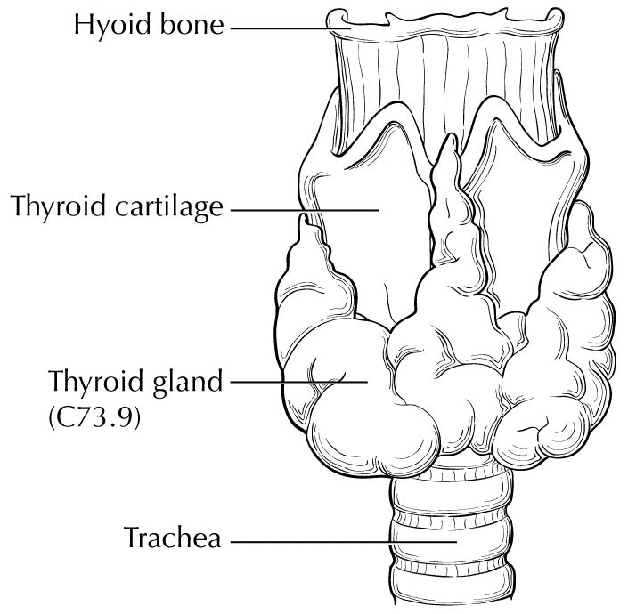
The thyroid gland ordinarily is composed of a right and a left lobe lying adjacent and lateral to the upper trachea and esophagus (Figure 93.1). An isthmus connects the two lobes, and in some cases, a pyramidal lobe is present, extending cephalad anterior to the thyroid cartilage.
Rarely, thyroid cancer may arise from thyroid follicular cells located outside the thyroid gland in locations such as the thyroglossal duct remnants, thyroid rests in the neck/upper mediastinum (thyrothymic tract), and ovaries (malignant struma ovarii).
Regional Lymph Nodes
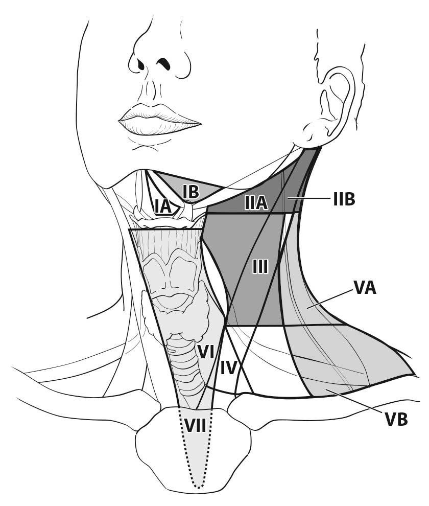
A seven-compartment nomenclature is commonly used to define anatomic lymph node compartment boundaries (Figure 93.2).24,25 The term central neck usually refers to levels VI and VII, whereas the lateral neck includes levels I, II, III, IV, and V. The first echelon of nodal metastasis most commonly includes the paralaryngeal, paratracheal, and prelaryngeal (Delphian) nodes adjacent to the thyroid gland.
Metastases also may involve the high (level IIA), mid- (level III), and lower jugular (level IV), and the supraclavicular (level V) and (less commonly) the upper deep jugular and spinal accessory lymph nodes (level IIB). Lymph node metastasis to submandibular and submental lymph nodes (level I) is rare. Upper mediastinal (level VII) nodal spread occurs frequently, both anteriorly and posteriorly. Retropharyngeal nodal metastasis may be seen, usually in the presence of extensive lateral cervical metastases. Bilateral nodal spread is common.
Clinical Classification
Most thyroid cancer patients present with asymptomatic nodules in the thyroid in the setting of normal thyroid function tests. Symptoms such as changes in the voice, dysphagia, or upper airway problems suggest more aggressive local disease. Distant metastases usually are identified as asymptomatic pulmonary nodules in high-risk patients but also may present as painful bone metastases or as masses causing local neurologic or vascular compromise.
Detailed guidance in preoperative assessments and management is provided in guidelines from the NCCN and the ATA.4,5 Preoperative staging for thyroid cancer typically includes neck ultrasound to evaluate the thyroid gland and central and lateral neck lymph node compartments. Fine-needle aspiration of suspicious thyroid nodules and/or abnormal-appearing lymph nodes should be undertaken preoperatively to obtain a definitive diagnosis and allow for appropriate surgical planning. FDG positron emission tomography (PET) scanning or neck computed tomography (CT)/magnetic resonance (MR) imaging is not recommended, except in patients in whom there is clinical suspicion of gross extrathyroidal extension or extensive, clinically apparent, cervical/mediastinal lymphadenopathy.
For staging purposes, the treatment date for most patients with differentiated thyroid cancer should be the date of thyroidectomy, because thyroid surgery is almost always the first step in treatment. Rarely, patients may receive external beam irradiation, chemotherapy, metastasectomy, or other neoadjuvant therapy as their initial treatment (before thyroid surgery or in patients who never undergo thyroid surgery). In these cases, the treatment date would correspond to the initiation of these other therapies, provided they begin before thyroid surgery.
The date of diagnosis should correspond to the first date of cytologic or histologic confirmation of thyroid cancer.
With regard to sizing of the primary tumor (T), this is almost always done based on the size of the largest differentiated thyroid cancer nodule within the thyroid gland, determined when the thyroid surgical specimen is processed for histologic examination. In situations in which the thyroid cancer is not surgically removed, the size of the primary lesion may be obtained by correlating cross-sectional imaging studies with biopsy results (by cytology or histology). These situations may become more common, as the 2015 ATA guidelines allow for an active surveillance management approach (observation instead of immediate surgery) in subcentimeter thyroid nodules that are either cytologically confirmed PTC or presumed to be thyroid cancer based on highly suspicious ultrasound characteristics.4 Although a highly suspicious ultrasound pattern carries a >70-90% likelihood that thyroid cancer is present,28-30 cytologic or histologic proof of disease is required before staging.
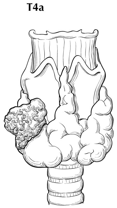
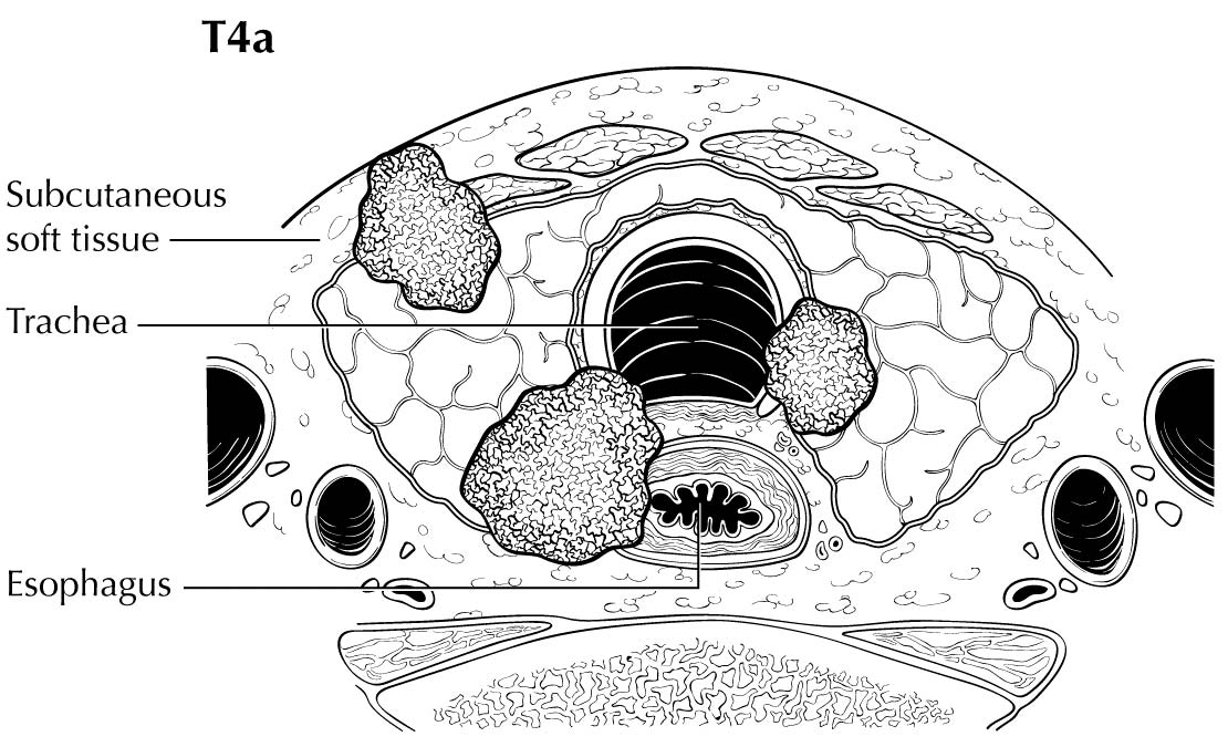
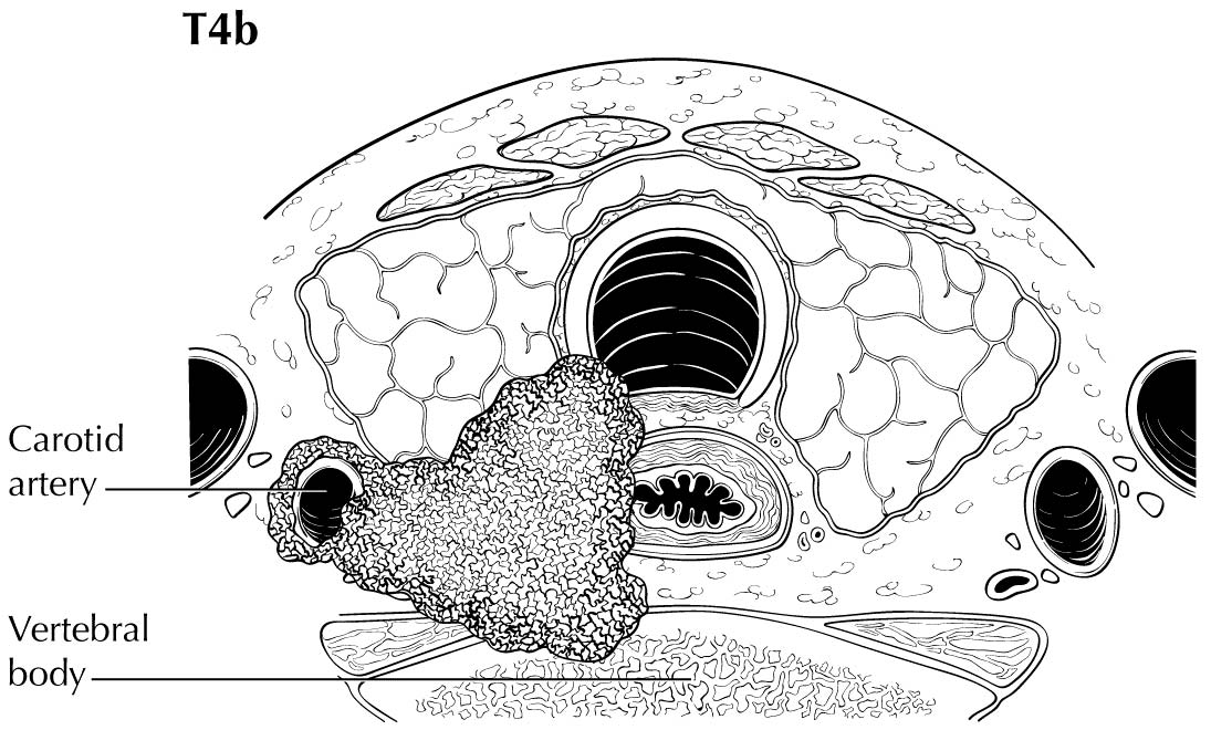
Extrathyroidal extension refers to the involvement of perithyroidal soft tissues by direct extension from the thyroid primary. Invasion outside the thyroid that can be identified clearly by imaging or intraoperative findings ranges from T3b disease (gross extrathyroidal extension involving only strap muscles) to T4a disease (demonstrating gross extrathyroidal extension invading the subcutaneous soft tissues, larynx, trachea, esophagus, muscle, or recurrent laryngeal nerve) to T4b disease (demonstrating gross extrathyroidal extension invading the prevertebral fascia or encasing the carotid artery or mediastinal vessels). The four infrahyoid muscles that either originate from or insert on the hyoid are often referred to as the strap muscles (including the sternohyoid, sternothyroid, thyrohyoid, and omohyoid muscles). Lesser degrees of extrathyroidal extension (minor) that are not clinically appreciated can be identified by microscopy as the tumor involves perithyroidal adipose tissue, strap muscles, nerves, or small vascular structures. Because the fibrous capsule of the thyroid is often incomplete, it is often difficult to determine whether the the boundary between thyroid cancer and fibroadipose tissue reflects an invasive process or simply the absence of a well-defined thyroid capsule in that area. For these reasons, and based on the lack of prognostic significance, the presence of minor extrathyroidal extension involving perithyroidal adipose tissue, strap muscles, nerves, or small vascular structures detected only by microscopy (not grossly evident) does not constitute T3b disease.
Clinical N1 disease (cN1) includes clinically apparent lymph node metastases (palpable or seen on imaging) that are either cytologically confirmed or highly suspicious for metastatic disease. Likewise, M1 status can be confirmed by cytologic/histologic assessment, documentation of RAI avidity of the metastatic lesion, or other imaging findings highly suspicious for distant metastasis in the proper clinical setting (e.g., inappropriately elevated serum thyroglobulin, ATA high-risk patient).
Imaging
Pretherapy Imaging
Preoperative neck ultrasonography to evaluate the thyroid gland as well as central and lateral neck lymph node chains is usually recommended as the initial staging procedure. Additional cross-sectional imaging with CT or MR imaging of the neck or distant sites is usually reserved for patients demonstrating clinical features of advanced disease, such as locally invasive primary tumor, clinically apparent multiple or bulky lymph node metastases, symptoms of distant metastases, or anaplastic thyroid cancers. Preoperative FDG-PET scanning is not routinely recommended but may be considered as part of initial staging in cases in which there is a reasonable likelihood of distant metastatic disease, such as in cases of poorly differentiated thyroid cancers, Hürthle cell cancers, and anaplastic thyroid cancers.4,5
Because of the high likelihood of both regional and distant metastases in anaplastic thyroid cancer, initial staging usually includes neck ultrasound; cross-sectional imaging of the head, neck, chest, abdomen, and pelvis with either CT or MR imaging; and/or FDG-PET scanning. At sites where PET scanning is performed using optimized PET/CT, the CT portion of the scan may supplant the need for additional anatomic imaging.4,5
These preoperative imaging examinations form the primary basis for preoperative clinical staging. Clinical T stage is based on the size of the primary tumor and an assessement of whether imaging demonstrates invasion of the tumor into the strap muscles, subcutaneous soft tissues, larynx, trachea, esophagus, recurrent laryngeal nerve, or prevertebral fascia or whether the tumor encases either the carotid artery or mediastinal vessels. The location of metastatic lymph nodes is used to define the clinical N stage (central neck vs. lateral neck disease). Most patients will be clinical M0, as routine use of cross-sectional or functional (RAI) imaging beyond the neck is not routinely performed, except in patients with locally advanced or anaplastic thyroid cancers.
One of the challenges with clinical staging is that nonspecific cervical lymphadenopathy is commonly found on routine ultrasonographic imaging and cannot be confidently classified as cN0 or cN1. In clinical practice, ultrasound-guided fine-needle aspiration of sonographically suspicious lymph nodes >=8 mm in smallest dimension is often performed if the results of the biopsy would alter initial management.4 Likewise, nonspecific pulmonary nodules also are quite common in the general population and usually cannot be confidently classified as benign or malignant findings before thyroid surgery.
Posttherapy ImagingMany patients undergo RAI scanning several weeks after thyroid surgery and at various time points during follow-up. These scans take advantage of the unique ability of most thyroid cells (both thyroid cancer and normal thyroid cells) to concentrate iodine. Although a focus of RAI uptake on the scan outside the thyroid bed usually indicates the presence of persistent or recurrent thyroid cancer, false positives do occur, which means that the RAI scans must be interpreted within the context of serum thyroglobulin and other patient risk factors for recurrence.
In most patients, neck ultrasonography is the primary imaging modality, with the testing interval based on initial risk stratification and the patient's response to therapy. Patients at high risk of regional or distant metastases also may be evaluated with cross-sectional imaging or FDG-PET scanning, depending on the serum thyroglobulin level and response to therapy classification.4
Because of the very high risk of recurrence and distant metastases, patients with anaplastic thyroid cancer require more frequent and extensive imaging. Cross-sectional imaging of the brain, neck, chest, abdomen, and pelvis is often performed at 1- to 3-month intervals for the first year of follow-up and then at 4- to 6-month intervals for an additional year. In addition, FDG-PET scanning is also considered at 3 to 6 months after initial therapy to identify persistent or recurrent disease.31
Radioiodine imaging is typically performed with either iodine-123 or iodine-131. Both are imaged with a conventional gamma camera, typically 24 to 48 hours after administration of the RAI. There is increasing interest in radioiodine imaging with an isotope of iodine that emits positrons, allowing the use of PET scanning for imaging. Iodine-124 has a half-life of 4.18 days, allowing for delayed imaging. PET scanning has a high sensitivity for detecting small-volume disease, and coacquisition with CT provides anatomic localization for therapy planning purposes. The quantitiative nature of PET scanning also allows for dosimetric therapy planning for radioiodine treatment using iodine-131, maximizing dose delivery to the tumor while limiting toxicity to bone marrow and other organs. This imaging technique has not yet achieved widespread acceptance, but it is being actively investigated at several sites.32
Pathological Classification
Pathological staging requires the use of all information obtained during clinical staging, as well as histologic study of the surgically resected specimen. The surgeon's description of gross extrathyroidal extension must also be included.
In this edition, the presence of minor extrathyroidal extension identified only on histologic examination and not apparent clinically is not used as a risk factor for staging. No distinction is made between tumors with and those without minor extrathyroidal extension. However gross extrathyroidal extension that can be identified clearly by imaging, intraoperative findings, or examination of tumor speciments is classified as T3b disease (gross extrathyroidal extension involving only strap muscles), T4a disease (gross extrathyroidal extension invading the subcutaneous soft tissues, larynx, trachea, esophagus, muscle, or recurrent laryngeal nerve), or T4b disease (gross extrathyroidal extension invading the prevertebral fascia or encasing the carotid artery or mediastinal vessels). Furthermore, because of the poorer survival outcomes associated with gross extrathyroidal extension, patients older than 55 years at diagnosis with T3b disease are classified as Stage II, those with T4a disease are classified as Stage III, and those with T4b disease are classified as Stage IV.
For staging purposes, any N includes pN0, pN1, pNX, cN0, or cN1 disease. Pathological confirmation of lymph node status is not required for staging purposes. Rather, patients with pNX disease who are cN0 are classified as cN0/pNX in the staging tables. As detailed in the section on the impact of regional lymph node metastasis on prognosis in differentiated thyroid cancer, subclinical (cN0) small-volume pN1 disease has little prognostic significance and is associated with outcomes that are very similar to those of pN0 disease. Because there is no requirement for a minimum number of lymph nodes to be sampled, pathological confirmation of one or more benign lymph nodes mandates a pN0 designation.
The identification of psammomatous calcifications within a cervical lymph node is considered metastatic disease and should be classified as N1 disease.
Complete assessment of N/M status may not be possible until after the RAI scans are complete, which often happens 1 to 3 months after initial surgery. Therefore, identification of metastatic disease (by any modality) within the first 4 months of thyroid surgery should be used to refine the N and M status.
Consistent with AJCC staging rules, the formal stage established during the first 4 months of follow-up does not change over time, even if the cancer progresses or recurs. However, the cancer may be restaged as new data become available during follow-up using the same approach and definitions applied during the initial staging. The lower case r is used to designate the restaging. In differentiated thyroid cancer, clinicians recognize both structural disease recurrence/progression (structural or functional evidence of disease) and biochemical disease recurrence/progression (abnormal thyroglobulin without structural or functional evidence of disease). Consistent with the approach to initial staging, restaging should be based only on the identification of structurally or functionally identifiable disease and not on the basis of abnormal biomarkers of disease (serum thyroglobulin or thyroglobulin antibodies).
Prognostic Factors Required for Stage Grouping
Unlike with most malignancies, age at diagnosis of thyroid cancer is almost always identified as an independent predictor of disease-specific survival (DSS) in the published staging systems. Poor outcomes in differentiated thyroid cancer were reported as early as 1979 in patients older than 45 years at diagnosis.33 The AJCC TNM staging system has incorporated a 45-year age cutoff as a major determinant of DSS since the AJCC Cancer Staging Manual, 2nd Edition was published in 1983. Most of the other clinicopathologic staging systems use an age cut point of between 40 and 50 years in their models.3 The MACIS system, designed as a postoperative risk stratification system, uses age as a continuous variable in patients more than 40 years old at diagnosis.34
Multiple studies confirmed that mortality from PTC increases progressively with advancing age, beginning at about age 35.35-43 Unfortunately, there is no single age cutoff that discretely allocates patients into separate risk categories. Many authors have recommended using nomograms,37,44 mathematical models,34,45 or multiple age categories37,46 to better reflect the continuous nature of the relationship between age at diagnosis and disease-specific mortality. Other authors have endorsed using an age cutoff of 55 years as the optimal single time point for prognostic models.47-51
A recent international multicenter retrospective study demonstrated that by moving the age cut point from 45 to 55 years, 17% of the patient population was downstaged to a lower risk category.37,52 Overall, 10% of patients who would have been classified as having advanced-stage disease based on the 45-year-old cut point (Stage III/IV) were downstaged to Stage I/II when a 55-year-old cut point was used, without affecting the survival curves in the lower risk categories. Furthermore, an age cutoff of 55 years produced a wider distribution in survival among the risk groups, ranging from 99.6% in Stage I to 70% in Stage IV, compared with the corresponding values of 99.6% and 79% when 45 years was used as the age cut point. Likewise, Ito et al.49 demonstrated effective risk stratification when comparing the iStage modifications with the Union for International Cancer Control (UICC) TNM system. Therefore, although it seems unlikely that raising the age will have a significant impact on the performance of the staging system, it does have the significant clinical benefit of preventing upstaging based only on age of diagnosis between 45 and 55 years in patients who otherwise would be considered low risk (Stage I or II).
The specific histologies are described in the pathology report. There are no pertinent cutoff values.
Additional Factors Recommended for Clinical Care
Extrathyroidal extension may range from gross extrathyroidal extension involving major structures (T3b, T4a, T4b) to minor extension through the thyroid capsule identified only on histologic examination.
Gross extrathyroidal extension is documented in the operative report, whereas minor extrathyroidal extension is found in the pathology report, described as extension of the primary tumor through the thyroid capsule and into surrounding structures.
Gross extrathyroidal extension (see definitions for T3b, T4a, and T4b disease) identified either preoperatively or intraoperatively is an important factor for staging, whereas minor extension through the thyroid capsule seen only on histologic examination is not used for staging.
Gross extrathyroidal extension in differentiated thyroid cancer increases disease persistence/recurrence and decreases survival.53-55 Most differentiated thyroid cancer staging systems incorporate gross extrathyroidal extension as a predictor of recurrence and/or death (AMES, MACIS, AJCC, UICC).3
The AJCC Cancer Staging Manual, 6th Edition first distinguished between minimal and gross extrathyroidal extension. Authors downstaged to T3 any tumor with minimal extrathyroidal extention (e.g., extention to sternothyroid muscle or perithyroidal soft tissue). Since this delineation in 2002, pathological and clinical thyroid cancer studies have attempted to define its relevance.
Pathologically, the thyroid has an incomplete capsule. The thyroid gland may contain adipose tissue and skeletal muscle under normal circumstances. According to the College of American Pathologists, "defining (minimal) extrathyroidal extension may be problematic and subjective." Ghossein and colleagues57,58 warned that the presence of adipose tissue, and muscle in some circumstances, in association with thyroid carcinoma should not be mistaken for extrathyroidal extension.56,57
During the past decade of clinical studies, nuances have been identified in the spectrum between minimal and gross extrathyroidal extension. Several recent studies demonstrated that microscopic extrathyroidal extension is not an independent prognostic factor for persistent/recurrent disease; disease-free survival is equivalent in patients with microscopic extrathyroidal extension and those with completely intrathyroidal tumors.5862 A study of T1/T2 well-differentiated thyroid carcinoma showed no difference in 10-year DSS or recurrence-free survival in those with microscopic extrathyroidal extension (who would have been upstaged on the basis of extrathyroidal extension alone).63 There appears to be agreement that minimal/microscopic extrathyroidal extension in small differentiated thyroid cancer portends an outcome equivalent to that seen with completely intrathyroidal tumors.
However, several retrospective studies suggest an association between minimal extrathyroidal extension and the presence of lymph node metastases/extranodal extension,59,61,64,65 concluding that minimal extrathyroidal extension is an indicator of disease biology in PTC. None demonstrated minimal extrathyroidal extension as an independent predictor of persistence/recurrence or survival. A recent large clinicohistopathologic analysis demonstrated that the presence of extrathyroidal extension in PTC is not associated with extranodal extension, whereas the number of positive lymph nodes is.66
Margin positivity in differentiated thyroid cancer may be considered similarly to extrathyroidal extension. There appears to be no difference in outcomes in patients with an R0 resection (microscopically negative margin) compared with those with an R1 resection (microscopically positive margin).60,67 However, patients with a grossly positive margin (R2, or incomplete, resection) have a significantly higher risk of recurrence and disease-specific death.
Clinical staging information is found in preoperative imaging and clinical examination reports, whereas pathological staging information is found in the pathology report. There are no pertinent cutoff values.
The combination of high-resolution imaging, extensive neck dissection, and meticulous histological examination results in the identification of regional lymph node metastases in up to 80% of patients with PTC.68 In many cases, the lymph node metastases are quite small (<1 cm), but up to 35% of patients may present with larger lymph node metastases.68,69 Regional lymph node metastases are also common in medullary and anaplastic thyroid cancers but are seen less often in FTCs and Hürthle cell thyroid cancers.
Most studies,59,70-77 but not all,34,78,79 have suggested that regional lymph node metastases have prognostic significance in differentiated thyroid cancer. The impact of lymph node metastasis on survival is most evident in older patients.59,72-74,76,77 Based on these data, previous AJCC staging systems used the presence of N1 disease to influence stage in patients older than 45 years at diagnosis.
The impact of lymph node metastases in younger patients has remained more controversial. However, recent studies based on the SEER and National Cancer Data Base (NCDB) datasets provided strong evidence that lymph node metastasis in patients younger than 45 years at diagnosis has a statistically significant impact on overall survival.70,76 However, this statistically significant difference translates to 20-year adjusted survival rates of 97% without lymph node metastasis and 96% with lymph node metastasis in patients less than 45 years of age at diagnosis.70
Unlike the data available with regard to the risk of structural disease recurrence, data correlating lymph node characteristics with survival are less well developed. Several studies demonstrated that lateral neck lymph node metastases are associated with compromised survival,74-76,80 which forms the basis for differentiating risk based on location of regional metastases (central vs. lateral neck compartments). However, because prophylactic neck dissections seldom are performed in the lateral neck, these observations are confounded by the fact that pathologically involved lateral neck lymph nodes are usually much larger than metastatic lymph nodes identified in the central neck (often incidentally removed or identified only by prophylactic neck dissection). Therefore, it is very difficult to differentiate the effect of location (central vs. lateral neck ) from the effect of size and number of lymph node metastases in these retrospective datasets. Some70 but not all71,75 have suggested that the number of involved lymph nodes may be related to survival. Using both the SEER and NCDB databases, controlling for important confounders, and restricting the analysis to patients younger than 45 years at diagnosis, Adam et al.70 demonstrated a statistically significant association between the number of involved lymph nodes and survival. In their analysis, mortality increased incrementally up to six involved lymph nodes, after which no further increase in mortality was seen with additional metastatic lymph nodes.
Multiple studies confirmed the association between extranodal extension and persistent/recurrent disease68,80-82 and demonstrated that extranodal extension is not infrequently seen in subcentimeter lymph node metastases.83 Some studies,64 but not all,66 demonstated a correlation between extranodal extension and extrathyroidal extension. Although several publications demonstrated statistically significant associations between extranodal extension and DSS,48,80,82,84,85 each of these studies was single center, rather small, and lacked long-term follow-up. Furthermore, the impact of extranodal extension on survival appears to be dependent on the clinical context, with more significant impacts seen in association with BRAF-mutant tumors82 or in the setting of lateral neck disease.48 Therefore, although the data strongly suggest that extranodal extension may have a strong association with DSS in differentiated thyroid cancer, currently available evidence does not rise to a level that justifies its inclusion as an independent survival variable.
Other investigators examined the impact of metastatic lymph node ratio (number of metastatic nodes/total number of nodes harvested) on prognosis. Metastatic lymph node ratio (>0.42) was associated with compromised DSS, but this significance was lost when lateral neck lymph node metastases were excluded.75 Similarly, metastatic lymph node ratio was not a significant predictor of survival in either young patients or older patients with metastatic lymph node involvement, even when the analysis was restricted to patients with six or more lymph nodes examined.71
No studies have adequately evaluated the impact of metastatic lymph node size on survival. Further complicating this analysis is the observation that some pathologists report the overall size of the lymph node, whereas others report the size of the metastatic foci within the lymph node. However, several clinical observations suggest that small-volume regional metastases likely have very little impact on overall survival. Extensive lymph node dissections can identify regional lymph node metastases in up to 80% of patients.68 Despite having a DSS rate of >99%, prophylactic neck dissections in cN0 papillary microcarcinoma patients may identify central neck regional metastases in 40-50% and lateral neck regional metastases disease in 45%.86-89 Lymph node metastasis identified in prophylactic neck dissections usually represents small-volume disease (95% <1 cm, a mean of two to three involved nodes).87,88 These data demonstrate that many patients classified as having N0 disease would prove to have small-volume pN1 disease if extensive neck dissections had been performed since staging is commonly based on histologic examination of three or fewer cervical lymph nodes71,75 or without histologic examination of any lymph nodes.70,77Hence, very similar recurrence risks and mortality outcomes are seen in patients classified as having cN0, pN0, or small volume pN1. When these observations are combined with the data showing low recurrence rates with small-volume lymph node metastases (five or fewer subcentimeter metastatic lymph nodes),68 it is clear that small-volume lymph node metastases have little prognostic impact on survival in differentiated thyroid cancer.
In summary, regional lymph node metastases appear to have strong prognostic significance in most differentiated thyroid cancer. Although statistically significant in all adult age groups, the clinical impact of N1 disease on survival is most apparent in older patients, but the magnitude of effect on overall survival appears much smaller if the N1 disease is not associated with T4a, T4b, or M1 disease. With regard to lymph node characteristics, lateral neck regional node disease carries a worse prognosis than central neck regional node disease, although it is not clear whether the poorer outcomes are related to location or to the size/number of metastatic lymph nodes. It seems likely that the number of involved lymph nodes also may correlate with survival, but additional studies are needed, particularly in the older population. Small-volume lymph node metastases have little impact on the risk of structural disease recurrence and DSS.
Using Age at Diagnosis, Extrathyroidal Extension, and Regional Lymph Node Status to Define Prognostic Stage Groups
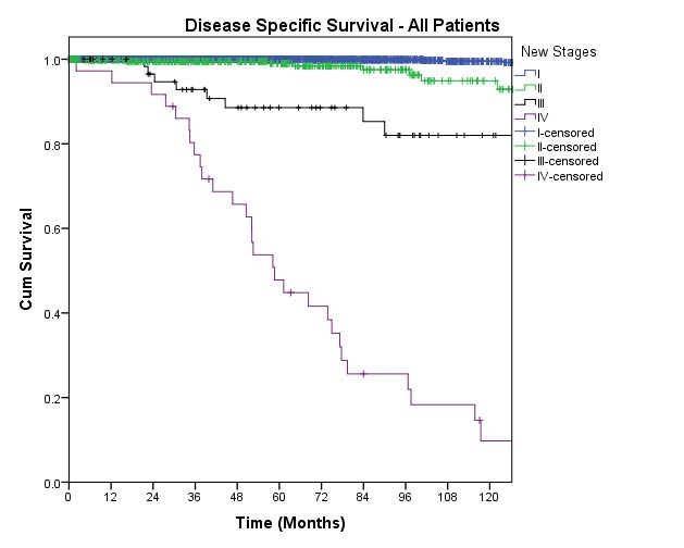
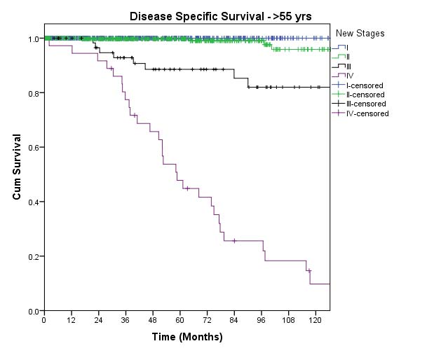
Previous editions of the AJCC thyroid cancer staging systems provided suboptimal separation with regard to the risk for disease-specific mortality between Stage I and Stage II disease. Furthermore, less than 20% of patients classified as having Stage III or IV disease will actually die of thyroid cancer. In most series, 5- to 10-year survival (variously reported as overall survival or relative survival) approximates 97-100% in Stage I and II disease and 88-95% in Stage III disease. Stage IV disease consistently predicts 10-year survival rates in the 50-75% range.52,90-92 However, within the AJCC Cancer Staging Manual, 7th Edition Stage IV group, significantly worse outcomes are seen with Stage IVc (M1 disease, any N, any T) compared with Stage IVa/b (T4a or T1-2, Na1b, M0), with 10-year survival rates of approximately 50% and 70%, respectively.76,92
In the 7th Edition, identification of central neck cervical lymph node metastases upstaged patients to Stage III if the primary tumor was T1-T3, whereas the presence of lateral neck disease (N1b) upstaged patients to Stage IVa if the primary tumor was T1-T4a. This stratification was based on previous studies demonstrating poorer survival in older patients with N1 disease and on data suggesting that N1b disease has a worse outcome than N1a disease.59,70-77,79 In most of these series, 10-year overall survival for all N0 patients was approximately 80-85%, whereas for N1 patients, it was 75-80%.72,77 The difference in overall survival between N0 and N1 patients is much smaller in young patients (<1-2%) than in older patients (5-10%).52,70,77,90-93
Furthermore, much of the mortality risk associated with N1 disease in older patients may be attributed to the common co-ocurrence of M1 and/or T4a/b disease in the setting of clinically significant N1b disease. For example, in older patients, 99% DSS is seen in N0 disease (only 1.5% have concurrent M1 disease), whereas a DSS of 92% is observed in patients with N1 disease in levels II to VA (5% M1) and 85% DSS is seen in patients with Vb/VII nodes involved (10% M1).93 Studies analyzing the impact of lymph node metastases in patients without M1 or T4b disease demonstrated that the mortality risk associated with T1-3N1bM0 disease or T4aN1M0 disease is minimal.39,52,74,94-97 For example, N1bM0 was associated with 85% 15-year relative survival.92 Additionally 96-97% DSS was reported in patients with T1-3N1M0, whereas much poorer outcomes were seen in T4a, any N, M0 (82% DSS) and T4aN1b disease (70% DSS).52,74,89,94
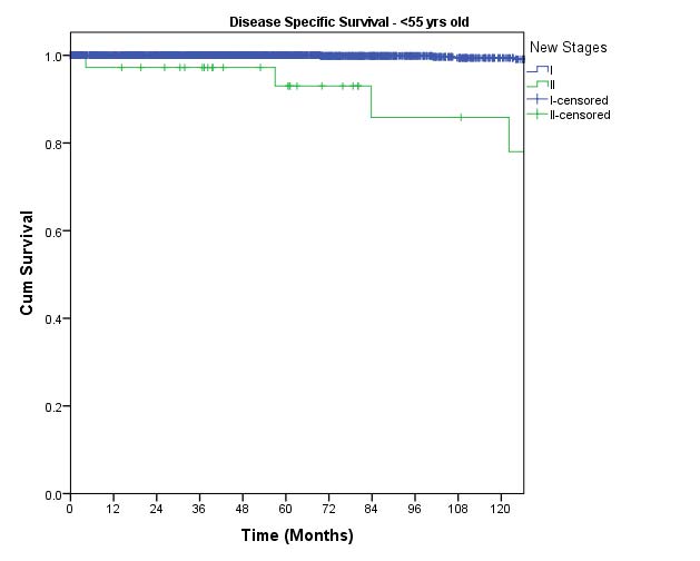
Although the preponderance of the data demonstrates that lymph node metastases may convey a statistically measurable increase in the risk of disease-specific mortality in the elderly, and probably to a lesser extent in younger adult patients, the actual magnitude of this risk is much smaller than the risk conveyed by either T4a/b or M1 disease. Therefore, in the AJCC Cancer Staging Manual, 8th Edition staging system, N1 disease is classified as Stage I in patients less than 55 years old at diagnosis and reclassified as Stage II disease in older patients. Because the survival of older patients with N1 disease in the absence of T4a/M1 disease should approximate 85-95%, these changes should define a cohort of patients who have outcomes worse than those of Stage I patients.
With regard to the 7th Edition Stage IV grouping, it is clear that two separate subgroups can be defined: patients with distant metastasis (IVC patients) and those with gross extrathyroidal extension (T4a/T4b patients) regardless of the lymph node status. Because all N1 disease is now classified as Stage II in older patients (but still as Stage I in younger patients), the previous Stage III would include only T3N0 disease, which would not be expected to have significant mortality. Therefore, the 8th Edition Stage III includes only the older T4a, any N, M0 patients and is expected to have a survival curve very similar to the 7th Edition Stage IVA curve. Finally, older patients with T4b, any N, M0, and all M1 disease (any T, any M) will define the 8th Edition Stage IV disease.
To validate the 8th Edition staging system, the proposed changes were analyzed using the previously published dataset from Memorial Sloan Kettering Cancer Center (re-analysis by Ian Ganly and Jatin Shah, Head and Neck Surgery, MSKCC).52
The location of metastatic lymph nodes, referring to either central (N1a) or lateral (N1b) cervical lymph node chains. The location of metastatic lymph nodes may be found in the operative notes and the pathology report. There are no pertinent cutoff values.
The number of lymph nodes histologically confirmed to have a metastatic foci of thyroid cancer. The number of involved lymph nodes is described in the pathology report. There are no pertinent cutoff values.
The number of lymph nodes histologically examined (including both metastatic and benign lymph nodes). The number of involved lymph nodes is described in the pathology report. There are no pertinent cutoff values.
The maximum diameter of the largest metastatic lymph node in millimeters. This will be described in the pathology report. There are no pertinent cutoff values.
The maximum diameter of the metastatic tumor foci within a lymph node in millimeters. This will be described in the pathology report. There are no pertinent cutoff values.
Microscopic or gross extension of the metastatic lymph node tumor foci outside the lymph node capsule. This will be described in the pathology report. There are no pertinent cutoff values.
Vascular invasion is defined as invasion of the thyroid cancer into vascular structures. This will be described in the pathology report. There are no pertinent cutoff values.
A serum tumor marker specific for thyroid tissue (benign or malignant). It is measured by a clinical laboratory using a variety of methods and is reported in the clinical record in nanograms per milliliter. There are no pertinent cutoff values.
A description of whether the surgeon provided a complete resection of all grossly visible disease. This is reported in the operative note and staged using residual tumor (R) classification. There are no pertinent cutoff values.
In addition to the specific histologic subtypes defined by the WHO Classification of Tumors table, several other histologic features may have prognostic importance, including perineural invasion, multifocality, and high mitotic index.4 These features are documented on the pathology report. There are no pertinent cutoff values.
Follicular cell-derived thyroid cancers display a spectrum of biological behavior ranging from clinically insignificant (papillary microcarcinomas in the elderly) to highly lethal (anaplastic thyroid cancer). When matched for stage, the prognosis of PTCs and FTCs are usually very similar. Poorer prognosis may be seen in some of the more aggressive variants, such as the poorly differentiated thyroid cancers, tall cell variants, hobnail variants, and columnar cell variants. Conversely, better prognosis may be seen in noninvasive encapsulated FTC and noninvasive encapsulated follicular variants of PTC.98,99 Hürthle cell carcinomas are classified as a variant of follicular carcinoma by the World Health Organization (WHO), but newer data suggesting differences in biological behavior and unique genetic alterations indicate that Hürthle cell cancers may be best classified as a distinct histologic tumor (rather than a subtype of FTC).100 Although anaplastic thyroid cancer is associated with 1-year survival rates of less than 10-20%, small, incidentally discovered anaplastic thyroid cancers that are completely resected may display a much better prognosis.31,101
Tumors harboring BRAF, TERT, and/or P53 mutations have the potential to be aggressive, with increased rates of recurrence and disease-specific mortality.102-107 Furthermore, BRAF V600E mutation appears to increase the risk of disease-specific mortality if identified in conjunction with lymph node metastases, distant metastases, AJCC Stage IV disease, and age >=45 years at diagnosis.105 More recent data suggest that tumors harboring multiple oncogenic mutations, although uncommon, may have a more aggressive phenotype.106,108 Because many of these mutations are tightly linked to aggressive histologic features, it is difficult to estimate the proportion of risk attributable to the actual mutation versus that attributable to the other clinicopathologic features. Although the 2015 ATA guidelines recognize the potential for improvement in risk stratification with regard to molecular profiling, they do not recommend molecular testing for initial staging because it is not yet clear how much incremental improvement in risk stratification would be gained.4
The increased use of more complete, in-depth genomic analyses has led to the discovery of targetable mutations in a small percentage of patients with aggressive thyroid cancers. Recently, promising results were seen with inhibitors of ALK fusion genes in medullary109 and anaplastic thyroid cancer110 and with a BRAF inhibitor in anaplastic thyroid cancer.111,112
Risk Assesment Models
The AJCC recently established guidelines that will be used to evaluate published statistical prediction models for the purpose of granting endorsement for clinical use.113 Although this is a monumental step toward the goal of precision medicine, this work was published only very recently. Therefore, the existing models that have been published or may be in clinical use have not yet been evaluated for this cancer site by the Precision Medicine Core of the AJCC. In the future, the statistical prediction models for this cancer site will be evaluated, and those that meet all AJCC criteria will be endorsed.
The following stratification criteria stem from the prognostic factor analyses suggested for use in thyroid cancer trials, depending on the specific objectives of the study, the cancer stage(s), and the population under study, including sample size:
The primary stratification for thyroid cancer clinical trials focused primarily on structurally progressive RAI-refractory patient populations will be based on tumor histology, AJCC stage, and age. However, as many of the clinical trial agents are molecularly targeted therapies, risk stratification and data analysis based on biomarker analysis of the tumor may also yield important insights.
Clinical T (cT)
cT Category | cT Criteria |
---|---|
cTX | Primary tumor cannot be assessed |
cT0 | No evidence of primary tumor |
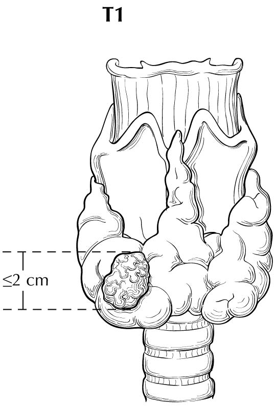
cT1 | Tumor less than or equal to 2 cm in greatest dimension limited to the thyroid |
cT1a | Tumor less than or equal to 1 cm in greatest dimension limited to the thyroid |
cT1b | Tumor greater than 1 cm but less than or equal to 2 cm in greatest dimension limited to the thyroid |
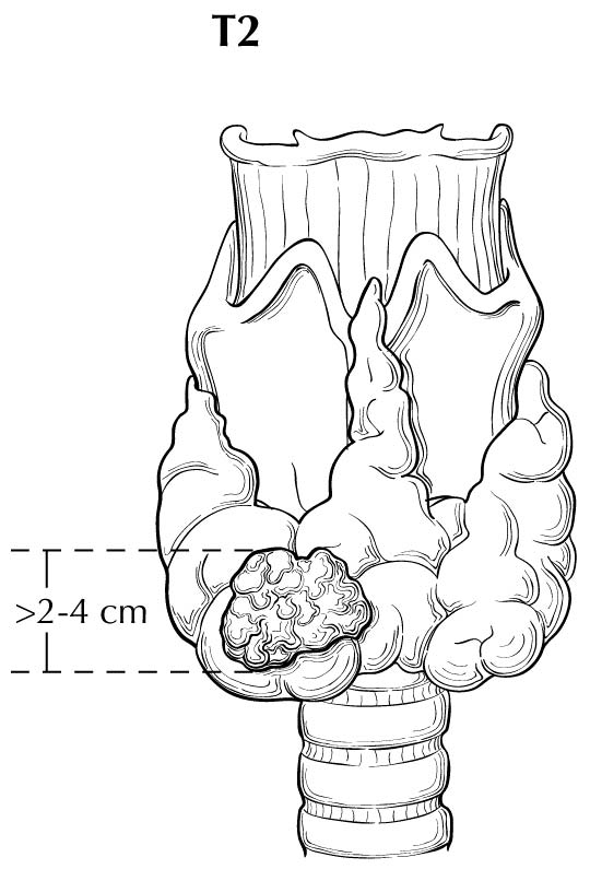
cT2 | Tumor greater than 2 cm but less than or equal to 4 cm in greatest dimension limited to the thyroid |
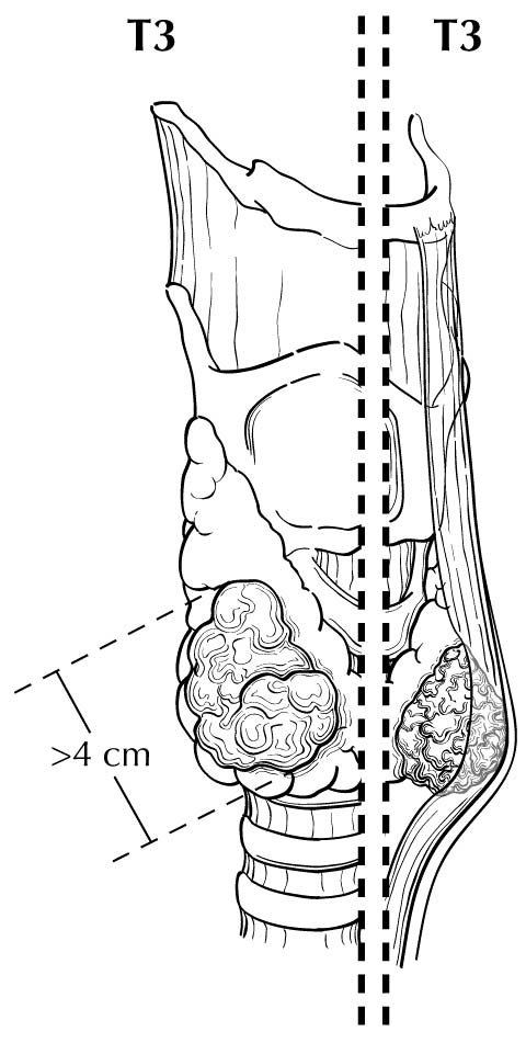
cT3 | Tumor greater than 4 cm limited to the thyroid, or gross extrathyroidal extension invading only strap muscles |
cT3a | Tumor greater than 4 cm limited to the thyroid |
cT3b | Gross extrathyroidal extension invading only strap muscles (sternohyoid, sternothyroid, thyrohyoid, or omohyoid muscles) from a tumor of any size |
cT4 | Includes gross extrathyroidal extension beyond the strap muscles |
cT4a | Gross extrathyroidal extension invading subcutaneous soft tissues, larynx, trachea, esophagus, or recurrent laryngeal nerve from a tumor of any size |
cT4b | Gross extrathyroidal extension invading prevertebral fascia or encasing the carotid artery or mediastinal vessels from a tumor of any size |
These definitions apply to Papillary, Follicular, Poorly Differentiated, Hürthle Cell and Anaplastic Thyroid Carcinoma. All categories may be subdivided: (s) solitary tumor and (m) multifocal tumor. The largest tumor determines the classification.
Pathological T (pT)
pT Category | pT Criteria |
---|---|
pTX | Primary tumor cannot be assessed |
pT0 | No evidence of primary tumor |
pT1 | Tumor less than or equal to 2 cm in greatest dimension limited to the thyroid |
pT1a | Tumor less than or equal to 1 cm in greatest dimension limited to the thyroid |
pT1b | Tumor greater than 1 cm but less than or equal to 2 cm in greatest dimension limited to the thyroid |
pT2 | Tumor greater than 2 cm but less than or equal to 4 cm in greatest dimension limited to the thyroid |
pT3 | Tumor greater than 4 cm limited to the thyroid, or gross extrathyroidal extension invading only strap muscles |
pT3a | Tumor greater than 4 cm limited to the thyroid |
pT3b | Gross extrathyroidal extension invading only strap muscles (sternohyoid, sternothyroid, thyrohyoid, or omohyoid muscles) from a tumor of any size |
pT4 | Includes gross extrathyroidal extension beyond the strap muscles |
pT4a | Gross extrathyroidal extension invading subcutaneous soft tissues, larynx, trachea, esophagus, or recurrent laryngeal nerve from a tumor of any size |
pT4b | Gross extrathyroidal extension invading prevertebral fascia or encasing the carotid artery or mediastinal vessels from a tumor of any size |
cTX | Primary tumor cannot be assessed |
cT0 | No evidence of primary tumor |
cT1 | Tumor less than or equal to 2 cm in greatest dimension limited to the thyroid |
cT1a | Tumor less than or equal to 1 cm in greatest dimension limited to the thyroid |
cT1b | Tumor greater than 1 cm but less than or equal to 2 cm in greatest dimension limited to the thyroid |
cT2 | Tumor greater than 2 cm but less than or equal to 4 cm in greatest dimension limited to the thyroid |
cT3 | Tumor greater than 4 cm limited to the thyroid, or gross extrathyroidal extension invading only strap muscles |
cT3a | Tumor greater than 4 cm limited to the thyroid |
cT3b | Gross extrathyroidal extension invading only strap muscles (sternohyoid, sternothyroid, thyrohyoid, or omohyoid muscles) from a tumor of any size |
cT4 | Includes gross extrathyroidal extension beyond the strap muscles |
cT4a | Gross extrathyroidal extension invading subcutaneous soft tissues, larynx, trachea, esophagus, or recurrent laryngeal nerve from a tumor of any size |
cT4b | Gross extrathyroidal extension invading prevertebral fascia or encasing the carotid artery or mediastinal vessels from a tumor of any size |
These definitions apply to Papillary, Follicular, Poorly Differentiated, Hürthle Cell and Anaplastic Thyroid Carcinoma. All categories may be subdivided: (s) solitary tumor and (m) multifocal tumor. The largest tumor determines the classification.
Neoadjuvant Clinical T (yT)
ycT Category | ycT Criteria |
---|---|
ycTX | Primary tumor cannot be assessed |
ycT0 | No evidence of primary tumor |
ycT1 | Tumor less than or equal to 2 cm in greatest dimension limited to the thyroid |
ycT1a | Tumor less than or equal to 1 cm in greatest dimension limited to the thyroid |
ycT1b | Tumor greater than 1 cm but less than or equal to 2 cm in greatest dimension limited to the thyroid |
ycT2 | Tumor greater than 2 cm but less than or equal to 4 cm in greatest dimension limited to the thyroid |
ycT3 | Tumor greater than 4 cm limited to the thyroid, or gross extrathyroidal extension invading only strap muscles |
ycT3a | Tumor greater than 4 cm limited to the thyroid |
ycT3b | Gross extrathyroidal extension invading only strap muscles (sternohyoid, sternothyroid, thyrohyoid, or omohyoid muscles) from a tumor of any size |
ycT4 | Includes gross extrathyroidal extension beyond the strap muscles |
ycT4a | Gross extrathyroidal extension invading subcutaneous soft tissues, larynx, trachea, esophagus, or recurrent laryngeal nerve from a tumor of any size |
ycT4b | Gross extrathyroidal extension invading prevertebral fascia or encasing the carotid artery or mediastinal vessels from a tumor of any size |
These definitions apply to Papillary, Follicular, Poorly Differentiated, Hürthle Cell and Anaplastic Thyroid Carcinoma. All categories may be subdivided: (s) solitary tumor and (m) multifocal tumor. The largest tumor determines the classification.
Neoadjuvant Pathological T (yT)
ypT Category | ypT Criteria |
---|---|
ypTX | Primary tumor cannot be assessed |
ypT0 | No evidence of primary tumor |
ypT1 | Tumor less than or equal to 2 cm in greatest dimension limited to the thyroid |
ypT1a | Tumor less than or equal to 1 cm in greatest dimension limited to the thyroid |
ypT1b | Tumor greater than 1 cm but less than or equal to 2 cm in greatest dimension limited to the thyroid |
ypT2 | Tumor greater than 2 cm but less than or equal to 4 cm in greatest dimension limited to the thyroid |
ypT3 | Tumor greater than 4 cm limited to the thyroid, or gross extrathyroidal extension invading only strap muscles |
ypT3a | Tumor greater than 4 cm limited to the thyroid |
ypT3b | Gross extrathyroidal extension invading only strap muscles (sternohyoid, sternothyroid, thyrohyoid, or omohyoid muscles) from a tumor of any size |
ypT4 | Includes gross extrathyroidal extension beyond the strap muscles |
ypT4a | Gross extrathyroidal extension invading subcutaneous soft tissues, larynx, trachea, esophagus, or recurrent laryngeal nerve from a tumor of any size |
ypT4b | Gross extrathyroidal extension invading prevertebral fascia or encasing the carotid artery or mediastinal vessels from a tumor of any size |
These definitions apply to Papillary, Follicular, Poorly Differentiated, Hürthle Cell and Anaplastic Thyroid Carcinoma. All categories may be subdivided: (s) solitary tumor and (m) multifocal tumor. The largest tumor determines the classification.
Definition of Regional Lymph Node (N)
Clinical N (cN)cN Category | cN Criteria |
---|---|
cNX | Regional lymph nodes cannot be assessed |
cN0 | No evidence of locoregional lymph node metastasis |
cN0a | One or more cytologically or histologically confirmed benign lymph nodes |
cN0b | No radiologic or clinical evidence of locoregional lymph node metastasis |
cN1 | Metastasis to regional nodes |
cN1a | Metastasis to level VI or VII (pretracheal, paratracheal, or prelaryngeal/Delphian, or upper mediastinal) lymph nodes. This can be unilateral or bilateral disease. |
cN1b | Metastasis to unilateral, bilateral, or contralateral lateral neck lymph nodes (levels I, II, III, IV, or V) or retropharyngeal lymph nodes |
pN Category | pN Criteria |
---|---|
pNX | Regional lymph nodes cannot be assessed |
pN0 | No evidence of locoregional lymph node metastasis |
pN0a | One or more cytologically or histologically confirmed benign lymph nodes |
pN1 | Metastasis to regional nodes |
pN1a | Metastasis to level VI or VII (pretracheal, paratracheal, or prelaryngeal/Delphian, or upper mediastinal) lymph nodes. This can be unilateral or bilateral disease. |
pN1b | Metastasis to unilateral, bilateral, or contralateral lateral neck lymph nodes (levels I, II, III, IV, or V) or retropharyngeal lymph nodes |
cNX | Regional lymph nodes cannot be assessed |
cN0 | No evidence of locoregional lymph node metastasis |
cN0a | One or more cytologically or histologically confirmed benign lymph nodes |
cN0b | No radiologic or clinical evidence of locoregional lymph node metastasis |
cN1 | Metastasis to regional nodes |
cN1a | Metastasis to level VI or VII (pretracheal, paratracheal, or prelaryngeal/Delphian, or upper mediastinal) lymph nodes. This can be unilateral or bilateral disease. |
cN1b | Metastasis to unilateral, bilateral, or contralateral lateral neck lymph nodes (levels I, II, III, IV, or V) or retropharyngeal lymph nodes |
ycN Category | ycN Criteria |
---|---|
ycNX | Regional lymph nodes cannot be assessed |
ycN0 | No evidence of locoregional lymph node metastasis |
ycN0a | One or more cytologically or histologically confirmed benign lymph nodes |
ycN0b | No radiologic or clinical evidence of locoregional lymph node metastasis |
ycN1 | Metastasis to regional nodes |
ycN1a | Metastasis to level VI or VII (pretracheal, paratracheal, or prelaryngeal/Delphian, or upper mediastinal) lymph nodes. This can be unilateral or bilateral disease. |
ycN1b | Metastasis to unilateral, bilateral, or contralateral lateral neck lymph nodes (levels I, II, III, IV, or V) or retropharyngeal lymph nodes |
ypN Category | ypN Criteria |
---|---|
ypNX | Regional lymph nodes cannot be assessed |
ypN0 | No evidence of locoregional lymph node metastasis |
ypN0a | One or more cytologically or histologically confirmed benign lymph nodes |
ypN1 | Metastasis to regional nodes |
ypN1a | Metastasis to level VI or VII (pretracheal, paratracheal, or prelaryngeal/Delphian, or upper mediastinal) lymph nodes. This can be unilateral or bilateral disease. |
ypN1b | Metastasis to unilateral, bilateral, or contralateral lateral neck lymph nodes (levels I, II, III, IV, or V) or retropharyngeal lymph nodes |
Definition of Distant Metastasis (M)- Clinical M (cN)
cM Category | cM Criteria |
---|---|
cM0 | No distant metastasis |
cM1 | Distant metastasis |
pM1 | Microscopic evidence of distant metastasis |
Definition of Distant Metastasis (M)- Pathological M (pN)
pM Category | pM Criteria |
---|---|
cM0 | No distant metastasis |
cM1 | Distant metastasis |
pM1 | Microscopic evidence of distant metastasis |
Definition of Distant Metastasis (M)- Neoadjuvant Clinical M (pY)
ycM Category | ycM Criteria |
---|---|
cM0 | No distant metastasis |
cM1 | Distant metastasis |
pM1 | Microscopic evidence of distant metastasis |
Definition of Distant Metastasis (M)- Neoadjuvant Pathological M (pY)
ypM Category | ypM Criteria |
---|---|
cM0 | No distant metastasis |
cM1 | Distant metastasis |
pM1 | Microscopic evidence of distant metastasis |
Clinical
When T is | and N is | and M is | Then the Clinical Prognostic Stage Group is |
---|---|---|---|
cT1 | cNX | cM0 | IVA |
cT1 | cN0 | cM0 | IVA |
cT1 | cN0a | cM0 | IVA |
cT1 | cN0b | cM0 | IVA |
cT1a | cNX | cM0 | IVA |
cT1a | cN0 | cM0 | IVA |
cT1a | cN0a | cM0 | IVA |
cT1a | cN0b | cM0 | IVA |
cT1b | cNX | cM0 | IVA |
cT1b | cN0 | cM0 | IVA |
cT1b | cN0a | cM0 | IVA |
cT1b | cN0b | cM0 | IVA |
cT2 | cNX | cM0 | IVA |
cT2 | cN0 | cM0 | IVA |
cT2 | cN0a | cM0 | IVA |
cT2 | cN0b | cM0 | IVA |
cT3a | cNX | cM0 | IVA |
cT3a | cN0 | cM0 | IVA |
cT3a | cN0a | cM0 | IVA |
cT3a | cN0b | cM0 | IVA |
cT1 | cN1 | cM0 | IVB |
cT1 | cN1a | cM0 | IVB |
cT1 | cN1b | cM0 | IVB |
cT1a | cN1 | cM0 | IVB |
cT1a | cN1a | cM0 | IVB |
cT1a | cN1b | cM0 | IVB |
cT1b | cN1 | cM0 | IVB |
cT1b | cN1a | cM0 | IVB |
cT1b | cN1b | cM0 | IVB |
cT2 | cN1 | cM0 | IVB |
cT2 | cN1a | cM0 | IVB |
cT2 | cN1b | cM0 | IVB |
cT3 | cN1 | cM0 | IVB |
cT3 | cN1a | cM0 | IVB |
cT3 | cN1b | cM0 | IVB |
cT3a | cN1 | cM0 | IVB |
cT3a | cN1a | cM0 | IVB |
cT3a | cN1b | cM0 | IVB |
cT3b | cNX | cM0 | IVB |
cT3b | cN0 | cM0 | IVB |
cT3b | cN0a | cM0 | IVB |
cT3b | cN0b | cM0 | IVB |
cT3b | cN1 | cM0 | IVB |
cT3b | cN1a | cM0 | IVB |
cT3b | cN1b | cM0 | IVB |
cT4 | cNX | cM0 | IVB |
cT4 | cN0 | cM0 | IVB |
cT4 | cN0a | cM0 | IVB |
cT4 | cN0b | cM0 | IVB |
cT4 | cN1 | cM0 | IVB |
cT4 | cN1a | cM0 | IVB |
cT4 | cN1b | cM0 | IVB |
cT4a | cNX | cM0 | IVB |
cT4a | cN0 | cM0 | IVB |
cT4a | cN0a | cM0 | IVB |
cT4a | cN0b | cM0 | IVB |
cT4a | cN1 | cM0 | IVB |
cT4a | cN1a | cM0 | IVB |
cT4a | cN1b | cM0 | IVB |
cT4b | cNX | cM0 | IVB |
cT4b | cN0 | cM0 | IVB |
cT4b | cN0a | cM0 | IVB |
cT4b | cN0b | cM0 | IVB |
cT4b | cN1 | cM0 | IVB |
cT4b | cN1a | cM0 | IVB |
cT4b | cN1b | cM0 | IVB |
cTX | cNX | cM1 | IVC |
cTX | cN0 | cM1 | IVC |
cTX | cN0a | cM1 | IVC |
cTX | cN0b | cM1 | IVC |
cTX | cN1 | cM1 | IVC |
cTX | cN1a | cM1 | IVC |
cTX | cN1b | cM1 | IVC |
cT0 | cNX | cM1 | IVC |
cT0 | cN0 | cM1 | IVC |
cT0 | cN0a | cM1 | IVC |
cT0 | cN0b | cM1 | IVC |
cT0 | cN1 | cM1 | IVC |
cT0 | cN1a | cM1 | IVC |
cT0 | cN1b | cM1 | IVC |
cT1 | cNX | cM1 | IVC |
cT1 | cN0 | cM1 | IVC |
cT1 | cN0a | cM1 | IVC |
cT1 | cN0b | cM1 | IVC |
cT1 | cN1 | cM1 | IVC |
cT1 | cN1a | cM1 | IVC |
cT1 | cN1b | cM1 | IVC |
cT1a | cNX | cM1 | IVC |
cT1a | cN0 | cM1 | IVC |
cT1a | cN0a | cM1 | IVC |
cT1a | cN0b | cM1 | IVC |
cT1a | cN1 | cM1 | IVC |
cT1a | cN1a | cM1 | IVC |
cT1a | cN1b | cM1 | IVC |
cT1b | cNX | cM1 | IVC |
cT1b | cN0 | cM1 | IVC |
cT1b | cN0a | cM1 | IVC |
cT1b | cN0b | cM1 | IVC |
cT1b | cN1 | cM1 | IVC |
cT1b | cN1a | cM1 | IVC |
cT1b | cN1b | cM1 | IVC |
cT2 | cNX | cM1 | IVC |
cT2 | cN0 | cM1 | IVC |
cT2 | cN0a | cM1 | IVC |
cT2 | cN0b | cM1 | IVC |
cT2 | cN1 | cM1 | IVC |
cT2 | cN1a | cM1 | IVC |
cT2 | cN1b | cM1 | IVC |
cT3 | cNX | cM1 | IVC |
cT3 | cN0 | cM1 | IVC |
cT3 | cN0a | cM1 | IVC |
cT3 | cN0b | cM1 | IVC |
cT3 | cN1 | cM1 | IVC |
cT3 | cN1a | cM1 | IVC |
cT3 | cN1b | cM1 | IVC |
cT3a | cNX | cM1 | IVC |
cT3a | cN0 | cM1 | IVC |
cT3a | cN0a | cM1 | IVC |
cT3a | cN0b | cM1 | IVC |
cT3a | cN1 | cM1 | IVC |
cT3a | cN1a | cM1 | IVC |
cT3a | cN1b | cM1 | IVC |
cT3b | cNX | cM1 | IVC |
cT3b | cN0 | cM1 | IVC |
cT3b | cN0a | cM1 | IVC |
cT3b | cN0b | cM1 | IVC |
cT3b | cN1 | cM1 | IVC |
cT3b | cN1a | cM1 | IVC |
cT3b | cN1b | cM1 | IVC |
cT4 | cNX | cM1 | IVC |
cT4 | cN0 | cM1 | IVC |
cT4 | cN0a | cM1 | IVC |
cT4 | cN0b | cM1 | IVC |
cT4 | cN1 | cM1 | IVC |
cT4 | cN1a | cM1 | IVC |
cT4 | cN1b | cM1 | IVC |
cT4a | cNX | cM1 | IVC |
cT4a | cN0 | cM1 | IVC |
cT4a | cN0a | cM1 | IVC |
cT4a | cN0b | cM1 | IVC |
cT4a | cN1 | cM1 | IVC |
cT4a | cN1a | cM1 | IVC |
cT4a | cN1b | cM1 | IVC |
cT4b | cNX | cM1 | IVC |
cT4b | cN0 | cM1 | IVC |
cT4b | cN0a | cM1 | IVC |
cT4b | cN0b | cM1 | IVC |
cT4b | cN1 | cM1 | IVC |
cT4b | cN1a | cM1 | IVC |
cT4b | cN1b | cM1 | IVC |
cTX | cNX | pM1 | IVC |
cTX | cN0 | pM1 | IVC |
cTX | cN0a | pM1 | IVC |
cTX | cN0b | pM1 | IVC |
cTX | cN1 | pM1 | IVC |
cTX | cN1a | pM1 | IVC |
cTX | cN1b | pM1 | IVC |
cT0 | cNX | pM1 | IVC |
cT0 | cN0 | pM1 | IVC |
cT0 | cN0a | pM1 | IVC |
cT0 | cN0b | pM1 | IVC |
cT0 | cN1 | pM1 | IVC |
cT0 | cN1a | pM1 | IVC |
cT0 | cN1b | pM1 | IVC |
cT1 | cNX | pM1 | IVC |
cT1 | cN0 | pM1 | IVC |
cT1 | cN0a | pM1 | IVC |
cT1 | cN0b | pM1 | IVC |
cT1 | cN1 | pM1 | IVC |
cT1 | cN1a | pM1 | IVC |
cT1 | cN1b | pM1 | IVC |
cT1a | cNX | pM1 | IVC |
cT1a | cN0 | pM1 | IVC |
cT1a | cN0a | pM1 | IVC |
cT1a | cN0b | pM1 | IVC |
cT1a | cN1 | pM1 | IVC |
cT1a | cN1a | pM1 | IVC |
cT1a | cN1b | pM1 | IVC |
cT1b | cNX | pM1 | IVC |
cT1b | cN0 | pM1 | IVC |
cT1b | cN0a | pM1 | IVC |
cT1b | cN0b | pM1 | IVC |
cT1b | cN1 | pM1 | IVC |
cT1b | cN1a | pM1 | IVC |
cT1b | cN1b | pM1 | IVC |
cT2 | cNX | pM1 | IVC |
cT2 | cN0 | pM1 | IVC |
cT2 | cN0a | pM1 | IVC |
cT2 | cN0b | pM1 | IVC |
cT2 | cN1 | pM1 | IVC |
cT2 | cN1a | pM1 | IVC |
cT2 | cN1b | pM1 | IVC |
cT3 | cNX | pM1 | IVC |
cT3 | cN0 | pM1 | IVC |
cT3 | cN0a | pM1 | IVC |
cT3 | cN0b | pM1 | IVC |
cT3 | cN1 | pM1 | IVC |
cT3 | cN1a | pM1 | IVC |
cT3 | cN1b | pM1 | IVC |
cT3a | cNX | pM1 | IVC |
cT3a | cN0 | pM1 | IVC |
cT3a | cN0a | pM1 | IVC |
cT3a | cN0b | pM1 | IVC |
cT3a | cN1 | pM1 | IVC |
cT3a | cN1a | pM1 | IVC |
cT3a | cN1b | pM1 | IVC |
cT3b | cNX | pM1 | IVC |
cT3b | cN0 | pM1 | IVC |
cT3b | cN0a | pM1 | IVC |
cT3b | cN0b | pM1 | IVC |
cT3b | cN1 | pM1 | IVC |
cT3b | cN1a | pM1 | IVC |
cT3b | cN1b | pM1 | IVC |
cT4 | cNX | pM1 | IVC |
cT4 | cN0 | pM1 | IVC |
cT4 | cN0a | pM1 | IVC |
cT4 | cN0b | pM1 | IVC |
cT4 | cN1 | pM1 | IVC |
cT4 | cN1a | pM1 | IVC |
cT4 | cN1b | pM1 | IVC |
cT4a | cNX | pM1 | IVC |
cT4a | cN0 | pM1 | IVC |
cT4a | cN0a | pM1 | IVC |
cT4a | cN0b | pM1 | IVC |
cT4a | cN1 | pM1 | IVC |
cT4a | cN1a | pM1 | IVC |
cT4a | cN1b | pM1 | IVC |
cT4b | cNX | pM1 | IVC |
cT4b | cN0 | pM1 | IVC |
cT4b | cN0a | pM1 | IVC |
cT4b | cN0b | pM1 | IVC |
cT4b | cN1 | pM1 | IVC |
cT4b | cN1a | pM1 | IVC |
cT4b | cN1b | pM1 | IVC |
Pathological
When T is | and N is | and M is | Then the Pathological Prognostic Stage Group is |
---|---|---|---|
pT1 | pNX | cM0 | IVA |
pT1 | pN0 | cM0 | IVA |
pT1 | pN0a | cM0 | IVA |
pT1a | pNX | cM0 | IVA |
pT1a | pN0 | cM0 | IVA |
pT1a | pN0a | cM0 | IVA |
pT1b | pNX | cM0 | IVA |
pT1b | pN0 | cM0 | IVA |
pT1b | pN0a | cM0 | IVA |
pT2 | pNX | cM0 | IVA |
pT2 | pN0 | cM0 | IVA |
pT2 | pN0a | cM0 | IVA |
pT3a | pNX | cM0 | IVA |
pT3a | pN0 | cM0 | IVA |
pT3a | pN0a | cM0 | IVA |
pT1 | pN1 | cM0 | IVB |
pT1 | pN1a | cM0 | IVB |
pT1 | pN1b | cM0 | IVB |
pT1a | pN1 | cM0 | IVB |
pT1a | pN1a | cM0 | IVB |
pT1a | pN1b | cM0 | IVB |
pT1b | pN1 | cM0 | IVB |
pT1b | pN1a | cM0 | IVB |
pT1b | pN1b | cM0 | IVB |
pT2 | pN1 | cM0 | IVB |
pT2 | pN1a | cM0 | IVB |
pT2 | pN1b | cM0 | IVB |
pT3 | pN1 | cM0 | IVB |
pT3 | pN1a | cM0 | IVB |
pT3 | pN1b | cM0 | IVB |
pT3a | pN1 | cM0 | IVB |
pT3a | pN1a | cM0 | IVB |
pT3a | pN1b | cM0 | IVB |
pT3b | pNX | cM0 | IVB |
pT3b | pN0 | cM0 | IVB |
pT3b | pN0a | cM0 | IVB |
pT3b | pN1 | cM0 | IVB |
pT3b | pN1a | cM0 | IVB |
pT3b | pN1b | cM0 | IVB |
pT4 | pNX | cM0 | IVB |
pT4 | pN0 | cM0 | IVB |
pT4 | pN0a | cM0 | IVB |
pT4 | pN1 | cM0 | IVB |
pT4 | pN1a | cM0 | IVB |
pT4 | pN1b | cM0 | IVB |
pT4a | pNX | cM0 | IVB |
pT4a | pN0 | cM0 | IVB |
pT4a | pN0a | cM0 | IVB |
pT4a | pN1 | cM0 | IVB |
pT4a | pN1a | cM0 | IVB |
pT4a | pN1b | cM0 | IVB |
pT4b | pNX | cM0 | IVB |
pT4b | pN0 | cM0 | IVB |
pT4b | pN0a | cM0 | IVB |
pT4b | pN1 | cM0 | IVB |
pT4b | pN1a | cM0 | IVB |
pT4b | pN1b | cM0 | IVB |
pTX | pNX | cM1 | IVC |
pTX | pN0 | cM1 | IVC |
pTX | pN0a | cM1 | IVC |
pTX | pN1 | cM1 | IVC |
pTX | pN1a | cM1 | IVC |
pTX | pN1b | cM1 | IVC |
pT0 | pNX | cM1 | IVC |
pT0 | pN0 | cM1 | IVC |
pT0 | pN0a | cM1 | IVC |
pT0 | pN1 | cM1 | IVC |
pT0 | pN1a | cM1 | IVC |
pT0 | pN1b | cM1 | IVC |
pT1 | pNX | cM1 | IVC |
pT1 | pN0 | cM1 | IVC |
pT1 | pN0a | cM1 | IVC |
pT1 | pN1 | cM1 | IVC |
pT1 | pN1a | cM1 | IVC |
pT1 | pN1b | cM1 | IVC |
pT1a | pNX | cM1 | IVC |
pT1a | pN0 | cM1 | IVC |
pT1a | pN0a | cM1 | IVC |
pT1a | pN1 | cM1 | IVC |
pT1a | pN1a | cM1 | IVC |
pT1a | pN1b | cM1 | IVC |
pT1b | pNX | cM1 | IVC |
pT1b | pN0 | cM1 | IVC |
pT1b | pN0a | cM1 | IVC |
pT1b | pN1 | cM1 | IVC |
pT1b | pN1a | cM1 | IVC |
pT1b | pN1b | cM1 | IVC |
pT2 | pNX | cM1 | IVC |
pT2 | pN0 | cM1 | IVC |
pT2 | pN0a | cM1 | IVC |
pT2 | pN1 | cM1 | IVC |
pT2 | pN1a | cM1 | IVC |
pT2 | pN1b | cM1 | IVC |
pT3 | pNX | cM1 | IVC |
pT3 | pN0 | cM1 | IVC |
pT3 | pN0a | cM1 | IVC |
pT3 | pN1 | cM1 | IVC |
pT3 | pN1a | cM1 | IVC |
pT3 | pN1b | cM1 | IVC |
pT3a | pNX | cM1 | IVC |
pT3a | pN0 | cM1 | IVC |
pT3a | pN0a | cM1 | IVC |
pT3a | pN1 | cM1 | IVC |
pT3a | pN1a | cM1 | IVC |
pT3a | pN1b | cM1 | IVC |
pT3b | pNX | cM1 | IVC |
pT3b | pN0 | cM1 | IVC |
pT3b | pN0a | cM1 | IVC |
pT3b | pN1 | cM1 | IVC |
pT3b | pN1a | cM1 | IVC |
pT3b | pN1b | cM1 | IVC |
pT4 | pNX | cM1 | IVC |
pT4 | pN0 | cM1 | IVC |
pT4 | pN0a | cM1 | IVC |
pT4 | pN1 | cM1 | IVC |
pT4 | pN1a | cM1 | IVC |
pT4 | pN1b | cM1 | IVC |
pT4a | pNX | cM1 | IVC |
pT4a | pN0 | cM1 | IVC |
pT4a | pN0a | cM1 | IVC |
pT4a | pN1 | cM1 | IVC |
pT4a | pN1a | cM1 | IVC |
pT4a | pN1b | cM1 | IVC |
pT4b | pNX | cM1 | IVC |
pT4b | pN0 | cM1 | IVC |
pT4b | pN0a | cM1 | IVC |
pT4b | pN1 | cM1 | IVC |
pT4b | pN1a | cM1 | IVC |
pT4b | pN1b | cM1 | IVC |
pTX | pNX | pM1 | IVC |
pTX | pN0 | pM1 | IVC |
pTX | pN0a | pM1 | IVC |
pTX | pN1 | pM1 | IVC |
pTX | pN1a | pM1 | IVC |
pTX | pN1b | pM1 | IVC |
pT0 | pNX | pM1 | IVC |
pT0 | pN0 | pM1 | IVC |
pT0 | pN0a | pM1 | IVC |
pT0 | pN1 | pM1 | IVC |
pT0 | pN1a | pM1 | IVC |
pT0 | pN1b | pM1 | IVC |
pT1 | pNX | pM1 | IVC |
pT1 | pN0 | pM1 | IVC |
pT1 | pN0a | pM1 | IVC |
pT1 | pN1 | pM1 | IVC |
pT1 | pN1a | pM1 | IVC |
pT1 | pN1b | pM1 | IVC |
pT1a | pNX | pM1 | IVC |
pT1a | pN0 | pM1 | IVC |
pT1a | pN0a | pM1 | IVC |
pT1a | pN1 | pM1 | IVC |
pT1a | pN1a | pM1 | IVC |
pT1a | pN1b | pM1 | IVC |
pT1b | pNX | pM1 | IVC |
pT1b | pN0 | pM1 | IVC |
pT1b | pN0a | pM1 | IVC |
pT1b | pN1 | pM1 | IVC |
pT1b | pN1a | pM1 | IVC |
pT1b | pN1b | pM1 | IVC |
pT2 | pNX | pM1 | IVC |
pT2 | pN0 | pM1 | IVC |
pT2 | pN0a | pM1 | IVC |
pT2 | pN1 | pM1 | IVC |
pT2 | pN1a | pM1 | IVC |
pT2 | pN1b | pM1 | IVC |
pT3 | pNX | pM1 | IVC |
pT3 | pN0 | pM1 | IVC |
pT3 | pN0a | pM1 | IVC |
pT3 | pN1 | pM1 | IVC |
pT3 | pN1a | pM1 | IVC |
pT3 | pN1b | pM1 | IVC |
pT3a | pNX | pM1 | IVC |
pT3a | pN0 | pM1 | IVC |
pT3a | pN0a | pM1 | IVC |
pT3a | pN1 | pM1 | IVC |
pT3a | pN1a | pM1 | IVC |
pT3a | pN1b | pM1 | IVC |
pT3b | pNX | pM1 | IVC |
pT3b | pN0 | pM1 | IVC |
pT3b | pN0a | pM1 | IVC |
pT3b | pN1 | pM1 | IVC |
pT3b | pN1a | pM1 | IVC |
pT3b | pN1b | pM1 | IVC |
pT4 | pNX | pM1 | IVC |
pT4 | pN0 | pM1 | IVC |
pT4 | pN0a | pM1 | IVC |
pT4 | pN1 | pM1 | IVC |
pT4 | pN1a | pM1 | IVC |
pT4 | pN1b | pM1 | IVC |
pT4a | pNX | pM1 | IVC |
pT4a | pN0 | pM1 | IVC |
pT4a | pN0a | pM1 | IVC |
pT4a | pN1 | pM1 | IVC |
pT4a | pN1a | pM1 | IVC |
pT4a | pN1b | pM1 | IVC |
pT4b | pNX | pM1 | IVC |
pT4b | pN0 | pM1 | IVC |
pT4b | pN0a | pM1 | IVC |
pT4b | pN1 | pM1 | IVC |
pT4b | pN1a | pM1 | IVC |
pT4b | pN1b | pM1 | IVC |
cTX | cNX | pM1 | IVC |
cTX | cN0 | pM1 | IVC |
cTX | cN0a | pM1 | IVC |
cTX | cN0b | pM1 | IVC |
cTX | cN1 | pM1 | IVC |
cTX | cN1a | pM1 | IVC |
cTX | cN1b | pM1 | IVC |
cT0 | cNX | pM1 | IVC |
cT0 | cN0 | pM1 | IVC |
cT0 | cN0a | pM1 | IVC |
cT0 | cN0b | pM1 | IVC |
cT0 | cN1 | pM1 | IVC |
cT0 | cN1a | pM1 | IVC |
cT0 | cN1b | pM1 | IVC |
cT1 | cNX | pM1 | IVC |
cT1 | cN0 | pM1 | IVC |
cT1 | cN0a | pM1 | IVC |
cT1 | cN0b | pM1 | IVC |
cT1 | cN1 | pM1 | IVC |
cT1 | cN1a | pM1 | IVC |
cT1 | cN1b | pM1 | IVC |
cT1a | cNX | pM1 | IVC |
cT1a | cN0 | pM1 | IVC |
cT1a | cN0a | pM1 | IVC |
cT1a | cN0b | pM1 | IVC |
cT1a | cN1 | pM1 | IVC |
cT1a | cN1a | pM1 | IVC |
cT1a | cN1b | pM1 | IVC |
cT1b | cNX | pM1 | IVC |
cT1b | cN0 | pM1 | IVC |
cT1b | cN0a | pM1 | IVC |
cT1b | cN0b | pM1 | IVC |
cT1b | cN1 | pM1 | IVC |
cT1b | cN1a | pM1 | IVC |
cT1b | cN1b | pM1 | IVC |
cT2 | cNX | pM1 | IVC |
cT2 | cN0 | pM1 | IVC |
cT2 | cN0a | pM1 | IVC |
cT2 | cN0b | pM1 | IVC |
cT2 | cN1 | pM1 | IVC |
cT2 | cN1a | pM1 | IVC |
cT2 | cN1b | pM1 | IVC |
cT3 | cNX | pM1 | IVC |
cT3 | cN0 | pM1 | IVC |
cT3 | cN0a | pM1 | IVC |
cT3 | cN0b | pM1 | IVC |
cT3 | cN1 | pM1 | IVC |
cT3 | cN1a | pM1 | IVC |
cT3 | cN1b | pM1 | IVC |
cT3a | cNX | pM1 | IVC |
cT3a | cN0 | pM1 | IVC |
cT3a | cN0a | pM1 | IVC |
cT3a | cN0b | pM1 | IVC |
cT3a | cN1 | pM1 | IVC |
cT3a | cN1a | pM1 | IVC |
cT3a | cN1b | pM1 | IVC |
cT3b | cNX | pM1 | IVC |
cT3b | cN0 | pM1 | IVC |
cT3b | cN0a | pM1 | IVC |
cT3b | cN0b | pM1 | IVC |
cT3b | cN1 | pM1 | IVC |
cT3b | cN1a | pM1 | IVC |
cT3b | cN1b | pM1 | IVC |
cT4 | cNX | pM1 | IVC |
cT4 | cN0 | pM1 | IVC |
cT4 | cN0a | pM1 | IVC |
cT4 | cN0b | pM1 | IVC |
cT4 | cN1 | pM1 | IVC |
cT4 | cN1a | pM1 | IVC |
cT4 | cN1b | pM1 | IVC |
cT4a | cNX | pM1 | IVC |
cT4a | cN0 | pM1 | IVC |
cT4a | cN0a | pM1 | IVC |
cT4a | cN0b | pM1 | IVC |
cT4a | cN1 | pM1 | IVC |
cT4a | cN1a | pM1 | IVC |
cT4a | cN1b | pM1 | IVC |
cT4b | cNX | pM1 | IVC |
cT4b | cN0 | pM1 | IVC |
cT4b | cN0a | pM1 | IVC |
cT4b | cN0b | pM1 | IVC |
cT4b | cN1 | pM1 | IVC |
cT4b | cN1a | pM1 | IVC |
cT4b | cN1b | pM1 | IVC |
Neoadjuvant Clinical
There is no Postneoadjuvant Clinical Therapy (ycTNM) stage group available at this time.
Neoadjuvant Pathological
When T is | and N is | and M is | Then the Postneoadjuvant Pathological Stage Group is |
---|---|---|---|
ypT1 | ypNX | cM0 | IVA |
ypT1 | ypN0 | cM0 | IVA |
ypT1 | ypN0a | cM0 | IVA |
ypT1a | ypNX | cM0 | IVA |
ypT1a | ypN0 | cM0 | IVA |
ypT1a | ypN0a | cM0 | IVA |
ypT1b | ypNX | cM0 | IVA |
ypT1b | ypN0 | cM0 | IVA |
ypT1b | ypN0a | cM0 | IVA |
ypT2 | ypNX | cM0 | IVA |
ypT2 | ypN0 | cM0 | IVA |
ypT2 | ypN0a | cM0 | IVA |
ypT3a | ypNX | cM0 | IVA |
ypT3a | ypN0 | cM0 | IVA |
ypT3a | ypN0a | cM0 | IVA |
ypT1 | ypN1 | cM0 | IVB |
ypT1 | ypN1a | cM0 | IVB |
ypT1 | ypN1b | cM0 | IVB |
ypT1a | ypN1 | cM0 | IVB |
ypT1a | ypN1a | cM0 | IVB |
ypT1a | ypN1b | cM0 | IVB |
ypT1b | ypN1 | cM0 | IVB |
ypT1b | ypN1a | cM0 | IVB |
ypT1b | ypN1b | cM0 | IVB |
ypT2 | ypN1 | cM0 | IVB |
ypT2 | ypN1a | cM0 | IVB |
ypT2 | ypN1b | cM0 | IVB |
ypT3 | ypN1 | cM0 | IVB |
ypT3 | ypN1a | cM0 | IVB |
ypT3 | ypN1b | cM0 | IVB |
ypT3a | ypN1 | cM0 | IVB |
ypT3a | ypN1a | cM0 | IVB |
ypT3a | ypN1b | cM0 | IVB |
ypT3b | ypNX | cM0 | IVB |
ypT3b | ypN0 | cM0 | IVB |
ypT3b | ypN0a | cM0 | IVB |
ypT3b | ypN1 | cM0 | IVB |
ypT3b | ypN1a | cM0 | IVB |
ypT3b | ypN1b | cM0 | IVB |
ypT4 | ypNX | cM0 | IVB |
ypT4 | ypN0 | cM0 | IVB |
ypT4 | ypN0a | cM0 | IVB |
ypT4 | ypN1 | cM0 | IVB |
ypT4 | ypN1a | cM0 | IVB |
ypT4 | ypN1b | cM0 | IVB |
ypT4a | ypNX | cM0 | IVB |
ypT4a | ypN0 | cM0 | IVB |
ypT4a | ypN0a | cM0 | IVB |
ypT4a | ypN1 | cM0 | IVB |
ypT4a | ypN1a | cM0 | IVB |
ypT4a | ypN1b | cM0 | IVB |
ypT4b | ypNX | cM0 | IVB |
ypT4b | ypN0 | cM0 | IVB |
ypT4b | ypN0a | cM0 | IVB |
ypT4b | ypN1 | cM0 | IVB |
ypT4b | ypN1a | cM0 | IVB |
ypT4b | ypN1b | cM0 | IVB |
ypTX | ypNX | cM1 | IVC |
ypTX | ypN0 | cM1 | IVC |
ypTX | ypN0a | cM1 | IVC |
ypTX | ypN1 | cM1 | IVC |
ypTX | ypN1a | cM1 | IVC |
ypTX | ypN1b | cM1 | IVC |
ypT0 | ypNX | cM1 | IVC |
ypT0 | ypN0 | cM1 | IVC |
ypT0 | ypN0a | cM1 | IVC |
ypT0 | ypN1 | cM1 | IVC |
ypT0 | ypN1a | cM1 | IVC |
ypT0 | ypN1b | cM1 | IVC |
ypT1 | ypNX | cM1 | IVC |
ypT1 | ypN0 | cM1 | IVC |
ypT1 | ypN0a | cM1 | IVC |
ypT1 | ypN1 | cM1 | IVC |
ypT1 | ypN1a | cM1 | IVC |
ypT1 | ypN1b | cM1 | IVC |
ypT1a | ypNX | cM1 | IVC |
ypT1a | ypN0 | cM1 | IVC |
ypT1a | ypN0a | cM1 | IVC |
ypT1a | ypN1 | cM1 | IVC |
ypT1a | ypN1a | cM1 | IVC |
ypT1a | ypN1b | cM1 | IVC |
ypT1b | ypNX | cM1 | IVC |
ypT1b | ypN0 | cM1 | IVC |
ypT1b | ypN0a | cM1 | IVC |
ypT1b | ypN1 | cM1 | IVC |
ypT1b | ypN1a | cM1 | IVC |
ypT1b | ypN1b | cM1 | IVC |
ypT2 | ypNX | cM1 | IVC |
ypT2 | ypN0 | cM1 | IVC |
ypT2 | ypN0a | cM1 | IVC |
ypT2 | ypN1 | cM1 | IVC |
ypT2 | ypN1a | cM1 | IVC |
ypT2 | ypN1b | cM1 | IVC |
ypT3 | ypNX | cM1 | IVC |
ypT3 | ypN0 | cM1 | IVC |
ypT3 | ypN0a | cM1 | IVC |
ypT3 | ypN1 | cM1 | IVC |
ypT3 | ypN1a | cM1 | IVC |
ypT3 | ypN1b | cM1 | IVC |
ypT3a | ypNX | cM1 | IVC |
ypT3a | ypN0 | cM1 | IVC |
ypT3a | ypN0a | cM1 | IVC |
ypT3a | ypN1 | cM1 | IVC |
ypT3a | ypN1a | cM1 | IVC |
ypT3a | ypN1b | cM1 | IVC |
ypT3b | ypNX | cM1 | IVC |
ypT3b | ypN0 | cM1 | IVC |
ypT3b | ypN0a | cM1 | IVC |
ypT3b | ypN1 | cM1 | IVC |
ypT3b | ypN1a | cM1 | IVC |
ypT3b | ypN1b | cM1 | IVC |
ypT4 | ypNX | cM1 | IVC |
ypT4 | ypN0 | cM1 | IVC |
ypT4 | ypN0a | cM1 | IVC |
ypT4 | ypN1 | cM1 | IVC |
ypT4 | ypN1a | cM1 | IVC |
ypT4 | ypN1b | cM1 | IVC |
ypT4a | ypNX | cM1 | IVC |
ypT4a | ypN0 | cM1 | IVC |
ypT4a | ypN0a | cM1 | IVC |
ypT4a | ypN1 | cM1 | IVC |
ypT4a | ypN1a | cM1 | IVC |
ypT4a | ypN1b | cM1 | IVC |
ypT4b | ypNX | cM1 | IVC |
ypT4b | ypN0 | cM1 | IVC |
ypT4b | ypN0a | cM1 | IVC |
ypT4b | ypN1 | cM1 | IVC |
ypT4b | ypN1a | cM1 | IVC |
ypT4b | ypN1b | cM1 | IVC |
ypTX | ypNX | pM1 | IVC |
ypTX | ypN0 | pM1 | IVC |
ypTX | ypN0a | pM1 | IVC |
ypTX | ypN1 | pM1 | IVC |
ypTX | ypN1a | pM1 | IVC |
ypTX | ypN1b | pM1 | IVC |
ypT0 | ypNX | pM1 | IVC |
ypT0 | ypN0 | pM1 | IVC |
ypT0 | ypN0a | pM1 | IVC |
ypT0 | ypN1 | pM1 | IVC |
ypT0 | ypN1a | pM1 | IVC |
ypT0 | ypN1b | pM1 | IVC |
ypT1 | ypNX | pM1 | IVC |
ypT1 | ypN0 | pM1 | IVC |
ypT1 | ypN0a | pM1 | IVC |
ypT1 | ypN1 | pM1 | IVC |
ypT1 | ypN1a | pM1 | IVC |
ypT1 | ypN1b | pM1 | IVC |
ypT1a | ypNX | pM1 | IVC |
ypT1a | ypN0 | pM1 | IVC |
ypT1a | ypN0a | pM1 | IVC |
ypT1a | ypN1 | pM1 | IVC |
ypT1a | ypN1a | pM1 | IVC |
ypT1a | ypN1b | pM1 | IVC |
ypT1b | ypNX | pM1 | IVC |
ypT1b | ypN0 | pM1 | IVC |
ypT1b | ypN0a | pM1 | IVC |
ypT1b | ypN1 | pM1 | IVC |
ypT1b | ypN1a | pM1 | IVC |
ypT1b | ypN1b | pM1 | IVC |
ypT2 | ypNX | pM1 | IVC |
ypT2 | ypN0 | pM1 | IVC |
ypT2 | ypN0a | pM1 | IVC |
ypT2 | ypN1 | pM1 | IVC |
ypT2 | ypN1a | pM1 | IVC |
ypT2 | ypN1b | pM1 | IVC |
ypT3 | ypNX | pM1 | IVC |
ypT3 | ypN0 | pM1 | IVC |
ypT3 | ypN0a | pM1 | IVC |
ypT3 | ypN1 | pM1 | IVC |
ypT3 | ypN1a | pM1 | IVC |
ypT3 | ypN1b | pM1 | IVC |
ypT3a | ypNX | pM1 | IVC |
ypT3a | ypN0 | pM1 | IVC |
ypT3a | ypN0a | pM1 | IVC |
ypT3a | ypN1 | pM1 | IVC |
ypT3a | ypN1a | pM1 | IVC |
ypT3a | ypN1b | pM1 | IVC |
ypT3b | ypNX | pM1 | IVC |
ypT3b | ypN0 | pM1 | IVC |
ypT3b | ypN0a | pM1 | IVC |
ypT3b | ypN1 | pM1 | IVC |
ypT3b | ypN1a | pM1 | IVC |
ypT3b | ypN1b | pM1 | IVC |
ypT4 | ypNX | pM1 | IVC |
ypT4 | ypN0 | pM1 | IVC |
ypT4 | ypN0a | pM1 | IVC |
ypT4 | ypN1 | pM1 | IVC |
ypT4 | ypN1a | pM1 | IVC |
ypT4 | ypN1b | pM1 | IVC |
ypT4a | ypNX | pM1 | IVC |
ypT4a | ypN0 | pM1 | IVC |
ypT4a | ypN0a | pM1 | IVC |
ypT4a | ypN1 | pM1 | IVC |
ypT4a | ypN1a | pM1 | IVC |
ypT4a | ypN1b | pM1 | IVC |
ypT4b | ypNX | pM1 | IVC |
ypT4b | ypN0 | pM1 | IVC |
ypT4b | ypN0a | pM1 | IVC |
ypT4b | ypN1 | pM1 | IVC |
ypT4b | ypN1a | pM1 | IVC |
ypT4b | ypN1b | pM1 | IVC |