Cancers Staged Using This Staging System
Malignancies that arise in the epithelial lining of the paranasal sinuses and nasal cavity, with the exception of lymphoma and sarcoma, are staged according to this system.
Cancers Not Staged Using This Staging System
These histopathologic types of cancer | Are staged according to the classification for | and can be found in chapter |
---|---|---|
Mucosal melanoma of the nasal cavity and paranasal sinuses | Mucosal melanoma of the head and neck | 14 |
Summary of Changes
Change | Details of Change | Level of Evidence |
---|---|---|
Anatomy - Primary Site(s) | Occult Primary Tumor: Staging of the patient who presents with EBV-unrelated and HPV-unrelated metastatic cervical lymphadenopathy is not included in this chapter. | IV |
Definition of Regional Lymph Node (N) | Separate N staging approaches have been described for HPV-related and HPV-unrelated cancers. | II1,2 |
Definition of Regional Lymph Node (N) | Separate N category approaches have been described for patients treated without cervical lymph node dissection (clinical N) and patients treated with cervical lymph neck dissection (pathologic N) | II2 |
Definition of Regional Lymph Node (N) | Extranodal extension (ENE) is introduced as a descriptor in all HPV-unrelated cancers. | II1 |
Definition of Regional Lymph Node (N) | ENE in HPV negative cancers: Only clinically and radiographically overt ENE [ENE(+)]should be used for cN. | II1 |
Definition of Regional Lymph Node (N) | ENE in HPV negative cancers: Any pathologically detected ENE is considered ENE(+) and is used for pN. | II1 |
Definition of Regional Lymph Node (N) | ENE in HPV-negative cancers: Presence of ENE is designated pN2a for a single ipsilateral node less than 3 cm and pN3b for all other node(s). | II1 |
Definition of Regional Lymph Node (N) | Classification of ENE: Clinically overt ENE is classified as ENEc and is considered ENE(+) for cN. | III3 |
Definition of Regional Lymph Node (N) | Classification of ENE: Pathologically detected ENE is classified as either ENEmi (less than or equal to 2 mm) or ENEma (greater than 2 mm) for data collection purposes only, but both are considered ENE(+) for definition of pN. | III3 |
WHO Classification of Tumors
This list includes histology codes and preferred terms from the WHO Classification of Tumors and the International Classification of Diseases for Oncology (ICD-O). Most of the terms in this list represent malignant behavior. For cancer reporting purposes, behavior codes /3 (denoting malignant neoplasms), /2 (denoting in situ neoplasms), and in some cases /1 (denoting neoplasms with uncertain and unknown behavior) may be appended to the 4-digit histology codes to create a complete morphology code.
Code | Description |
---|---|
8000 | Neoplasm, malignant |
8010 | Carcinoma, NOS |
8010 | Carcinoma in situ, NOS |
8013 | Large cell neuroendocrine carcinoma |
8020 | Sinonasal undifferentiated carcinoma |
8023 | NUT carcinoma |
8041 | Small cell neuroendocrine carcinoma |
8051 | Verrucous squamous cell carcinoma |
8052 | Papillary squamous cell carcinoma |
8070 | Squamous cell carcinoma |
8071 | Keratinizing squamous cell carcinoma |
8072 | Non-keratinizing squamous cell carcinoma |
8074 | Spindle cell squamous cell carcinoma |
8075 | Acantholytic squamous cell carcinoma |
8082 | Lymphoepithelial carcinoma |
8083 | Basaloid squamous cell carcinoma |
8140 | Non-intestinal-type adenocarcinoma |
8144 | Intestinal-type adenocarcinoma |
8200 | Adenoid cystic carcinoma |
8240 | Well-differentiated neuroendocrine carcinoma |
8249 | Moderately differentiated neuroendocrine carcinoma |
8310 | Clear cell carcinoma |
8430 | Mucoepidermoid carcinoma |
8525 | Polymorphous adenocarcinoma |
8560 | Adenosquamous carcinoma |
8562 | Epithelial-myoepithelial carcinoma |
8941 | Carcinoma ex pleomorphic adenoma |
8982 | Myoepithelial carcinoma |
Histology is not ideal for clinical care, as the staging system was not developed using these cases. Data collectors may use this code only if there is not enough information in the medical record to document a more specific diagnosis.
El-Naggar AK, Chan JKC, Grandis JR, Takata T, Slootweg PJ, eds. World Health Organization Classification of Head and Neck Tumours. Lyon: IARC; 2017. Used with permission.
International Agency for Research on Cancer, World Health Organization. International Classification of Diseases for Oncology. ICD-O-3-Online.http://codes.iarc.fr/home. Accessed September 29, 2017. Used with permission.
Malignancies that arise in the epithelial lining of the paranasal sinuses and nasal cavity, with the exception of lymphoma and sarcoma, are staged according to this system. Many histologic subtypes exist and also are a factor in determining prognosis.4 T category is identical to the American Joint Committee on Cancer (AJCC) Cancer Staging Manual, 7th Edition (7th Edition). As in other head and neck sites, the N category has been expanded to incorporate extranodal extension (ENE).5
The role of ENE in prognosis of head and neck cancers is profound. Accounting for this important prognostic feature was considered critical in revising staging. Most of the data supporting ENE as an adverse prognostic factor are based on histopathological characterization of ENE, especially the distinction between microscopic and macroscopic or gross ENE.3,6,7As per the uncertain rule of the AJCC/Union for International Cancer Control (UICC) staging, which mandates that the lower category should always be assigned in ambiguous cases, a case should be categorized as ENE(-) unless there is unquestionable ENE. For clinical ENE, the known inability of current imaging modalities to define ENE accurately mandated that stringent criteria be met prior to assigning a clinical diagnosis of ENE.8 However, unambiguous evidence of gross ENE on clinical examination (e.g., invasion of skin, infiltration of musculature/dense tethering to adjacent structures, or cranial nerve, brachial plexus, sympathetic trunk, or phrenic nerve invasion with dysfunction) supported by strong radiographic evidence permit classification of disease as ENE(+). Pathological ENE also will be clearly defined in this chapter. Again, if there is doubt or uncertainty of the presence of ENE, the case should be categorized as ENE(-).
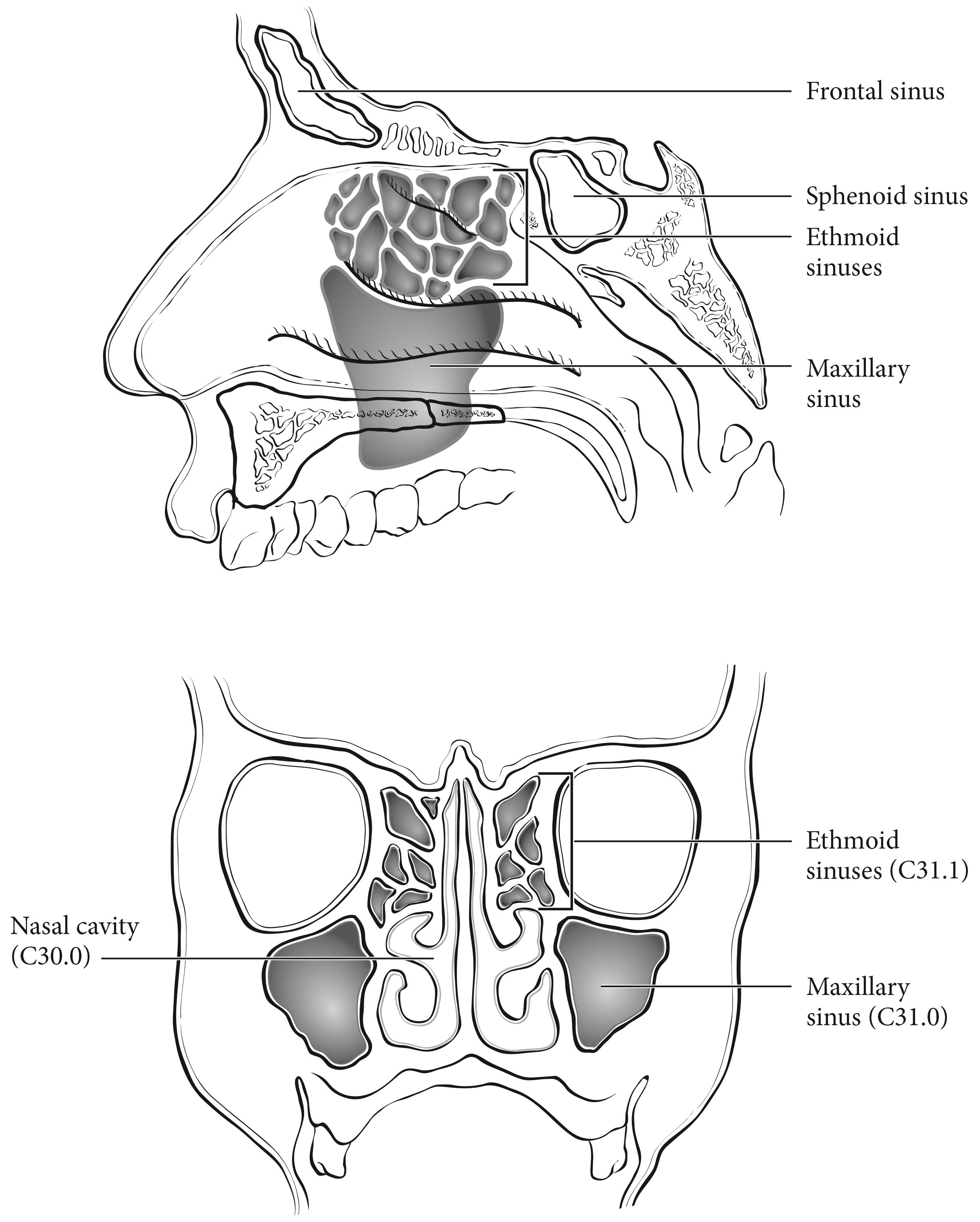
Cancer of the maxillary sinus is the most common of the sinonasal malignancies. Ethmoid sinus and nasal cavity cancers are equal in frequency, but considerably less common than maxillary sinus cancers. Tumors of the sphenoid and frontal sinuses are rare.
Primary Site(s)
The location and the extent of the mucosal lesion within the maxillary sinus have prognostic significance. Historically, a plane connecting the medial canthus of the eye to the angle of the mandible, represented by Ohngren's line, is used to divide the maxillary sinus into an anteroinferior portion (infrastructure), which is associated with a good prognosis, and a posterosuperior portion (suprastructure), which has a poor prognosis (Figure 14.1). The poorer outcome associated with suprastructure cancers reflects early invasion by these tumors to critical structures, including the orbit, skull base, pterygoid plates, and infratemporal fossa.
For the purpose of staging, the nasoethmoidal complex is divided into two sites: nasal cavity and ethmoid sinuses. The ethmoids are further subdivided into two subsites: left and right, separated by the nasal septum (perpendicular plate of ethmoid). The nasal cavity is divided into four subsites: the septum, floor, lateral wall, and the edge of naris to mucocutaneous junction.
Site | Subsite(s) |
---|---|
Maxillary sinus | Left/right |
Nasal cavity | Septum |
Floor | |
Lateral wall | |
Edge of naris to mucocutaneous junction | |
Ethmoid sinus | Left/right |
Regional lymph node spread from cancer of nasal cavity and paranasal sinuses is relatively uncommon. Involvement of buccinator, prevascular facial, submandibular, upper jugular, and (occasionally) retropharyngeal nodes may occur with advanced maxillary sinus cancer, particularly those extending beyond the sinus walls to involve adjacent structures, including soft tissues of the cheek, upper alveolus, palate, and buccal mucosa or overlying skin. Ethmoid sinus cancers are less prone to regional lymphatic spread. When only one side of the neck is involved, it should be considered ipsilateral. Bilateral spread may occur with advanced primary cancer, particularly with spread of the primary beyond the midline.
Clinical Classification
The assessment of primary maxillary sinus, nasal cavity, and ethmoid tumors is based on inspection and palpation, including examination of the orbits, nasal and oral cavities, and nasopharynx, and neurologic evaluation of the cranial nerves. Nasal endoscopy with rigid or fiberoptic flexible instruments is recommended for inspection and biopsy.
Neck nodes are assessed by palpation. In clinical evaluation, the maximum size of any nodal mass should be measured. Biopsy, when indicated, is done with a fine needle and not open approach and is included in clinical classification if done. The three categories of clinically involved nodes for the nasal cavity and paranasal sinus are N1, N2, and N3. Midline nodes are considered ipsilateral nodes. Superior mediastinal lymph nodes are considered regional lymph nodes (level VII). In addition to the components to describe the N category, regional lymph nodes should also be described according to the level of the neck that is involved. Provide a description or map of the regional lymph nodes and node groups that the cancer affects. Unambiguous evidence of gross ENE (i.e., defined as invasion of skin, infiltration of musculature/fixation to adjacent structures on clinical examination, cranial nerve, brachial plexus, sympathetic trunk or phrenic nerve invasion with dysfunction) is a sufficiently high threshold to classify these as clinical ENE(+). Examinations for distant metastases include appropriate imaging, blood chemistries, blood count, and other routine studies as indicated. Biopsy is typically needed to confirm metastasis, although a risk-to-benefit ratio is always weighed and is included in clinical stage as pM1 if done.
Imaging studies showing amorphous spiculated margins of involved nodes or involvement of internodal fat resulting in loss of normal oval-to-round nodal shape strongly suggest extracapsular (extranodal) tumor spread. No imaging study (as yet) can identify microscopic foci in regional nodes or distinguish between small reactive nodes and small malignant nodes without central radiographic inhomogeneity.
Imaging
Imaging is beneficial for lesions that cannot be fully assessed on clinical examination, locally advanced disease, or symptomatic patients. Computed tomography (CT) and magnetic resonance (MR) imaging are complementary imaging studies for staging patients with cancers involving the nasal cavity and/or the paranasal sinuses. There is no indication for plain films or positron emission tomography (PET)-CT for staging of the primary site. PET-CT may be helpful for assessing nodal metastases; however, lymph node staging is discussed in Chapter 6.
CT is superior to MR imaging for identifying bone erosion of thin walls and septa of the paranasal sinuses. CT is superior to MR imaging for identifying involvement of the hard palate. Either CT or MR imaging may be performed for tumor extending outside of the nose and/or paranasal sinuses to involve the adjacent structures, including the orbital apex (T4b). MR imaging, especially with T2-weighted images, is helpful for tumor mapping and for distinguishing between tumor extension and obstructed secretions. Tumors may obstruct the frontal recess or sphenoethmoidal recess, which may result in obstruction of the frontal or sphenoid sinus, respectively. Differential between tumor extension and proteinaceous obstructed secretions is best performed with MR imaging, which will help accurately identify T4a tumors. Both CT and MR imaging may be used to evaluate for posterior spread to the pterygopalantine fossa. However, MR imaging is superior to CT for evaluating for retrograde perineural spread along V2 through foramen rotundum or V3. CT is superior to MR imaging for early cortical involvement, but MR imaging is superior to CT for bone marrow invasion. MR imaging also is superior to CT for identifying dural involvement or other types of intracranial extension (T4b).
Pathological Classification
Complete resection of the primary site and/or regional nodal dissections, followed by pathological examination of the resected specimen(s), allows the use of this designation for pT and/or pN, respectively. Specimens that are resected after radiation or chemotherapy need to be identified and considered in context, and use yp instead of p. pT is derived from the invasion of bone, orbit, dura, and presence of disease in multiple subsites. Pathological staging represents additional and important information and should be included as such in staging, but it does not supplant clinical staging as the primary staging scheme.
For pN, a selective neck dissection will ordinarily include 10 or more lymph nodes, and a radical or modified radical neck dissection will ordinarily include 15 or more lymph nodes. Negative pathological examination of a smaller number of nodes still mandates a pN0 designation.
Definition of ENE and Description of Its Extent
All surgically resected metastatic nodes should be examined for the presence and extent of ENE. The precise definition of ENE has varied in the literature over the course of time. The American College of Pathologists defines ENE as extension of metastatic tumor, present within the confines of the lymph node, through the lymph node capsule into the surrounding connective tissue, with or without associated stromal reaction.
ENE detected on histopathologic examination is designated as ENEmi (microscopic ENE less than or equal to 2 mm) or ENEma (major ENE greater than 2 mm). Both ENEmi and ENEma qualify as ENE(+) for definition of pN. These descriptors of ENE will not be required for current pN definition, but data collection is recommended to allow standardization of data collection and future analysis.
Prognostic Factors Required for Stage Grouping
Beyond the factors used to assign T, N, or M categories, no additional prognostic factors are required for stage grouping.
Additional Factors Recommended for Clinical Care
ENE is defined as extension of metastatic tumor, present beyond the confines of the lymph node, through the lymph node capsule into the surrounding connective tissue, with or without associated stromal reaction. Unambiguous evidence of gross ENE extension (i.e., defined as invasion of skin, infiltration of musculature/fixation to adjacent structures on clinical examination, cranial nerve, brachial plexus, sympathetic trunk, or phrenic nerve invasion with dysfunction) is a sufficiently high threshold to classify these as clinical ENE(+).
In addition to the importance of the TNM factors outlined previously, the overall health of the patient clearly influences outcome. An ongoing effort to better assess prognosis using both tumor and nontumor-related factors is underway. Chart abstraction will continue to be performed by cancer registrars to obtain important information regarding specific factors related to prognosis. These data will then be used to further hone the predictive power of the staging system in future revisions.
Comorbidity can be classified by specific measures of additional medical illnesses.9 Accurate reporting of all illnesses in the patient's medical record is essential to the assessment of these parameters. General performance measures are helpful in predicting survival. The AJCC strongly recommends the clinician report performance status using the Eastern Cooperative Oncology Group (ECOG), Zubrod, or Karnofsky performance measures, along with standard staging information. An interrelationship between each of the major performance tools exists.
Zubrod/ECOG Performance Scale | |||
---|---|---|---|
0 | Fully active, able to carry out all predisease activities without restriction (Karnofsky 90-100) | ||
1 | Restricted in physically strenuous activity but ambulatory and able to carry out work of a light or sedentary nature. For example, light housework, office work (Karnofsky 70-80) | ||
2 | Ambulatory and capable of all self-care but unable to carry out any work activities. Up and about more than 50% of waking hours (Karnofsky 50-60) | ||
3 | Capableof only limited self-care, confined to bed or chair 50% or more of waking hours(Karnofsky 30-40) | ||
4 | Completely disabled. Cannot carry out self-care. Totally confined to bed (Karnofsky 10-20) | ||
5 | Death (Karnofsky 0) |
Lifestyle factors such as tobacco and alcohol abuse negatively influence survival. Accurate recording of smoking in pack-years and alcohol in number of days drinking per week and number of drinks per day will provide important data for future analysis. Nutrition is important to prognosis and will be indirectly measured by weight loss of greater than 5% of body weight in the previous 6 months.10 Depression adversely impacts quality of life and survival. Notation of a previous or current diagnosis of depression should be recorded in the medical record.11
The role of tobacco as a negative prognostic factor is well established. However, exactly how this could be codified in the staging system is less clear. At this time, smoking is known to have a deleterious effect on prognosis but is hard to accurately apply to the staging system.
Smoking history should be collected as an important element of the demographics and may be included in Prognostic Groups in the future. For practicality, the minimum standard should classify smoking history as never, less than or equal to 10 pack-years, greater than 10 but less than or equal to 20 pack-years, or greater than 20 pack-years.
The authors have not identified any emerging factors for clinical care.
The AJCC recently established guidelines that will be used to evaluate published statistical prediction models for the purpose of granting endorsement for clinical use.12 Although this is a monumental step toward the goal of precision medicine, this work was published only very recently. Therefore, the existing models that have been published or may be in clinical use have not yet been evaluated for this cancer site by the Precision Medicine Core of the AJCC. In the future, the statistical prediction models for this cancer site will be evaluated, and those that meet all AJCC criteria will be endorsed.
Definition of Primary Tumor (T)
T Category | T Criteria |
---|---|
TX | Primary tumor cannot be assessed |
Tis | Carcinoma in situ |
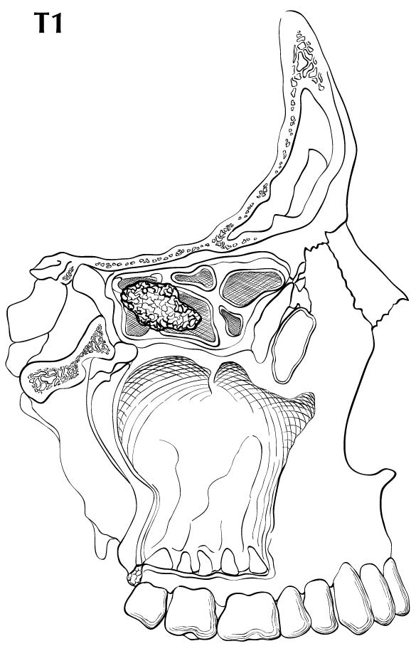
T1 | Tumor restricted to any one subsite, with or without bony invasion |
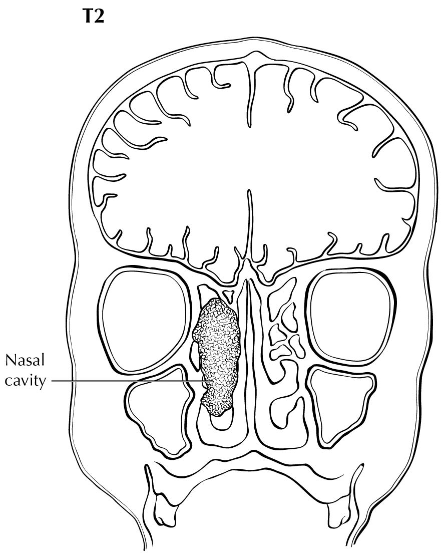
T2 | Tumor invading two subsites in a single region or extending to involve an adjacent region within the nasoethmoidal complex, with or without bony invasion |
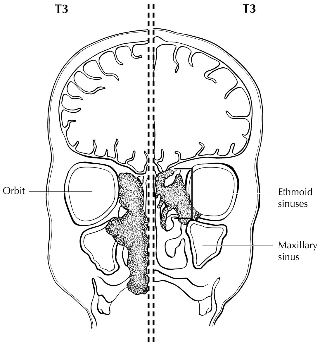
T3 | Tumor extends to invade the medial wall or floor of the orbit, maxillary sinus, palate, or cribriform plate |
T4 | Moderately advanced or very advanced local disease |
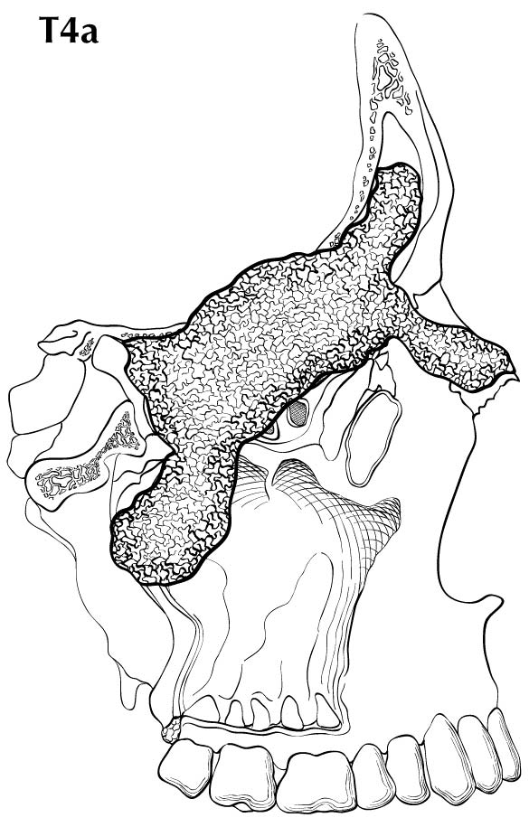
T4a | Moderately advanced local disease. Tumor invades any of the following: anterior orbital contents, skin of nose or cheek, minimal extension to anterior cranial fossa, pterygoid plates, sphenoid or frontal sinuses. |
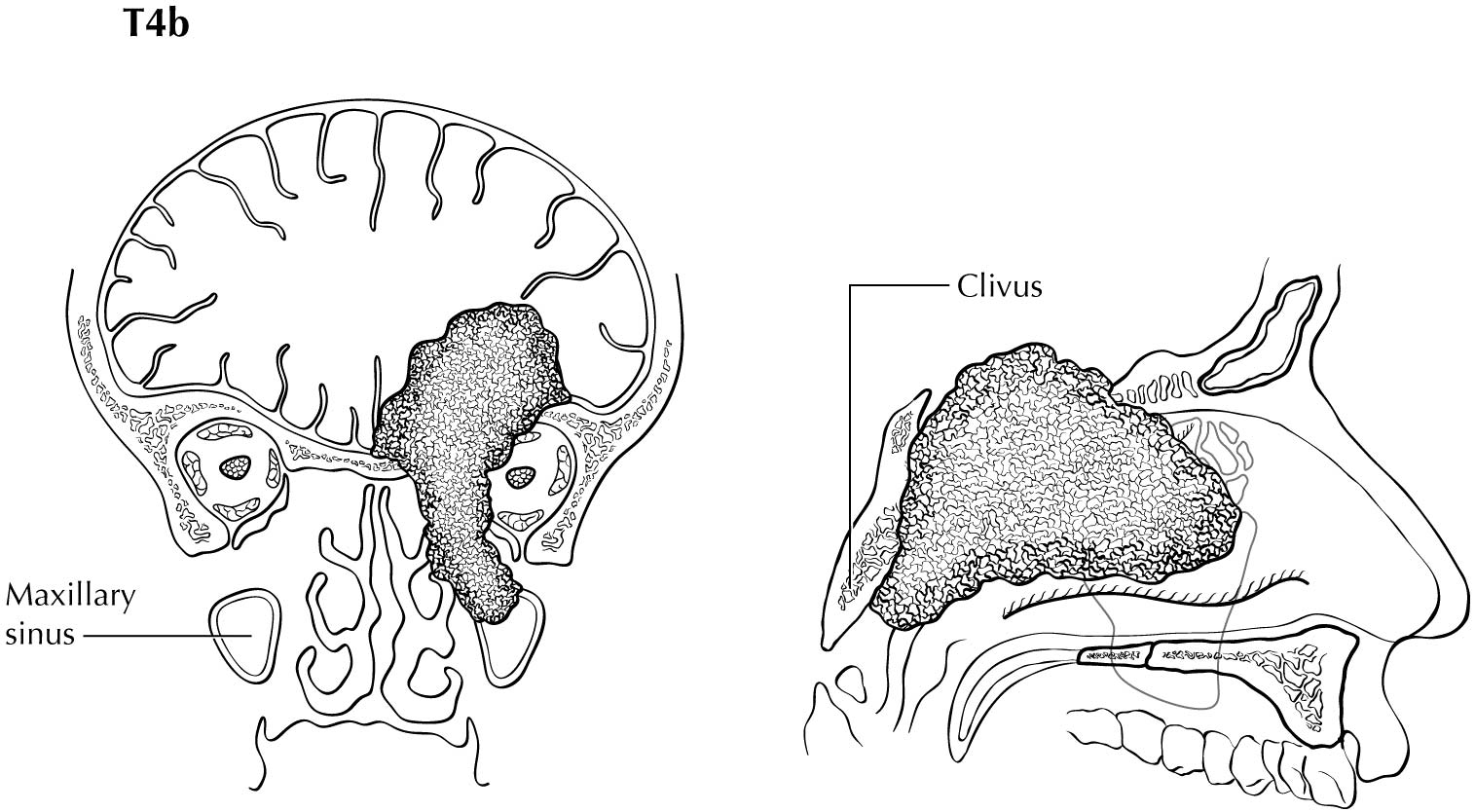
T4b | Very advanced local disease. Tumor invades any of the following: orbital apex, dura, brain, middle cranial fossa, cranial nerves other than (V2), nasopharynx, or clivus. |
Definition of Regional Lymph Node (N)
Clinical N (cN)N Category | N Criteria |
---|---|
NX | Regional lymph nodes cannot be assessed |
N0 | No regional lymph node metastasis |
N1 | Metastasis in a single ipsilateral lymph node, 3 cm or smaller in greatest dimension and ENE(-) |
N2 | Metastasis in a single ipsilateral node larger than 3 cm but not larger than 6 cm in greatest dimension and ENE(-); or metastases in multiple ipsilateral lymph nodes, none larger than 6 cm in greatest dimension and ENE(-); or in bilateral or contralateral lymph nodes, none larger than 6 cm in greatest dimension and ENE(-) |
N2a | Metastasis in a single ipsilateral node larger than 3 cm but not larger than 6 cm in greatest dimension and ENE(-) |
N2b | Metastases in multiple ipsilateral nodes, none larger than 6 cm in greatest dimension and ENE(-) |
N2c | Metastases in bilateral or contralateral lymph nodes, none larger than 6 cm in greatest dimension and ENE(-) |
N3 | Metastasis in a lymph node larger than 6 cm in greatest dimension and ENE(-); or metastasis in any node(s) with clinically overt ENE(+) |
N3a | Metastasis in a lymph node larger than 6 cm in greatest dimension and ENE(-) |
N3b | Metastasis in any node(s) with clinically overt ENE (ENEc) |
A designation of U or L may be used for any N category to indicate metastasis above the lower border of the cricoid (U) or below the lower border of the cricoid (L). Similarly, clinical and pathological ENE should be recorded as ENE(-) or ENE(+).
N Category | N Criteria |
---|---|
NX | Regional lymph nodes cannot be assessed |
N0 | No regional lymph node metastasis |
N1 | Metastasis in a single ipsilateral lymph node, 3 cm or smaller in greatest dimension and ENE(-) |
N2 | Metastasis in a single ipsilateral lymph node, 3 cm or smaller in greatest dimension and ENE(+); or larger than 3 cm but not larger than 6 cm in greatest dimension and ENE(-); or metastases in multiple ipsilateral lymph nodes, none larger than 6 cm in greatest dimension and ENE(-); or in bilateral or contralateral lymph node(s), none larger than 6 cm in greatest dimension and ENE(-) |
N2a | Metastasis in single ipsilateral node 3 cm or less in greatest dimension and ENE(+); or a single ipsilateral node larger than 3 cm but not larger than 6 cm in greatest dimension and ENE(-) |
N2b | Metastases in multiple ipsilateral nodes, none larger than 6 cm in greatest dimension and ENE(-) |
N2c | Metastases in bilateral or contralateral lymph node(s), none larger than 6 cm in greatest dimension and ENE(-) |
N3 | Metastasis in a lymph node larger than 6 cm in greatest dimension and ENE(-); or in a single ipsilateral node larger than 3 cm in greatest dimension and ENE(+); or multiple ipsilateral, contralateral or bilateral nodes, any with ENE(+); or a single contralateral node of any size and ENE(+) |
N3a | Metastasis in a lymph node larger than 6 cm in greatest dimension and ENE(-) |
N3b | Metastasis in a single ipsilateral node larger than 3 cm in greatest dimension and ENE(+); or multiple ipsilateral, contralateral or bilateral nodes, any with ENE(+); or a single contralateral node of any size and ENE(+) |
A designation of U or L may be used for any N category to indicate metastasis above the lower border of the cricoid (U) or below the lower border of the cricoid (L). Similarly, clinical and pathological ENE should be recorded as ENE(-) or ENE(+).
Definition of Distant Metastasis (M)
M Category | M Criteria |
---|---|
M0 | No distant metastasis |
M1 | Distant metastasis |
The predominant cancer is squamous cell carcinoma. The staging guidelines are applicable to all forms of carcinoma, including those arising from minor salivary glands. Other nonepithelial tumors such as those of lymphoid tissue, soft tissue, bone, and cartilage (i.e., lymphoma and sarcoma) are not included. Histologic confirmation of diagnosis is required. Histopathologic grading of squamous carcinoma is recommended. The grade is subjective and uses a descriptive, as well as numerical, form (i.e., well differentiated, moderately differentiated, and poorly differentiated), depending on the degree of closeness to or deviation from squamous epithelium in mucosal sites. Also recommended where feasible is a quantitative evaluation of depth of invasion of the primary tumor and the presence or absence of vascular invasion and perineural invasion. Although the grade of tumor does not enter into the staging of the tumor, it should be recorded. The pathological description of any lymphadenectomy specimen should describe the size, number, and position of the involved node(s) and the presence or absence of ENE.
HISTOLOGIC GRADE (G)
G | G Definition |
---|---|
GX | Grade cannot be assessed |
G1 | Well differentiated |
G2 | Moderately differentiated |
G3 | Poorly differentiated |