section name header

Basic Information

AUTHOR: Brian Block, MD

Definition

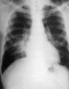
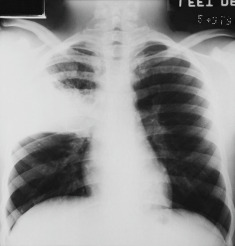
Mycoplasma pneumonia is an infection of the lung parenchyma caused by a small rod-shaped bacterium, Mycoplasma pneumoniae.

Synonyms

Primary atypical pneumonia

Walking pneumonia

Mycoplasma pneumonia

ICD-10CM CODE
J15.7Pneumonia due to Mycoplasma pneumoniae
Epidemiology & Demographics
Incidence (in U.S.)

  • Frequent cause of community-acquired pneumonia (CAP); particularly in children and adults <40 yr
  • Causes periodic outbreaks with high secondary attack rate among household contacts because it is easily transmitted1
  • Many cases probably resolve without coming to medical attention2
  • Asymptomatic carriage thought to be common
Predominant Sex

Equal distribution

Predominant Age

  • School-age children and young adults (ages 5 to 20 yr)3
Peak Incidence

  • Occurs throughout the year but outbreaks are more common in late summer and fall
  • Transmitted by respiratory droplets
Genetics

Familial disposition:

  • None known
  • May be more severe in patients with sickle cell anemia
Physical Findings & Clinical Presentation

  • Common symptoms include fever, cough, headache, and otalgia
  • Exam findings include rhonchi or rales, conjunctivitis, lymphadenopathy
  • Rash occurs in 10% to 25% of infected individuals, ranging from self-limited morbilliform eruptions to life-threatening rashes including Stevens-Johnson syndrome4
  • Some patients develop autoimmune hemolytic anemia due to autoantibodies (cold agglutinins)
  • Neurologic manifestations are also possible but less common (mono- or polyneuritis, transverse myelitis, cranial nerve palsies)
  • Table E1 summarizes the clinical manifestations of Mycoplasma pneumoniae
Etiology

  • Spreads person-to-person via respiratory droplets with an incubation period of 1 to 3 wk

Diagnosis

Differential Diagnosis

  • Typical bacterial pneumonia (S. pneumoniae, H. influenza)
  • Other causes of “atypical” pneumonia (Legionella, Chlamydophila)
  • Viral pneumonia (e.g., influenza, RSV, COVID-19)
  • Noninfectious causes of respiratory distress
Workup

  • Thorough history and physical examination
  • Chest x-ray (Fig. E1)
Laboratory Tests
  1. White blood cells (WBCs):
    1. WBC count >10,000/mm3 in approximately one fourth of patients
    2. Leukopenia rare
  2. Cold agglutinins:
    1. Detected in approximately 50% to 70% of all patients within 1 to 2 wk of infection5
    2. Neither sensitive nor specific, so are of limited diagnostic utility. Cold agglutinins are also found in:
      1. Lymphoproliferative diseases
      2. Influenza
      3. Mononucleosis
      4. Adenovirus infections
      5. Legionella pneumonia
  3. PCR has replaced serology as the test of choice to confirm the diagnosis
  4. Serology and culture are other means of diagnosis, but culture is technically difficult, and results may take weeks
  5. Mycoplasma cannot be seen on Gram stain because they lack a cell wall
Imaging Studies

  • Classically patchy, reticular opacities with lower-lobe predominance
  • Radiographic abnormalities frequently out of proportion to physical findings (Fig. E2)
  • Pleural effusions are seen in approximately 30% of patients, typically small
  • Potential findings in late-stage of infection or after acute infection resolves:
    1. Lung abscess
    2. Pneumatoceles
    3. Lobar collapse
    4. Hyperlucent lung syndrome (aka Swyer-James or postinfectious bronchiolitis)

Treatment

Chronic Rx

  • Effective antimicrobial therapy does not eliminate the organism from the respiratory secretions, which may remain positive for weeks.
  • Serum antibody response does not necessarily provide lifelong immunity.
Disposition

  • Clinical improvement is almost universal within 10 days.
  • Chest imaging generally improves within 5 to 8 wk.
  • Person-to-person spread can be minimized by avoiding open coughing, especially in enclosed areas.
  • Azithromycin prophylaxis has efficacy in close contacts.7
Referral

  • To infectious disease specialist for patients not responding to treatment, for severe infection (including multilobar involvement), or for severe extrapulmonary manifestations
  • To pulmonologist, if diagnosis is unclear or alternate diagnosis being considered

TABLE E1 Clinical Manifestations of Mycoplasma Pneumoniae Infection

Respiratory tractPharyngitis, Laryngitis, Acute Bronchitis, Bronchopneumonia
Skin and mucosaMaculopapular and vesicular exanthema, urticaria, purpura, erythema nodosum, erythema multiforme, Stevens-Johnson syndrome
Central nervous systemMeningitis, meningoencephalitis, acute psychosis, cerebellitis, Guillain-Barré syndrome
Other organ involvementPancreatitis, diabetes mellitus, nonspecific reactive hepatitis, subacute thyroiditis
Autoimmune and other phenomenaHemorrhagic bullous myringitis, hemolytic anemia, pericarditis, thromboembolism

Some association remains uncertain.

From Cohen J, Powderly WG: Infectious diseases, ed 2, St Louis, 2004, Mosby.

Figure E1 Radiographic findings in Mycoplasma pneumoniae pneumonia are nonspecific.

Bilateral bronchopneumonia occurred in this patient.

From Mason RJ et al: Murray and Nadel’s textbook of respiratory medicine, ed 5, Philadelphia, 2010, Saunders.

Figure E2 Localized airspace opacification resulting from Mycoplasma pneumoniae.

From Specht N [ed]: Practical guide to diagnostic imaging, St Louis, 1998, Mosby.

Pearls & Considerations

Comments

  • Outbreaks occur in military recruits, group homes, nursing homes, and in the community.
  • Infection control: In the hospital these patients should be on droplet precautions.
  • Chest x-ray resolution is complete by 8 wk in approximately 90% of patients.
Related Content

Mycoplasma Pneumonia (Patient Information)

Pneumonia, Bacterial (Related Key Topic)

Related Content

  1. Hanukoglu A. : Pulmonary involvement in Mycoplasma pneumoniae infection in familiesInfection. ;14:1-6, 1986.
  2. Infectious Diseases Society of America/American Thoracic Society consensus guidelines on the management of community-acquired pneumonia in adultsClin Infect Dis. ;44(suppl 2):S27-S72, 2007.
  3. Krafft C., Christy C. : Mycoplasma pneumonia in children and adolescentsPediatr Rev. ;41(1):12-19, 2020.
  4. Tay Y.K. : Mycoplasma pneumoniae infection is associated with Stevens-Johnson syndrome, not erythema multiforme (von Hebra)J Am Acad Dermatol. ;35(5 Pt 1), 1996.
  5. Bajantri B. : Mycoplasma pneumoniae: a potentially severe infectionJ Clin Med Res. ;10(7):535-544, 2018.
  6. Diaz M.H. : Investigations of Mycoplasma pneumoniae infections in the United States: trends in molecular typing and macrolide resistance from 2006 to 2013J Clin Microbiol. ;53(1):124-130, 2015.
  7. Gray G.C. : Randomized, placebo-controlled clinical trial of oral azithromycin prophylaxis against respiratory infections in a high-risk, young adult populationClin Infect Dis. ;33(7):983-989, 2001.