AUTHORS: Christina Nestlerode, DO and Robert Neff, MD
Cancers of the uterus include tumors that originate from epithelial or mesenchymal tissue. Tumors that arise from the epithelium are referred to as adenocarcinomas, whereas tumors that arise from mesenchymal tissue (i.e., connective, muscular, vascular) are referred to as sarcomas. These tumors may be found in various locations of the uterus including the endometrium or myometrium.
Historically, endometrial adenocarcinomas were divided into two categories: Type 1 and Type 2. Type 1 endometrial carcinomas refer to endometrioid subtypes, whereas Type 2 endometrial carcinomas refer to serous, clear cell, and mixed Mullerian subtypes (see Endometrial Cancer). Recent molecular studies have shown carcinosarcomas (MMMT) to be closely related to epithelial tumors compared to sarcomas.
This chapter will focus on sarcomas of the uterus arising from the endometrial stroma or myometrium.
|
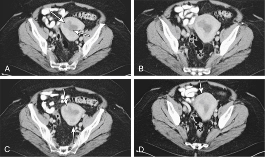
Incidence of all uterine cancer is 28.1 per 100,000 women per yr with 4.9 deaths per 100,000 women as of 2018. In the most recent update from SEER Annual Report to the Nation, uterine cancer showed the highest increase frequency in death of all cancers among U.S. women. Endometrial cancer remains the most common uterine malignancy in the U.S; however, sarcomas of the uterus are rare. Sarcomas account for approximately 3% to 8% of all cancers of the uterine corpus. Sarcomas are associated with a poor prognosis compared to endometrial cancer.
Diagnosis can be made histologically by biopsy for abnormal bleeding. Workup includes biopsy (in the office or operating room, in conjunction with hysteroscopy) and imaging (see Imaging Studies below). Surgical removal of the uterus is the most common way to diagnose a uterine sarcoma. Histologic criteria include mitotic index, cellular atypia, loss of polarity, and necrosis.
Figure 1 A 50-yr-old patient with uterine sarcoma.
A, Axial contrast-enhanced computed tomographic (CT) image. Low-attenuation lobulated and infiltrating soft tissue fills the endometrial canal (arrows), extending into the myometrium. B, Axial contrast-enhanced CT image showing same as A. Low-attenuation, lobulated, and infiltrating soft tissue fills the endometrial canal, extending into the myometrium. C, Axial contrast-enhanced CT image. Low-attenuation lobulated and infiltrating soft tissue fills the endometrial canal. Subtle myometrial invasion is seen anterior and posterior (arrows). D, Axial contrast-enhanced CT image. Low-attenuation lobulated and infiltrating soft tissue fills the endometrial canal (arrow), showing same. Subtle myometrial invasion is seen fundally.
From Fielding JR et al: Gynecologic imaging, Philadelphia, 2011, Saunders.
Staging of endometrial carcinoma and uterine adenosarcoma is summarized in Tables 1 and 2.
TABLE 2 International Federation of Gynecology and Obstetrics 2009 Staging for Uterine Sarcoma (Including Leiomyosarcoma and Endometrial Stromal Sarcoma)
Stage | |||
---|---|---|---|
I | Tumor limited to the uterus | ||
IA | Tumor 5 cm or less in greatest dimension | ||
IB | Tumor more than 5 cm | ||
II | Tumor extends beyond the uterus, within the pelvis | ||
IIA | Adnexal involvement | ||
IIB | Extrauterine pelvic tissue involvement | ||
III | Involvement of abdominal tissues | ||
IIIA | 1 site | ||
IIIB | >1 site | ||
IIIC | Regional lymph node metastasis | ||
IV | |||
IVA | Tumor invades bladder or rectum | ||
IVB | Distant metastases |
From Niederhuber JE: Abeloffs clinical oncology, ed 6, Philadelphia, 2020, Elsevier.
TABLE 1 International Federation of Gynecology and Obstetrics 2009 Staging for Endometrial Carcinoma (Including Carcinosarcoma)
Stage | |||
---|---|---|---|
I | Tumor confined to uterine corpus including endocervical glands | ||
IA | Tumor confined to endometrium or invades <50% of the myometrium | ||
IB | Tumor invades 50% or more the myometrium | ||
II | Tumor invades cervical stroma but confined to uterus | ||
III | Tumor involving the serosa, adnexa, vagina, or parametria | ||
IIIA | Tumor invades uterine serosa or adnexa | ||
IIIB | Involvement of vagina or parametrium | ||
IIIC1 | Metastasis to pelvic lymph nodes | ||
IIIC2 | Metastasis to paraaortic lymph nodes | ||
IV | Tumor involves bladder and/or bowel mucosa or distant metastasis | ||
IVA | Invasion of bladder or bowel mucosa | ||
IVB | Distant metastases including intraabdominal metastasis, inguinal lymph nodes, or both |
From Niederhuber JE: Abeloffs clinical oncology, ed 6, Philadelphia, 2020, Elsevier.
A gynecologic oncologist should manage uterine sarcoma.
Key points in the management of uterine sarcoma are described in Box 3.
BOX 3 Uterine Sarcoma: Key Points
From Greer IA et al: Mosbys color atlas and text of obstetrics and gynecology, London, 2001, Harcourt.