section name header

Basic Information

AUTHORS: Emily Sauck, DO, MBA and Anthony Sciscione, DO

Definition

Endometrial cancer, also called endometrial carcinoma (EC), is cancer of the endometrium, which is the lining of the uterus. Traditionally, EC was divided into two types: Type 1, which are estrogen-driven, and type 2, which are not estrogen driven. However, EC is now more commonly subdivided into different types based on histology-how the cells appear under the microscope (Table 1).

TABLE 1 Pathogenetic Subsets of Endometrial Carcinoma

ParameterType IType II
Age50s-60s60s-70s
ObesityCommonUncommon
Estrogenic stimuliCommonUncommon
EndometriumAnovulatoryAtrophic
PrecursorEndometrial intraepithelial neoplasiaPresumed EmGD
TransitionSlowUnknown
TypeEndometrioidPapillary serous or mixed
Molecular geneticsMSI, PTEN mutation; loss of PAX2p53 mutation, 1p deletions; loss of PAX2
FamilialHereditary nonpolyposis colonic cancer syndrome
SpreadLymph nodesPeritoneum
Concurrent ovarianCommonUncommon
PrognosisGoodPoor

EmGD, Endometrial glandular dysplasia; MSI, microsatellite instability.

From Crum CP et al: Diagnostic gynecologic and obstetric pathology, ed 3, Philadelphia, 2018, Elsevier.

Histologic types are summarized in Box 1 and include adenocarcinoma, uterine carcinosarcoma, squamous cell carcinoma, small cell carcinoma, transitional carcinoma, and serous carcinoma, with most endometrial cancers being adenocarcinomas and endometrioid cancer being the most common type of adenocarcinoma.

BOX 1 Endometrial Primary Adenocarcinomas

  • Typical endometrioid adenocarcinoma
  • Adenocarcinoma with squamous elements
  • Clear cell carcinoma
  • Serous carcinoma
  • Secretory carcinoma
  • Mucinous carcinoma
  • Squamous carcinoma

From Gershenson DM et al: Comprehensive gynecology, ed 8, Philadelphia, 2022, Elsevier.

Synonyms

Uterine cancer (some forms)

Carcinoma of the endometrium

EC

ICD-10CM CODES
C54.1Malignant neoplasm of endometrium
C54.9Malignant neoplasm of corpus uteri, unspecified
C55Malignant neoplasm of uterus, part unspecified
Epidemiology & Demographics
Incidence

  • In 2021, 66,570 new cases of uterine cancer are predicted in the U.S. The rate of new cases of EC was 28.1 per 100,000 based on 2014 to 2018 cases. Incidence was greater among white and black women compared with American Indian/Alaska Native, Hispanic, and Asian Pacific Islander women. It is the most common gynecologic malignancy in the U.S. and the most common type of cancer that affects the female reproductive organs.
  • Unlike most cancers in the U.S., endometrial cancer is rising in both incidence and associated mortality.
Predominant Sex & Age

Median age at diagnosis: 63 yr

Median age of death from EC: 70 yr

Risk Factors

Age: Most cases are diagnosed in postmenopausal women, with a median age of 63 yr.

Estrogen exposure/hormone imbalance: Whether from early menarche, late menopause, diabetes, nulliparity, tamoxifen use, polycystic ovary syndrome, or unopposed estrogen therapy, the more exposure the endometrium has to estrogen, the more a woman’s risk of developing EC increases.

  • Obesity: Body mass index of 25 of greater is a major risk factor for EC.
  • Genetics: Lynch syndrome increases the risk of EC (and ovarian, colon, and other types of cancers). Cowden syndrome: Relative risks can be found in Table 2.

TABLE 2 Risk Factors for Endometrial Cancer

FactorRelative Risk
Overweight (lbs):
  • 20-50
3.0
  • 50+
10.0
Nulliparous:
  • vs. one child
2.0
  • vs. five children
5.0
Late menopause (>52 vs. 49 yr)2.4
Diabetes mellitus2.7
Unopposed estrogen therapy6.0
Tamoxifen therapy2.0
Sequential oral contraceptives7.0
Combination oral contraceptives0.5
Cowden syndrome (PTEN mutation)Three- to fivefold increased risk
Hereditary nonpolyposis colonic cancer syndrome40%-60% lifetime risk
Family member with endometrial cancer3.4

From Crum CP et al: Diagnostic gynecologic and obstetric pathology, ed 3, Philadelphia, 2018, Elsevier.

Physical Findings & Clinical Presentation

  • Abnormal uterine bleeding or postmenopausal bleeding in 90%
  • Pyometra or hematometra
  • Abnormal Pap smear: Endometrial cells, atypical glandular cells, or adenocarcinoma
  • Incidental finding at hysterectomy
Etiology

Endogenous or exogenous chronic unopposed estrogen stimulation of the endometrium

Previously termed adenoacanthoma or adenosquamous carcinoma.

Diagnosis

Differential Diagnosis

  • Endometrial atypical hyperplasia
  • Other genital tract malignancy
  • Uterine polyps
  • Atrophic vaginitis
  • Granulosa cell tumor
  • Fibroid uterus
  • Adenomyosis
Workup

  • Complete history and physical examination
  • Endometrial biopsy or dilation and curettage (Table 3)
  • Assessment of operative risk
  • Staging (Tables 4 and 5)
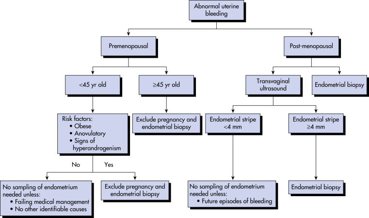
  • Fig. 1 is a diagnostic algorithm for diagnosing endometrial carcinoma for women with abnormal uterine bleeding.
Figure 1 Diagnostic Algorithm to Diagnose Endometrial Carcinoma for Women with Abnormal Uterine Bleeding

!!flowchart!!

From Niederhuber JE: Abeloff’s clinical oncology, ed 6, Philadelphia, 2020, Elsevier.

TABLE 4 National Comprehensive Cancer Network Treatment Guidelines for Endometrial Carcinoma After Comprehensive Surgical Staging

Stage IA
Grade 1 without ARFObserve
Grade 1 with ARFObserve or VBT
Grade 2 or 3 without ARFObserve or VBT
Grade 2 or 3 with ARFObserve or VBT and/or pelvic RT
Stage IB
Grade 1 without ARFObserve
Grade 1 with ARFObserve or VBT
Grade 2 without ARFObserve or VBT
Grade 2 with ARFObserve or VBT and/or pelvic RT
Grade 3 without ARFObserve or VBT and/or pelvic RT
Grade 3 with ARFObserve or VBT and/or pelvic RT ± chemotherapy
Stage II
Grade 1VBT and/or pelvic RT
Grade 2Pelvic RT and VBT
Grade 3Pelvic RT and VBT ± chemotherapy
Stage IIIAChemotherapy ± pelvic RT or tumor-directed RT ± chemotherapy or pelvic RT ± VBT
Stage IIIB-IIICChemotherapy and/or tumor-directed RT
Stage IVA-IVBChemotherapy ± RT

ARF, Adverse risk factors (age, positive lymphovascular space invasion, tumor size, lower uterine or cervical involvement); RT, radiation therapy; VBT, vaginal brachytherapy.

From Niederhuber JE: Abeloff’s clinical oncology, ed 6, Philadelphia, 2020, Elsevier.

TABLE 5 Revised FIGO Staging for Endometrial Cancer (adopted 2009)

StagesCharacteristic
ITumor confined to the corpus uteri
IANo or less than half myometrial invasion
IBInvasion equal to or more than half of the myometrium
IITumor invades cervical stroma but does not extend beyond the uterus
IIILocal or regional spread of the tumor
IIIATumor invades serosa of the corpus uteri or the adnexa
IIIBVaginal or parametrial involvement
IIICMetastases to pelvic or paraaortic lymph nodes
IIIC1Positive pelvic nodes
IIIC2Positive paraaortic lymph nodes with or without positive pelvic lymph nodes
IVTumor invades bladder or bowel mucosa, or distant metastasis
IVATumor invasion of bladder or bowel mucosa
IVBDistant metastases, including intraabdominal or inguinal lymph nodes

FIGO, Fédération Internationale de Gynécologie et d’Obstétrique (International Federation of Gynecology and Obstetrics).

G1, G2, or G3.

Endocervical glandular involvement should be considered only as stage I and no longer as stage II.

Positive cytology has to be reported separately without changing the stage.

From Lobo RA et al: Comprehensive gynecology, ed 7, Philadelphia, 2017, Elsevier.

TABLE 3 Differential Diagnosis of Endometrial Carcinoma (Curettings)

ParameterMimickingDifferential Diagnosis
Gland architectureCancerTelescoping artifact; stromal collapse breakdown; sectioning artifacts
BenignMicroglandular mucinous carcinoma; surface endometrioid carcinoma
Nuclear atypiaCancerSurface or glandular repair; Arias-Stella changes (hormonal therapy); radiation effect
Papillary changesCancerExfoliation artifact; stromal breakdown with papillary changes; papillary syncytial changes
BenignPapillary mucinous carcinoma

From Crum CP et al: Diagnostic gynecologic and obstetric pathology, ed 3, Philadelphia, 2018, Elsevier.

Laboratory Tests

  • Complete blood count
  • Prothrombin time and partial thromboplastin time if bleeding is heavy
  • Chemistry profile including liver function tests
  • Consider CA-125 level
Imaging Studies

  • Chest x-ray
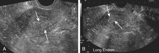
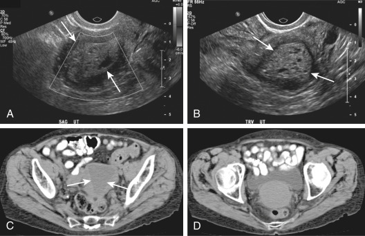
  • Computed tomography scan if concern for metastatic disease, and/or pelvic ultrasound (Fig. E2)
  • Transvaginal ultrasound (Fig. E3) in postmenopausal women with vaginal bleeding

Figure E2 A 48-yr-old woman with endometrial carcinoma.

A, Endovaginal ultrasound (US) showing thickened, heterogeneous, cystic, and vascular hyperechoic tissue filling the endometrial cavity (arrows). B, Second sagittal US image showing the same (arrows).

From Fielding JR et al: Gynecologic imaging, Philadelphia, 2011, Saunders.

Figure E3 A 56-yr-old woman with endometrial carcinoma.

A, Sagittal ultrasound (US) image showing thickened cystic echogenic soft tissue filling the endometrial cavity (arrows). B, Axial US image showing thickened cystic echogenic soft tissue filling the endometrial cavity (arrows). C, Noncontrast-enhanced axial computed tomographic (CT) image showing low-attenuation tissue filling the endometrial canal (arrows) in a postmenopausal patient. Note fundal thinning. D, Noncontrast-enhanced axial CT image showing cervical soft tissue fullness.

From Fielding JR et al: Gynecologic imaging, Philadelphia, 2011, Saunders.

Treatment

Nonpharmacologic Therapy

  • Surgery is the mainstay of treatment, with or without adjuvant radiation and/or chemotherapy, depending on tumor histology, stage, and grade. Laparoscopic surgery for early-stage EC is as safe and effective as laparotomy. Robotic laparoscopy procedures have increased significantly in recent yrs for this indication.
  • Surgery generally consists of pelvic washings, total hysterectomy and bilateral salpingo-oophorectomy, selective pelvic and periaortic lymphadenectomy, and omental biopsy depending on stage, grade, and histology.
  • Brachytherapy and/or teletherapy are added in an advanced stage.
  • Chemotherapy (carboplatin, paclitaxel) may be used for patients with high-risk endometrial cancer. Hormonal therapy (progestins alone or in combination with tamoxifen, selective estrogen-receptor modulators, aromatase inhibitors, synthetic steroid derivatives, and gonadotropin-releasing hormone analogues) may also be considered in cases where palliation, rather than cure, is the main intent of treatment, or in patients with multiple coexisting medical conditions who may not be surgical candidates.
  • Hormonal therapy, commonly a levonorgestrel intrauterine device, is an option for some young women with early-stage, low-grade EC who wish to preserve fertility. This choice should be discussed with a gynecologic oncologist.
Acute General Rx

  • A thorough workup should be completed before any therapy for EC.
  • Surgery hysterectomy with bilateral salpingo-oophorectomy is the treatment of choice.
Chronic Rx

  • Physical and pelvic examination every 3 mo for 2 yr, then every 6 mo for 2 yr, and annually thereafter with imaging as clinically indicated
  • Hormone replacement (combination) a consideration in low-risk patients (stage I or early stage II)
Disposition

  • Survival is generally defined by the stage of the disease and histology.
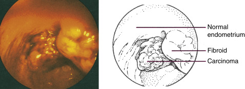
  • The majority of cases present early, and the 5-yr survival is generally good (Fig. E4).
  • Some histologic types (clear cell, papillary serous) have worse survival rates, as they tend to be more aggressive with higher rates of metastatic disease at the time of diagnosis.

Figure E4 Stage I endometrial carcinoma.

A small carcinoma can be seen adjacent to a uterine fibroid in this hysteroscopy photograph. Occasionally, a tumor this small may be missed on curettage.

From Skarin AT: Atlas of diagnostic oncology, ed 4, St Louis, 2010, Mosby.

Pearls & Considerations

Any woman with postmenopausal bleeding or abnormal uterine bleeding with risk factors for endometrial cancer needs evaluation by a gynecologist and either endometrial biopsy and/or pelvic ultrasound. When endometrial cancer is diagnosed, the patient should be cared for by a gynecologic oncologist and undergo surgical staging in a minimally invasive procedure when possible.

Related Content

Endometrial Cancer (Patient Information)

Abnormal Uterine Bleeding (Related Key Topic)

Uterine Malignancy (Related Key Topic)

Suggested Readings

    1. Braun M.M. : Diagnosis and management of endometrial cancerAm Fam Physician. ;93(6):468-474, 2016.
    2. Henley S.J. : Annual report to the nation on the status of cancer, part I: national cancer statisticsCancer. ;126:2225-2249, 2020.
    3. Kwon J.S. : Improving survival after endometrial cancer: the big pictureJ Gyn Oncol. ;26(3):227-231, 2015.
    4. Endometrial cancerObstet Gynecol. ;125:1006-1026, 2015.