section name header

Basic Information ⬇

AUTHOR: Roop Dutta, MD

Definition

Arrhythmogenic right ventricular dysplasia (ARVD) is a cardiomyopathy characterized by replacement of the normal myocardium with fibrous or fibrofatty tissue, mainly of the right ventricle but also occasionally with involvement of the lateral or posterior left ventricle. Historically, these macroscropic pathologic changes appeared in the “triangle” of dysplasia, which involves the right ventricular inflow tract, outflow tract, and apex. It is defined clinically by palpitations, syncope, and potentially life-threatening right-sided ventricular arrhythmias and, at later stages, heart failure.1

Synonyms

Arrhythmogenic right ventricular cardiomyopathy

ARVC

ICD-10CM CODE
I42.8Arrhythmogenic ventricular dysplasia
Epidemiology & Demographics
Prevalence

The current prevalence of ARVD in adults is 1:2000 to 5000 persons. It is one of the leading causes of arrhythmic cardiac arrest in young people and athletes.

Predominant Sex & Age

Mean age, 31 yr (range, 12 to 50 yr), predominantly male

Risk Factors

Family history of ARVD (present in nearly 50% of affected patients)

Genetics:

  • Autosomal dominant (most common) with variable penetrance and polymorphic phenotypic expression
  • Autosomal recessive (rarely, e.g., Naxos disease)
  • Several different gene mutations in desmosomal proteins
  • Gene mutations can be identified in 50% of affected individuals
Physical Findings & Clinical Presentation

  • ARVD can present with a wide range of clinical symptoms and severity from palpitations, syncope, and chest discomfort to ventricular tachycardia, sudden cardiac arrest, and signs of right ventricular failure such as dyspnea, edema, and fatigue. Patients may be clinically asymptomatic for many years.
  • Cardiac arrest after exertion or at rest may be the initial presentation.
  • Physical examination will be normal in most patients. Widely split S2 is an important diagnostic clue.
Etiology

ARVD is characterized by progressive replacement of mainly the right ventricular myocardium with fatty or fibrofatty tissue after apoptotic myocardial cell death caused by mutations of desmosomal proteins with isolated resudial cardiac cells. Historically, these macroscropic pathologic changes appeared in the “triangle” of dysplasia, which involves the right ventricular inflow tract, outflow tract, and apex. However, it is now recognized that there is sparing of the right ventricular apex early in disease. There can be involvement of the posterolateral LV with sparing of the LV septum.2

Diagnosis ⬆ ⬇

A major criterion equals 2 points; a minor criterion equals 1 point. The diagnosis of ARVD is considered definite if the patient has 4 points and probable with 3 points. See Table 1 for diagnostic criteria.

TABLE 1 Global or Regional Dysfunction and Structural Alterations

Major
2D echo criteria
Regional RV akinesia, dyskinesia, or aneurysm and one of the following measured at end diastole:
PLAX RVOT ≥32 mm or
PSAX RVOT ≥36
Fractional area change ≤33%
MRI criteria
Regional RV akinesia or dyskinesia or dyssynchronous RV contraction and one of the following:
Ratio of RV end-diastolic volume to BSA >100, <110 ml/m2 (male) or >100 ml/m2
RV ejection fraction >40% ≤45%
RV angiography criteria
Regional RV akinesia, dyskinesia, or aneurysm
Minor
2D echo criteria
Regional RV akinesia or dyskinesia or dyssynchronous RV contraction and one of the following measured at end diastole:
PLAX RVOT ≥29 <32 mm or
PSAX RVOT ≥32 <36
Fractional area change >33% ≤40%
MRI criteria
Regional RV akinesia or dyskinesia or dyssynchronous RV contraction and one of the following:
Ratio of RV end-diastolic volume to BSA ≥110 ml/m2 (male) or ≥100 ml/m2
RV ejection fraction ≤40%
Tissue characterization of wall
Major
Residual myocytes <60% by morphometric analysis (or <50% if estimated) with fibrous replacement of the RV free wall myocardium in >1 sample, with or without fatty replacement of tissue on endomyocardial biopsy
Minor
Residual myocytes 60%-75% by morphometric analysis (or 50%-65% if estimated), with fibrous replacement of the RV free wall myocardium in >1 sample with or without fatty replacement of tissue on endomyocardial biopsy
Repolarization abnormalities
Major
Inverted T waves in right precordial leads (V1, V2, and V3) or beyond in individuals >14 yr of age (in the absence of complete RBBB QRS ≥120 ms)
Minor
Inverted T waves in V1 and V2 in individuals >14 yr of age (in the absence of complete RBBB) or in V4, V5, and V6
Inverted T waves in leads V1, V2, V3, and V4 in individuals >14 yr of age in the presence of a complete RBBB
Depolarization or conduction abnormalities
Major
Epsilon wave (reproducible low-amplitude signals between end of QRS complex to onset of T wave) in the right precordial leads (V1-V3)
Minor
Late potentials by SAECG in ≥1 of 3 parameters in the absence of a QRSd of ≥110 ms on standard ECG
Filtered QRS ≥114 ms
Duration of terminal QRS <40 mV ≥38 ms
Root-mean-square voltage of terminal 40 ms ≤20 μV
Terminal activation duration ≥55 ms measured from the nadir of the end of the QRS, including R′, in V1, V2, or V3 in absence of complete RBBB
Arrhythmias
Major
Nonsustained or sustained VT of LBBB morph with superior axis
Minor
Nonsustained or sustained VT of RVOT configuration, LBBB morph with inferior axis or of unknown axis
>500 PVCs per 24 hr (Holter)
Family history
Major
ARVD/C in first-degree relative who meets Task Force criteria
ARVD/C confirmed pathologically at autopsy or surgery in first-degree relative
Identification of pathogenic mutation categorized as associated or probably associated with ARVD/C in the patient under evaluation
Minor
History of ARVD/C in first-degree relative in whom it is not possible to determine whether the family member meets Task Force criteria
Premature sudden death (<35 yr of age) caused by suspected ARVD/C in a first-degree relative
ARVD/C confirmed pathologically or by current Task Force criteria in second-degree relative

A major criterion equals 2 points, a minor criterion 1 point. The diagnosis of arrhythmogenic right ventricular dysplasia (ARVD) is considered definite if the patient has 4 points and probable with 3 points. BSA, Body surface area; ECG, electrocardiogram; MRI, magnetic resonance imaging; PLAX, parasternal long axis; PSAX, parasternal short axis; PVCs, premature ventricular contractions; RBBB, right bundle branch block; RV, right ventricle; RVOT, right ventricular outflow tract; SAECG, signal-averaged electrocardiogram; 2D, two dimensional; VT, ventricular tachycardia.

From Marcus IM: Diagnosis of arrhythmogenic right ventricular cardiomyopathy/dysplasia proposed modification of the Task Force criteria, Circulation 121:1533-1541, 2010.

Differential Diagnosis

  • Cardiomyopathy with involvement of the right ventricle
  • Uhl anomaly: Rare anomaly that presents mainly in childhood with signs and symptoms of right heart failure and characterized by a “paper-thin” right ventricle resulting from death of the myocytes throughout the right ventricle
  • Idiopathic RV tachycardia
  • Sarcoidosis
  • Right ventricular infarction
Workup

  • Initial workup includes history with focus on sudden death in the family, resting ECG, 24-Holter ECG, signal-averaged ECG, and imaging studies with echocardiography and cardiac MRI.
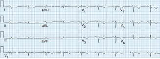
  • ECG will have diagnostic findings in 50% to 90% of patients with ARVD, including T-wave inversions in anterior precordial leads V1 to V6, epsilon waves, and a QRS duration longer than 110 ms in V1 or >40 ms longer in V1 than V6 (Fig. 1).
  • Ventricular tachycardia (VT) with a characteristic left bundle branch block pattern and frequent PVC’s (>500 in 24 hr) might be detected by 24-hr Holter monitoring.
  • An abnormal signal-averaged ECG is a minor diagnostic criteria.
  • Echocardiography will show right ventricular dilation with regional wall motion abnormalities, increased diameter of the right ventricular outflow tract, aneurysms, and depressed RV function that varies with the severity of the disease.
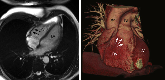
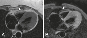
  • MRI (Fig. 2) is a noninvasive method to detect structural abnormalities (fibrofatty changes) and regional dysfunction. Cardiac MRI (CMR) is the most sensitive method to detect ARVD, but it has high false-positive rates. Cardiac CT angiogram (Fig. E3) will reveal thinning and aneurysmal dilation of the RV anterior wall and outflow tract.
  • If the routine tests are not conclusive, endomyocardial biopsy and electrophysiologic testing can be considered. However, biopsies and radionuclide ventriculography are rarely performed in the U.S.
Figure 2 Arrhythmogenic Right Ventricular Cardiomyopathy

Spin-Echo Cardiovascular Magnetic Resonance Without (A) and with (B), a Fat-Suppression Prepulse. There is Bright Signal in the Free Wall of the Right Ventricle that Suppresses with Fat Suppression (Arrows).

From Selke FW et al: Sabiston & Spencer surgery of the chest, ed 9, Philadelphia, 2016, Elsevier.

Figure 1 Arrhythmogenic Right Ventricular Cardiomyopathy-Epsilon Wave

This 12-Lead Electrocardiogram Tracing with Lead Vfn11 Rhythm Strip Shows Sinus Rhythm with T-Wave Inversion over the Right Precordial Leads. In Addition, There is an Epsilon Wave (Small Deflection at the End of the QRS Complex), Evident in Lead V1, Which is Characteristic of Arrhythmogenic Right Ventricular Cardiomyopathy.

From Olshansky B et al: Arrhythmia essentials, ed 2, Philadelphia, 2017, Elsevier.

Figure E3 Right Ventricular Aneurysm

This Cardiac Computed Tomography Angiogram Shows Thinning and Aneurysmal Dilation of the RV Anterior Wall and Outflow Tract (Arrows) in a Patient with Arrhythmogenic Right Ventricular Dysplasia, Cardiomyopathy, and Ventricular Tachycardia. Ao, Aorta; LV, Left Ventricle; PA, Pulmonary Artery; RV, Right Ventricle.

Courtesy of Dr. Nasar Nallamothu, Prairie Cardiovascular Consultants, Springfield, IL. From Issa Z et al: Clinical arrhythmology and electrophysiology, ed 2, Philadelphia, 2012, Saunders.

Treatment ⬆ ⬇

There is no curative treatment available. The treatment goals are focused on preventing sudden cardiac death, symptomatic treatment of right heart failure, and pharmacologic and invasive treatment of arrhythmias. Therapy with cardio-selective beta-blockers, such as metoprolol succinate, is recommended in both those with a history of ventricular arrythmias and those with a history of ventricular arrythmias for prophylaxis. Family members with a negative phenotype (either healthy gene carriers or those with an unknown genotype) do not need any specific treatment other than sports restriction; however, lifelong clinical assessment with the use of noninvasive tests at least every 2 yr is warranted.

Nonpharmacologic Therapy

  • Avoidance of activity, especially moderate and high instensity exercise, that may trigger ventricular tachycardia and may lead to disease progression.
  • For those with a definitive diagnosis of ARVD, implantable cardiodefibrillator (ICD) implantation needs to be considered if they are survivors of sudden cardiac arrest, have symptomatic ventricular tachycardia or ventricular fibrillation, have documented asymptomatic ventricular tachycardia or fibrillation, or have a left ventricular ejection fraction of 35% or less. Patients with unexplained syncope, advanced disease, documented ventricular arrhythmias, or a family history of sudden cardiac death or who have been resuscitated from cardiac arrest are at high risk. A subcutaneous ICD might be an alternative option instead of transvenous implantation.3,4
  • Radiofrequency catheter ablation is used in cases of refractory VT or frequent tachycardia after defibrillator placement.
  • Cardiac transplantation.
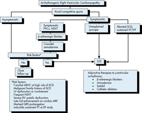
  • Fig. 4 describes a management algorithm for ARVD.
Figure 4 Arrhythmogenic Right Ventricular Cardiomyopathy

Arvc, Arrhythmogenic Right Ventricular Cardiomyopathy; EP, Electrophysiology; Gd, Gadolinium; ICD, Implantable Cardiodefibrillator; Nsvt, Nonsustained Ventricular Tachycardia; PVCs, Premature Ventricular Contractions; SCD, Sequential Compression Device; VF, Ventricular Fibrillation; VT, Ventricular Tachycardia.

!!flowchart!!

From Olshansky B et al: Arrhythmia essentials, ed 2, Philadelphia, 2017, Elsevier.

Pharmacologic Treatment

Antiarrhythmic therapy with sotalol (first-line treatment) or amiodarone, often in combination with beta-blockers, is used for tachycardia suppression.

Referral

  • Early cardiology and electrophysiology referral
  • Consider referring for genetic counseling

Pearls & Considerations ⬆ ⬇

Prevention

All first-degree relatives should be tested if ARVD is confirmed. Sports activity increases the risk of sudden cardiac death among adolescents and young adults with ARVC. The estimated overall mortality ranges from 0.08% to 3.6% per year.

Related Content ⬆

    1. Corrado D., Link M.S., Calkins H. : Arrhythmogenic right ventricular cardiomyopathyN Engl J Med. ;376(1):61-72, 2017.doi:10.1056/NEJMra1509267
    2. Te Riele A.S. : Mutation-positive arrhythmogenic right ventricular dysplasia/cardiomyopathy: the triangle of dysplasia displacedJ Cardiovasc Electrophysiol. ;24(12):1311-1320, 2013.doi:10.1111/jce.12222
    3. Schinkel A.F. : Implantable cardioverter defibrillators in arrhythmogenic right ventricular dysplasia/cardiomyopathy: patient outcomes, incidence of appropriate and inappropriate interventions, and complicationsCirc Arrhythm Electrophysiol. ;562-568, 2013.
    4. Bhonsale A. : Incidence and predictors of implantable cardioverter-defibrillator therapy in patients with arrhythmogenic right ventricular dysplasia/cardiomyopathy undergoing implantable cardioverter-defibrillator implantation for primary preventionJ Am Coll Cardiol. ;58:1485-1496, 2011.