AUTHOR: Russell J. McCulloh, MD


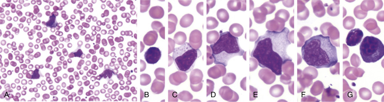
Infectious mononucleosis (IM) is a symptomatic infection most commonly caused by Epstein-Barr virus (EBV) and characterized by a classic triad of fever, tonsillar pharyngitis, and lymphadenopathy (predominantly cervical).1 Fatigue is also a very common finding. IM was first coined in 1920 to describe a syndrome characterized as an acute infectious process accompanied by atypical large peripheral blood lymphocytes.
| ||||||||||||||||||||||||
500 cases/100,000 persons/yr; worldwide, approximately 90% lifetime prevalence by age 30 yr.
Symptomatic infection most common between the ages of 15 and 24 yr. The age at which primary EBV infection is acquired is potentially increasing in developed countries.2
Young children rarely develop clinical signs of IM. Infection during childhood is more common in lower socioeconomic groups and may vary by geographic location or social factors, such as crowding, sharing a bedroom, maternal education, day care attendance, and school catchment area.
- Following an incubation period of 4 to 7 wk, there are two common presentations.1-3 The first presentation represents the prodromal period with gradual onset of fever, chills, malaise, and anorexia for several days. This is followed by the second presentation of the classic triad of pharyngitis, fever, and lymphadenopathy. These patients often report the worst sore throat of their life. Pharyngitis (Fig. E1) is typically the most severe symptom and is characterized by white tonsillar exudates that may spread to the tongue. Up to half of patients may have palatal petechiae.
- Lymphadenopathy (nonnecrotic) can be diffuse, but most commonly occurs in both the anterior and posterior triangles of the neck.
- Splenomegaly may be palpable, most commonly during the second week of illness. Most patients have some degree of splenomegaly on ultrasound assessment. Hepatomegaly with some degree of hepatitis is also common. In 75% of cases there is some increase in alanine aminotransferase (ALT).3
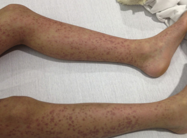
- Rash (Fig. E2) is uncommon but will occur in nearly all patients who receive ampicillin or amoxicillin due to a transient penicillin hypersensitivity.
- IM is usually a self-limited illness (2 to 4 wk), but symptoms of malaise and fatigue may last months before resolving.1
- At times, IM can present as fever and adenopathy without pharyngitis.
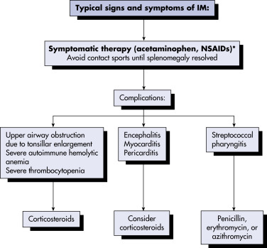
- Although acute complications may be severe, they are uncommon and tend to resolve completely. Reported complications include cholestatic liver disease, chronic hepatitis, or even liver failure; hemolytic anemia; splenic rupture; or airway compromise.3,4
- Splenic rupture is rare, with an incidence rate <1%, but it is the most feared.4 It should be suspected in anyone with confirmed or suspected IM who presents with acute abdominal or chest pain. Most cases occur in the first 3 wk of symptoms.
- Airway compromise, as evidenced by stridor, cyanosis, and/or tachypnea, is reported in 1% to 3% of cases and is an indication for hospitalization.
- Systemic corticosteroids are indicated for treating those at risk or with impending airway obstruction, and usually lead to improvement in 12 to 36 hr. Acute tonsillectomy may be pursued if steroids are not sufficient to mitigate the risk for obstruction.5
- Late complications may include lymphoproliferative cancers (Burkitt lymphoma and Hodgkin lymphoma), multiple sclerosis, rheumatoid arthritis, and chronic active EBV infection (CAEBV).3
- EBV may also cause hemophagocytic disease, alternatively referred to as EBV-associated hemophagocytic lymphohistiocytosis (EBV-HLH), characterized by fever, splenomegaly, and cytopenias with high ferritin and soluble CD25.
- Box 1 and Table 1 summarize the features of IM in immunocompetent patients.
TABLE 1 Clinical Manifestations of Infectious Mononucleosis in Children and Adults
| Frequency (%) | |||
|---|---|---|---|
| Sign or Symptom | Age <4 yr | Age 4-16 yr | Adults (Range) |
| Lymphadenopathy | 94 | 95 | 93-100 |
| Fever | 92 | 100 | 63-100 |
| Sore throat or tonsillopharyngitis | 67 | 75 | 70-91 |
| Exudative tonsillopharyngitis | 45 | 59 | 40-74 |
| Splenomegaly | 82 | 53 | 32-51 |
| Hepatomegaly | 63 | 30 | 6-24 |
| Cough or rhinitis | 51 | 15 | 5-31 |
| Rash | 34 | 17 | 0-15 |
| Abdominal pain or discomfort | 17 | 0 | 2-14 |
From Hoffman R et al: Hematology, basic principles and practice, ed 7, Philadelphia, 2018, Elsevier.
BOX 1 Summary of Features of Infectious Mononucleosis (IM) in Immunocompetent Patients
From McPherson RA, Pincus MR: Henry’s clinical diagnosis and management by laboratory methods, ed 23, Philadelphia, 2017, Elsevier.
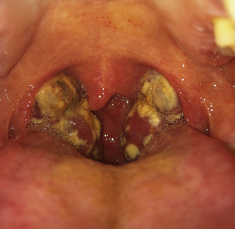
Courtesy Dr. Lauren Kjolhede, Baylor College of Medicine, Children’s Hospital of San Antonio, San Antonio, TX. In Hoffman R et al: Hematology, basic principles and practice, ed 7, Philadelphia, 2018, Elsevier.
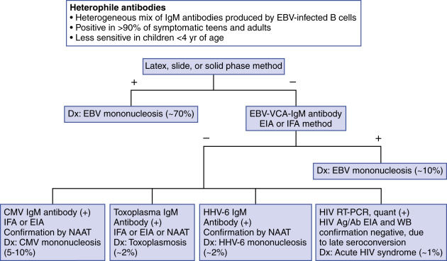
The most common cause of IM (90%) is primary infection with EBV.1 Cytomegalovirus (CMV) is the most common cause of the other 10% of IM, but CMV infection often occurs in infancy or early childhood and is minimally symptomatic. Other causes include human herpes virus-6, herpes simplex virus-1, and HIV. Primary EBV infection during childhood also often causes few or no symptoms; persistent fatigue and recurrent/persistent fevers are the most common reasons parents bring symptomatic children to medical care.
EBV infects epithelial cells and resting B cells of the oropharynx. It is then found in saliva, making it transmittable through coughing, sharing drink/food, and kissing; thus the pseudonym “kissing disease.” IM is more prevalent during adolescence when these types of close contact increase. How it is transmitted in younger children is less clear.
EBV levels peak during the active phase of infection but can persist in the oropharynx for up to 18 mo. Transmission may also occur sexually because EBV can be isolated in cervical epithelial cells and male seminal fluid. It has also been shown to be transmitted by blood transfusion, solid organ transplantation, or hematopoietic cell transplantation.