section name header

Basic Information

AUTHOR: Glenn G. Fort, MD, MPH

Definition

Epstein-Barr virus infection refers to a disease caused by Epstein-Barr virus (EBV), a human herpesvirus.

Synonyms

Infectious mononucleosis (IM)

Kissing disease

ICD-10CM CODES
B27.80Other infectious mononucleosis without complication
B27.81Other infectious mononucleosis with polyneuropathy
B27.82Other infectious mononucleosis with meningitis
B27.89Other infectious mononucleosis with other complication
B27.90Infectious mononucleosis, unspecified without complication
B27.91Infectious mononucleosis, unspecified with polyneuropathy
B27.92Infectious mononucleosis, unspecified with meningitis
B27.99Infectious mononucleosis, unspecified with other complication
Epidemiology & Demographics
Incidence (In U.S.)

5 cases/100,000 persons per yr of IM

Predominant Sex

Neither, although peak incidence occurs about 2 yr earlier in women

Predominant Age

  • Clinical evidence of IM: Occurs most commonly at ages 15 to 24 yr
  • EBV infection: Occurs earlier in life in lower socioeconomic groups
Physical Findings & Clinical Presentation

  • Most EBV infections either are asymptomatic or cause a nonspecific viral illness.
  • Incubation period is 1 to 2 mo, possibly followed by a prodrome of anorexia, low-grade fever, malaise, headache, and chills; after several days, clinical triad of pharyngitis, moderate to high fever, and adenopathy may appear, accompanied by fatigue and malaise.
  • Pharyngitis is usually the most severe symptom; white or necrotic exudates are common.
  • Symmetrical lymphadenopathy is most prominent in the posterior more than anterior cervical region but may be diffuse.
  • Splenomegaly (50% of cases) is possible, most commonly during the second week of illness.
  • Maculopapular or morbilliform rash is uncommon but will often occur in patients who receive ampicillin (Fig. E1). Patients may have palatal petechiae, periorbital, or palpebral edema. Mucocutaneous oral hairy leukoplakia (OHL), which is associated with intense EBV replication and the action of EBV-encoded proteins such as latent membrane protein-1, may occur.
  • Possible IM presentation: Fever and adenopathy without pharyngitis.
  • Nausea, vomiting, and anorexia are frequent in patients with IM, probably reflecting mild hepatitis encountered in 90% of infected individuals.
  • Although complications such as spleen rupture, airway obstruction, and malignancy may be severe and fatal, they are uncommon and tend to resolve completely.
  • Hematologic involvement includes hemolytic or aplastic anemia, thrombocytopenia, thrombotic thrombocytopenic purpura/hemolytic-uremic syndrome, and disseminated intravascular coagulation (DIC). Pneumonia, myocarditis, pancreatitis, mesenteric adenitis, myositis, and glomerulonephritis may occur as well. Nervous system involvement includes Guillain-Barré syndrome, facial nerve palsy, meningoencephalitis, aseptic meningitis, transverse myelitis, peripheral neuritis, and optic neuritis.
  • IM is usually a self-limited illness. Acute symptoms resolve in 1 to 2 wk, but symptoms of malaise and fatigue often persist for months.
  • EBV is related to lymphoproliferative syndromes in transplant recipients and in AIDS patients.
  • Increasing evidence showing an association between EBV infection and African Burkitt, B-cell or T-cell lymphoma, and nasopharyngeal carcinoma.

Figure E1 Patient with Infectious Mononucleosis and Ampicillin-Induced Rash

Maculopapular Rash Extends over the Trunk and Extremities. Rash Frequently Has a Violaceous Hue and is Often Accompanied by Pruritus.
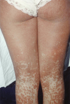
From Bennett JE et al: Mandell, Douglas, and Bennett’s principles and practice of infectious diseases, ed 8, Philadelphia, 2015, Saunders.

Etiology

  • EBV is a ubiquitous virus.
  • Infection during childhood is much less likely to cause significant illness.
  • Frequency of IM in late adolescence is attributed to the onset of social contact between the sexes.
  • Close personal contact is usually necessary for transmission, although EBV is occasionally transmitted by blood transfusion; transfer via saliva while kissing may be responsible for many cases.

Diagnosis

Differential Diagnosis

  • Heterophile-negative IM caused by cytomegalovirus (CMV)
  • Although clinical presentation similar, CMV more frequently follows transfusion
  • Bacterial and viral causes of pharyngitis
  • Toxoplasmosis
  • Acute retroviral syndrome of HIV
  • Lymphoma
  • Lyme disease
Workup

Heterophile antibody and CBC with blood smear. Table E1 describes frequently determined EBV-specific antibodies.

TABLE E1 Frequently Determined EBV-Specific Antibodies

Antibody SpecificityPositive in IM (%)Time of Appearance in IMPersistenceComments
Viral Capsid Antigen
VCA-IgM100At clinical presentation4-8 wkHighly sensitive and specific; of major diagnostic utility
VCA-IgG100At clinical presentationLifelongUseful for documentation of past EBV infection
Early Antigen
Anti-D70Peaks 3-4 wk after onset3-6 moCorrelates with disease severity; seen in NPC patients
Anti-RLow2 wk to several months after onset2 mo to >3 yrOccasionally seen with unusually severe cases; seen in African Burkitt lymphoma patients
EBNA1003-4 wk after onsetLifelongPresence excludes primary EBV infection

EBNA, EBV nuclear antigen; EBV, Epstein-Barr virus; Ig, immunoglobulin; IM, infectious mononucleosis; NPC, nasopharyngeal carcinoma; VCA, viral capsid antigen.

Adapted from Schooley RT: Epstein-Barr virus (infectious mononucleosis). In Mandell GL et al (eds): Principles and practice of infectious diseases, Philadelphia, 2010, Churchill Livingstone.

Laboratory Tests (Box E1

BOX E1 Laboratory Diagnosis of Epstein-Barr Virus.

  1. White blood cell counts typically range from 10,000 to 20,000/μL.
  2. Marked lymphocytosis and atypical lymphocytes.
  3. Often elevated serum transaminase, alkaline phosphatase, and bilirubin levels.
  4. Diagnostic assay: Heterophile agglutination test (monospot).
    • Measures immunoglobulin M antibodies (not specific EBV to antigens, produced by EBV-stimulated plasma B cells) 40%-60% positive in first week; 80%-90% positive at one month after onset.1
    • Sensitivity of 81%-95% for the commercial tests in a general population.2
    • Negative predictive value of 78%-88% has been shown in children.3
    • High positive predictive value, greater than 95%.4
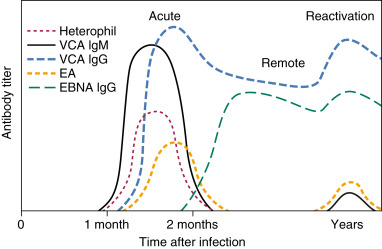
    • EBV-specific antibody titers (Fig. E2).
  5. Confirmation of acute or convalescent EBV infection.
  6. Perform in children under 4 yr and atypical or persistent symptoms with a negative heterophile antibody.
    • Methods based on the polymerase chain reaction (PCR) have a high sensitivity for EBV viral DNA but are expensive and used clinically only with suspected EBV-associated encephalitis.5

From Flint PW et al: Cummings otolaryngology, head and neck surgery, ed 7, Philadelphia 2021, Elsevier.

  • Increased WBC common, with a relative lymphocytosis of more than 50% and neutropenia identified.
  • Hallmark of IM: Atypical lymphocytes of more than 10% (not pathognomonic) are found.
  • Mild thrombocytopenia is present.
  • Falling hematocrit signals the possibility of splenic rupture or immune hemolytic anemia.
  • Elevated hepatocellular enzymes and cryoglobulins are found in most cases.
  • Heterophile antibody:
    1. As measured by the monospot test, may be positive at presentation or may appear later in the course of illness. Its accuracy is 71% to 90% but has a 25% false-negative rate in the first week of illness.
    2. A negative test should be repeated in 1 wk if clinical suspicion is high.
    3. A false-positive test has been reported with primary HIV infection.
  • Viral capsid antigen (VCA) IgG and IgM (Fig. E2) are rarely used for diagnosis but better value in children because heterophile antibody is negative in most children younger than 8 yr.
  • PCR DNA for CMV is the test of choice in transplant recipients who develop lympho-proliferative syndromes.

Figure E2 Response Time in Diagnosis of Epstein-Barr Virus Infection

Ea, Early Antigen; Ebna, EBV Nuclear Antigen; Ig, Immunoglobulin; Vca, Viral Capsid Antigen.

From Flint PW et al: Cummings otolaryngology, head and neck surgery, ed 7, Philadelphia 2021, Elsevier.

Imaging Studies

Chest radiograph examination:

  • May rarely show infiltrates
  • Possible elevated left hemidiaphragm with splenic rupture

Treatment

Nonpharmacologic Therapy

  • Supportive including rest
  • Splenectomy if rupture occurs
  • Transfusions for severe anemia or thrombocytopenia
Acute General Rx

  • Pharmacologic therapy is not indicated in uncomplicated illness.
  • Avoid aspirin due to the risk of Reye syndrome.
  • Avoid ampicillin and amoxicillin as their use can frequently precipitate a nonallergic rash. Azithromycin has also been implicated.
  • Use of steroids is suggested in patients who have severe thrombocytopenia, hemolytic anemia, impending airway obstruction resulting from enlarged tonsils, or fulminant liver failure. Prednisone 60 to 80 mg PO qd for 3 days, then tapered over 1 to 2 wk.
  • Although it may reduce initial viral shedding, there is little evidence to support the use of antiviral agents such as acyclovir in the management of IM.
Chronic Rx

An extremely rare, chronic form of IM with persistent fevers and fatigue has been described and should be differentiated from chronic fatigue syndrome, which is not related to EBV.

Disposition

Eventual resolution of all symptoms

Referral

If more than mild illness

Pearls & Considerations

Comments

Avoidance of contact sports during the first month of illness because splenic rupture can occur even in the absence of clinically detectable splenomegaly in 0.1% to 0.5% of patients.

Related Content

Epstein-Barr Virus Infection (Patient Information)

Mononucleosis (Related Key Topic)

Related Content

  1. Shanahan JF, Davis KJ (eds): Harrison's principles of internal medicine, ed 20, New York, 2018, McGraw-Hill Education.
  2. Bruu A.L. : Evaluation of 12 commercial tests for detection of Epstein-Barr virus-specific and heterophile antibodiesClin Diagn Lab Immunol. ;7(3):451-456, 2000.
  3. Fisher R.G.B.T. : MOFFET'S pediatric infectious diseases: a problem-orientated approach 4Lippincott Williams & Wilkins-Philadelphia, 2005.
  4. Linderholm M. : Comparative evaluation of nine kits for rapid diagnosis of infectious mononucleosis and Epstein-Barr virus-specific serologyJ Clin Microbiol. ;32(1):259-261, 1994.
  5. Marshall-Andon T., Heinz P. : How to use … the Monospot and other heterophile antibody testsArch Dis Child Educ Pract Ed. ;102(4):188-193, 2017.
  6. Bjornevik K. : Longitudinal analysis reveals high prevalence of Epstein-Barr virus associated with multiple sclerosisScience. ;375(6578):296-301, 2022.
  7. Shepard R.J. : Exercise and the athlete with infectious mononucleosisClin J Sport Med. ;27:168-178, 2017.
  8. Thompson D.F., Ramos C.L. : Antibiotic-induced rash in patients with infectious mononucleosisAnn Pharmacother. ;51:154-162, 2017.
  9. Womack J., Jimenez M. : Common questions about infectious mononucleosisAm Fam Physician. ;91(6):372-376, 2015.