section name header

Basic Information

AUTHORS: Michael Lawrenz Co, MD, MSc and Daniel R. Frisch, MD

Definition

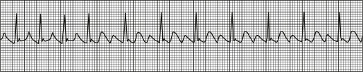
Typical atrial flutter is the term commonly applied to the atrial macroreentrant circuit that circulates around the tricuspid annulus in the right atrium. The critical isthmus of the circuit is the tissue between the inferior vena cava and the tricuspid annulus, and a more precise name for this arrhythmia is cavotricuspid isthmus-dependent atrial flutter, or CTI flutter. Because of its anatomic and physiologic stability, the result is regular atrial depolarizations, typically at a rate of 250 to 350 beats/min. Regular macroreentrant atrial arrhythmias at this rate that do not use the CTI are referred to as atypical atrial flutter. Because of the stability of atrial flutter, conduction through the atrioventricular node (AVN) is often predictable at a common mathematical denominator. For example, when the flutter rate is 300 beats/min, 2:1 conduction results in a ventricular rate of 150 beats/min. By extension, 3:1 conduction results in a ventricular rate of 100 beats/min, 4:1 in a rate of 75 beats/min, and 5:1 in a rate of 60 beats/min. If the regular atrial impulses conduct at a variable rate through the AVN, the result may be an irregular QRS pattern. However, a common denominator atrial interval is often present.

Table 1 summarizes the different types of atrial flutter and distinguishing features on scalar electrocardiography.

ICD-10CM CODES
I48.3Typical atrial flutter
I48.4Atypical atrial flutter
I48.92Unspecified atrial flutter

TABLE 1 Characteristics of Different Types of Atrial Flutter and Distinguishing Features on Scalar Electrocardiography

TypeReentrant CircuitECG PatternLead V1/V6
Typical counterclockwiseTricuspid annulus dependent on the CTISawtooth flutter wave; negative in II, III, and aVFPositive V1, negative V6
Typical clockwiseTricuspid annulus dependent on the CTI“Inverse sawtooth”; positive and often notched in II, III, and aVFBroad and negative in V1 (often notched)Positive in V6
Lower loop reentryCTIUsually similar to typical counterclockwise CTI flutter except subtle loss of terminal positive deflection in leads II, III, and aVFUsually similar to typical counterclockwise
Upper loop reentrySuperior vena cava and upper crista terminalisSimilar to typical clockwise flutterSimilar to typical clockwise flutter
Right atrial free wallAround areas of scar in lateral or posterior right atrium (caused by previous atrial surgery or spontaneously)VariableTypically negative or biphasic with terminal negative deflection in V1
Septal atrial flutterAtrial septum, typically after previous surgeryVariableUsually biphasic or isoelectric in V1
Mitral annular flutterAround mitral annulus, often slow zone of block around PV interval; frequently occurs in setting of left atrial surgery or ablationVariable; I, III, and aVF, often positive but low amplitudeUsually positive in V1 (or rarely isoelectric) and often broad
Postatrial fibrillation ablation/maze flutterVariable; circuit involves previous ablations or scar in left atriumVariableVariable

aVF, Augmented vector foot; CTI, cavotricuspid isthmus; PV, pulmonary vein.

From Zipes DP: Braunwald’s heart disease, a textbook of cardiovascular medicine, ed 11, Philadelphia, 2019, Elsevier.

Epidemiology & Demographics

  • Atrial flutter is the second most common sustained atrial tachyarrhythmia after atrial fibrillation, with an estimated 200,000 new cases annually in the U.S.
  • Atrial flutter is common in patients with congestive heart failure, chronic obstructive pulmonary disease (COPD), pulmonary vascular disorders such as pulmonary embolism, or during the first week after open heart surgery.
  • Atrial flutter occurs more frequently with advancing age (5/100,000 age <50 vs. 587/100,000 age >80 yr) and 2.5 times more frequently in men than in women.
  • Patients taking antiarrhythmics for chronic suppression of atrial fibrillation may present with atrial flutter.
  • Atrial flutter is typically seen in patients with underlying structural heart disease and is uncommon in children or young adults without congenital heart disease.
  • More than 50% of patients with atrial flutter will develop atrial fibrillation in 3 yr, and more than 80% will develop atrial fibrillation within 5 yr. This is important when considering treatment options for atrial flutter, especially anticoagulation.
Classification

Historically, the Wells classification designated atrial flutter as type I and type II. However, it is now recognized that tachycardias satisfying either of the definitions for type I or type II can be caused by reentrant circuits or by rapid focal atrial tachycardia, and this classification is infrequently used. Designating atrial flutter based on whether or not it is CTI dependent is more useful because of the management options (i.e., ablation). Type I CTI-dependent atrial flutter, also known as common atrial flutter or typical atrial flutter, has an atrial rate of 250 to 350 beats/min. The reentrant loop circles the tricuspid valve in the right atrium, passing through the CTI, a body of fibrous tissue in the lower atrium between the inferior vena cava and the tricuspid valve. CTI flutter can revolve around the tricuspid annulus in either direction (counterclockwise or clockwise) when viewing the tricuspid annulus en face.

  • Counterclockwise atrial flutter is the more common type (75%). The flutter waves are “sawtooth” and negative on the surface ECG leads II, III, and aVF; positive in V1; and negative in V6 (Fig. E1).
  • Clockwise atrial flutter is less common (25%): The reentry loop cycles in the opposite direction; thus the flutter waves are upright in leads II, III, and aVF; negative in V1; and positive in V6 (Fig. E2).
  • Atypical atrial flutter is defined by absence of CTI dependence and may occur in patients with prior cardiac surgery, congenital heart disease, or prior radiofrequency ablation (especially after left atrial ablation for atrial fibrillation) or may be idiopathic. One ECG feature is the lack of discordance of the flutter wave polarity between the inferior leads (leads II, III, and aVF) and V1. Flutter circuits in the left atrium (such as mitral annular flutter) often have upright flutter waves in all precordial leads.

Figure E1 Counterclockwise atrial flutter with 2:1 atrioventricular conduction.

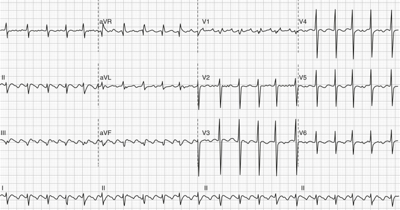
Note the negative flutter waves in leads II, III, and F, positive in V1 and negative in V6. The second flutter wave can be seen overlying the QRS in the inferior leads and at the end of the QRS in lead V1. aVF, Augmented vector foot; aVL, augmented vector left; aVR, augmented vector right.

Figure E2 Clockwise atrial flutter with predominant 2:1 atrioventricular conduction.

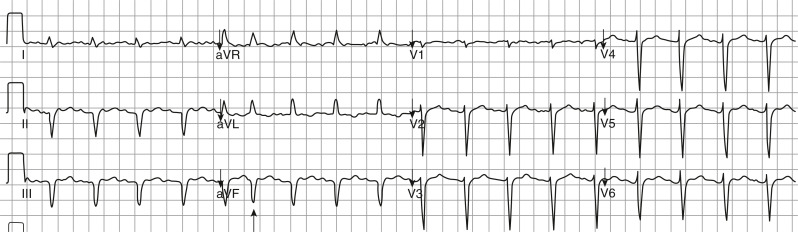
Note the positive flutter waves in leads II, III, and F, negative in V1 and positive in V6. The overall ventricular rate is slower than in Fig. E1 due to a slow flutter rate of approximately 200 bpm. One beat is conducted 1:1 (arrow). aVF, Augmented vector foot; aVL, augmented vector left; aVR, augmented vector right.

Physical Findings & Clinical Presentation

  • Palpitations
  • Dizziness, light-headedness, syncope, or near syncope
  • Angina
  • Congestive heart failure
  • Embolic phenomena from intracardiac thrombus
Etiology

  • Age-related degenerative changes
  • Rheumatic heart disease
  • Congenital heart disease
  • Left ventricular dysfunction or congestive heart failure
  • Acute myocardial infarction (rarely)
  • Thyrotoxicosis
  • Pulmonary embolism
  • Mitral valve disease
  • Cardiac surgery
  • Chronic obstructive pulmonary disease
  • Obesity
  • Pericarditis
  • Pulmonary hypertension
  • Antiarrhythmic therapy use in patients with atrial fibrillation
  • Other causes of right atrial enlargement
  • Systemic infections (such as COVID-19)1

Diagnosis

Differential Diagnosis

  • Atrial fibrillation
  • Atrial tachycardia
  • Supraventricular tachycardia:
    1. Atrioventricular node reentry
    2. Orthodromic reciprocating tachycardia (using a concealed bypass tract)
    3. Junctional ectopic tachycardia
    4. Wolff-Parkinson-White syndrome
  • Sinus tachycardia
Workup

  • ECG (Fig. 3)
  • Laboratory evaluation
  • Assessment of CHA2DS2-VaSc score (Table 2)

TABLE 2 CHA2DS2-VASc Risk Score for Prediction of Stroke Risk in Atrial Fibrillation

Risk FactorPoints
CHF/LV dysfunction1
Hypertension1
Age 75 yr2
Diabetes mellitus1
Stroke/TIA/embolism2
Vascular disease1
Age 64-74 yr1
Sex category (female)1
Maximum score9

CHF, Congestive heart failure; LV, left ventricular; TIA, transient ischemic attack.

Figure 3 Rhythm strip of atrial flutter.

From Cameron P: Textbook of adult emergency medicine, ed 5, Philadelphia, 2020, Elsevier.

Laboratory Tests

  • Thyroid function studies
  • Serum electrolytes, including renal and hepatic tests (anticipating antiarrhythmic therapy use)
  • CBC and PT/INR values (anticipating anticoagulation therapy)
Imaging Studies

  • ECG:
    1. Absence of P waves
    2. Regular, “sawtooth,” or “F” (flutter) wave pattern without an isoelectric baseline in leads II, III, and aVF (seen most commonly with counterclockwise typical CTI-flutter). The discordance of the polarity of the flutter waves between the inferior leads and V1 is a constant finding
    3. There is rarely 1:1 atrioventricular (AV) conduction in atrial flutter (unless pre-excitation is present). Rather, AV conduction is usually in a 2:1 (see Fig. E1), 3:1, or 4:1 fashion, with corresponding usual ventricular rates of 150, 100, or 75 beats/min, respectively (assuming an atrial rate of 300 beats/min). With high vagal tone or AV block, ventricular rates may be slow in atrial flutter
  • Holter monitoring or event recorder to assess for paroxysmal atrial flutter or rate control or to identify the arrhythmia if symptoms are nonspecific or to identify triggering events
  • Echocardiography (for new diagnoses) to evaluate for structural heart disease (atrial enlargement, left and right ventricular size, thickness, and function; atrial size, and valve function)
  • Transesophageal echocardiography: Consider in the setting of cardioversion in the absence of an appropriate duration of anticoagulation to ascertain the absence of intracardiac (e.g., left atrial appendage) thrombi
  • Electrophysiologic studies: Required for a precise diagnosis and for ablation
  • Pulmonary vascular imaging (e.g., a CT scan with contrast) if a pulmonary embolism is suspected

Treatment

Acute Rx Of Unstable Patient

  • Treatment choices are based on clinical circumstances. Fig. 4 describes an acute treatment of atrial flutter algorithm.2 Table 3 summarizes atrial flutter therapy.
  • In cases of electrocardiographic uncertainty, atrial flutter may be differentiated from SVT through vagal maneuvers that slow AV conduction (e.g., the Valsalva maneuver, carotid sinus massage, or the use of adenosine). Vagal maneuvers may reveal flutter waves but will not terminate atrial flutter.
  • Direct current cardioversion is the treatment of choice for unstable patients or those with debilitating symptoms. This may be successful with energies as low as 25 joules, but because 100 joules is virtually always successful, this may be a reasonable initial shock strength. If the electrical shock results in atrial fibrillation, a second shock at a higher energy level is used to restore normal sinus rhythm. Sedation of a conscious patient is highly recommended. After cardioversion, patients will need anticoagulation for at least one month due to resultant atrial stunning from the presence of atrial flutter. Subsequent anticoagulation will depend on their stroke risk (CHA2DS2-VASc score; Table 2).
  • Overdrive pacing in the atrium may also terminate atrial flutter. This method is especially useful in patients who have recently undergone cardiac surgery and still have temporary atrial pacing wires and in patients who have an implanted pacemaker or defibrillator with an atrial lead.
Figure 4 Acute Treatment of Atrial Flutter

Anticoagulation as Per Guideline is Mandatory. †for Rhythms that Break or Recur Spontaneously, Synchronized Cardioversion, or Rapid Atrial Pacing is Not Appropriate. IV, Intravenous.

!!flowchart!!

Reproduced with permission from Page RL et al: 2015 ACC/AHA/HRS guideline for the management of adult patients with supraventricular tachycardia, JACC 67[13]:e27-e115, 2016. In Olshansky B et al: Arrhythmia essentials, ed 2, Philadelphia, 2017, Elsevier.

TABLE 3 Atrial Flutter Therapy

Acute therapy for poorly tolerated AFL or continuous rapid ventricular rate
  • If prolonged (i.e., >48-72 hr), anticoagulation is required as cardioversion may be associated with thromboembolic risk. Anticoagulation guidelines for cardioversion are the same as for atrial fibrillation and may indicate need for a TEE for prolonged episodes. Adenosine and carotid massage can be used to help diagnose AFL masquerading as sinus rhythm.
  • First line: Electrical cardioversion under sedation with anticoagulation as necessary. Consider the length of the episode.
  • Second line:Ibutilide or procainamide may be attempted for conversion before electrical cardioversion attempts. Ibutilide may be 60%-70% effective if AFL has been present for <48 hr.
  • Alternate: Rapid atrial pacing (esophageal, epicardial, or endocardial, depending on the situation). To pace terminate, pace for 10-15 sec at a rate of 10%-20% faster than rate of flutter. If ineffective, burst pace 10 beats/min faster at a time for 10-15 sec at a time until conversion to AF or sinus rhythm. When AF occurs, it is usually short lived and terminates spontaneously within 24 hr. If persistent, electrical cardioversion can be attempted with or without antiarrhythmic drugs. Atrial fibrillation cannot be pace terminated. However, slower AFL (rate <350 bpm) of any flutter wave morphology can often be pace terminated.
  • Oral drug loading alone to terminate AFL is rarely useful.
  • If recurrent episodes, use class IC or III antiarrhythmic drugs until steady state is achieved, then attempt cardioversion. These drugs (particularly 1C drugs) may stabilize the flutter circuit. It may also create another form of AFL-“1C” AFL-from AF. Ablation remains first-line therapy, especially if AFL is isthmus dependent.
  • Consider ablation for cases of AFL. However, despite AFL ablation, AF may occur, especially in individuals with underlying structural heart disease.
  • AV nodal ablation, although not preferable, could be considered when ventricular rate control cannot be achieved and flutter cannot be ablated (e.g., in the presence of a left atrial appendage clot), or if symptomatic, refractory, and/or if associated with tachycardia-induced cardiomyopathy. This option may be considered in cases where individuals have multiple forms of nonablatable AFL or AF and especially for those who do have nonisthmus-dependent AFL.
Chronic prevention
  • Consider radiofrequency catheter ablation early; it has become first-line therapy.
  • Drug therapy alone for pure AFL flutter is usually not effective.
  • If drug therapy is chosen, be guided by the presence/absence of structural heart disease.
  • If structural heart disease without CHF: Sotalol (often initiated in the hospital), dofetilide, amiodarone.
  • If structural heart disease with CHF: Amiodarone, dofetilide.
  • If no structural heart disease: Propafenone, flecainide, sotalol, dofetilide, or amiodarone, but propafenone or flecainide may need concomitant AV nodal blocking drugs to prevent 1:1 conduction.
  • If class I or III drugs are used, first control the ventricular response rate with an AV nodal blocking drug. Otherwise, the vagolytic effects of class IA drugs can enhance AV nodal conduction, and both 1A and 1C drugs can lead to AFL with 1:1 AV conduction.
Nonresponders with severe symptoms
  • If typical AFL, radiofrequency ablation within the cavotricuspid isthmus is highly effective.
  • Atypical AFL is more difficult to ablate and depends on the location of reentrant circuit. Success rates are lower than that for typical AFL. It is more difficult when there is congenital heart disease, valve disease, or prior surgery in which significant areas of scar are present.
  • Ventricular rate control, antiarrhythmic drugs, or AV node ablation (less preferable) can be performed for atypical, nonablatable AFL.
  • If AV node ablation and pacing is performed and AFL is intermittent, mode-switching function should be programmed “ON.”
MI
  • If hemodynamic intolerance or ongoing refractory myocardial ischemia, emergent cardioversion. AFL may increase MVO2 due to rapid ventricular rate, causing further ischemia, diastolic dysfunction, and pulmonary congestion and edema.
  • If recurrent, IV amiodarone or procainamide.
  • Consider temporary antitachycardia pacing if recurrent and poorly tolerated.
Preoperative
  • For cardiac surgery, convert AFL to NSR if adequate anticoagulation has been achieved, or ensure that ventricular response is well controlled.
  • If surgery is elective and AFL is chronic, antiarrhythmic drugs or catheter ablation may be considered. However, anticoagulation should be continued at least 4 wk after conversion of longer-term (>48 hr) AFL prior to elective surgery.
  • For more urgent surgery in which anticoagulation cannot be used, consider rate control without cardioversion.
  • For short-duration (<48 hr) AFL, DCC can be performed (may consider therapeutic anticoagulation before electrical cardioversion with surgical consultation as to risk).
Postoperative
  • AFL occurs in 10%-20% of all patients after cardiac surgery; it typically occurs with AF. Incidence peaks at days 2-3. It is more common in older patients. It rarely occurs after other types of surgery. The AFL may resolve spontaneously; however, the rhythm can increase the length of hospital stay, exacerbate heart failure, slow the recovery process, and cause symptoms.
  • Control rate with β-adrenergic blocker if no CHF or bronchospastic disease and LVEF >40%. Diltiazem is often successful as a second-line drug, but use with caution in patients with a reduced LVEF. Digoxin for rate control is less effective but may be considered, particularly in patients with poor LV function.
  • IV amiodarone may be useful for persistent and poorly tolerated AFL; amiodarone or other antiarrhythmic drugs may be helpful for recurrent episodes.
  • Electrical cardioversion or atrial pace termination (if atrial pacing leads are present) is often successful, especially when employed early after the AFL onset.
  • Discontinue inotropic drugs, if possible.

AF, Atrial fibrillation; AFL, atrial flutter; AV, atrioventricular; CHF, congestive heart failure; DCC, direct current cardioversion; IV, intravenous; LV, left ventricle; LVEF, left ventricular ejection fraction; MI, myocardial infarction; MVO2, myocardial oxygen consumption; NSR, normal sinus rhythm; TEE, transesophageal echocardiography.

From Olshansky B et al: Arrhythmia essentials, ed 2, Philadelphia, 2017, Elsevier.

Acute Rx Of Stable Patient

  • Proceed with either rate control or rhythm control strategy. Data is inconclusive on which strategy is more effective, but in general, atrial flutter is more difficult to rate-control than atrial fibrillation. Atrial flutter may spontaneously convert to normal sinus rhythm with rate control strategy.
  • For a rate control strategy: First-line agents are calcium channel blockers and beta-blockers. Digoxin and amiodarone may be used in those with borderline hypotension, decompensated congestive heart failure, and those unable to tolerate first-line agents.
  • Avoid calcium channel blockers if known EF <40%.
  • Digoxin has a narrow therapeutic window, and thus caution is recommended in patients with kidney impairment.
  • Amiodarone use is typically a rhythm control agent but also has utility in rate control. It carries the risk of cardioverting a patient not on anticoagulation.
  • A rhythm control strategy can be employed using either electrical or pharmacologic cardioversion.
  • While electrical cardioversion is often successful, its effects are not durable. The rate of recurrence of atrial flutter with cardioversion alone is difficult to determine because most published data combine atrial flutter with atrial fibrillation. However, the recurrence rate is substantial, perhaps 50% at 1 yr.
  • For pharmacologic cardioversion, intravenous ibutilide is a first-line medication in patients with normal systolic function and QT intervals. The success rate is approximately 60%, and it is more effective than procainamide, sotalol, or amiodarone. Oral flecainide (300 mg once) or oral propafenone (600 mg) may be used as well. Cardiac telemetry monitoring is required for 4 to 6 hours after giving antiarrhythmic medications at these doses in patients receiving this therapy for the first time.
Chronic Rx

  • Chronic treatment of atrial flutter is indicated to prevent symptoms and adverse remodeling of the heart (arrhythmia induced cardiomyopathy, AIC). This may either be in the form of rate control or rhythm control strategy although the latter is preferred because successful rhythm control can be achieved in >90% of cases through catheter ablation.
  • Radiofrequency ablation is often offered as a first-line treatment of typical atrial flutter but is also highly effective for those with recurrent episodes. It has been shown to improve health-related quality of life.
  • In those patients who fail, opt not to, or not candidates for catheter ablation, electrical cardioversion followed by pharmacologic therapy to maintain sinus rhythm may be considered.
  • Elective outpatient cardioversion or ablation can be performed either immediately preceded by transesophageal echocardiography (TEE) to evaluate the left atrium and the left atrial appendage for thrombus or after a period of at least 3 weeks of documented therapeutic anticoagulation before cardioversion. At least 4 weeks of anticoagulation should be administered after cardioversion, if not longer, depending on the overall thromboembolic risk of the patient as determined by the CHA2DS2-VASc score.
  • Pharmacologic options to maintain sinus rhythm include dofetilide, amiodarone, flecainide, propafenone, or sotalol. The choice of antiarrhythmic therapy is, in part, dictated by the presence or absence of underlying structural heart disease. It is important to keep in mind that antiarrhythmic drugs carry the risk of toxicity and can also be proarrhythmic.
  • In addition to atrial flutter control, stroke risk mitigation is also a part of atrial flutter management and prophylactic anticoagulation should also be addressed depending on stroke risk, guided by the CHA2DS2-VASc score. Antithrombotic therapy is recommended as in atrial fibrillation.3 Anticoagulation should be considered for patients whose CHA2DS2-VaSc score is 2 in men or 3 in women. Options for oral anticoagulants include warfarin (target INR 2-3) or direct oral anticoagulants (DOACs, e.g., dabigatran, rivaroxaban, apixaban, or edoxaban), although the latter is now recommended over the former (except for cases of moderate to severe mitral stenosis or mechanical heart valves). For patients with a CHA2DS2-VaSc score of 1 (men) or 2 (women), prescribing an oral anticoagulant to reduce stroke risk may be considered (previous recommendation for aspirin was removed).4 Left atrial appendage closure may also be an option in patients at high risk for bleeding with anticoagulation.
Disposition

More than 85% of patients convert to regular sinus rhythm after cardioversion. Ablation success rates exceed 90%.

Referral

Refer patients who are considered for rhythm control of atrial flutter to a cardiac electrophysiologist, especially patients who are candidates for radiofrequency ablation.

Pearls & Considerations

Comments

  • The surface ECG is the best tool for recognizing atrial flutter and distinguishing atrial flutter from atrial fibrillation. The inferior leads (II, III, aVF) are the best for seeing the characteristic sawtooth pattern.
  • Ablation for typical atrial flutter is highly effective, straightforward, and relatively safe. It should be considered for patients with recurrent episodes and even for a first-ever episode.
  • Patients with atrial flutter carry a significant risk for subsequent development of atrial fibrillation.
  • Anticoagulation should be considered for all patients whose CHA2DS2-VaSc score is 2 (men) or 3 (women). For patients with a CHA2DS2-VaSc score of 1 (men) or 2 (women), risks and benefits should be considered on an individual basis.
  • Although anticoagulation recommendations for atrial flutter are identical to those for atrial fibrillation, studies suggest that the absolute risk of stroke is lower from atrial flutter than from fibrillation.
Related Content

Atrial Flutter (Patient Information)

Atrial Fibrillation (Related Key Topic)

Related Content

    1. Saha S.A. : COVID-19 and cardiac arrhythmias: a contemporary reviewCurr Treat Options Cardiovasc Med. ;24(6):87-107, 2022.doi:10.1007/s11936-022-00964-3
    2. Frisch D.R. : Algorithm for cavo-tricuspid isthmus flutter on surface ECGs: the ACTIONS studyOpen Hear. ;8(1):8-12, 2021.doi:10.1136/openhrt-2020-001431
    3. January C.T. : 2014 AHA/ACC/HRS guideline for the management of patients with atrial fibrillationJAC. ;1-125, 2014.doi:10.1016/j.jacc.2014.03.021
    4. January C.T. : 2019 AHA/ACC/HRS focused update of the 2014 AHA/ACC/HRS guideline for the management of patients with atrial fibrillation: a report of the American College of Cardiology/American Heart Association Task Force on clinical practice guidelines and the Heart Rhythm Society in collaboration with the Society of Thoracic SurgeonsCirculation. ;140(2):e125-e151, 2019.doi:10.1161/CIR.0000000000000665