section name header

Basic Information ⬇

AUTHORS: Rebecca A. Abelman, MD and Katerina L. Byanova, MD, MS and Laurence Huang, MD, FCCP, ATSF

Definition

Pneumocystis jirovecii pneumonia (PJP) or Pneumocystis pneumonia (PCP) is a respiratory infection caused by the fungal pathogen P. jirovecii (formerly known as P. carinii).

Synonyms

Pneumocystis jirovecii pneumonia

PJP

Pneumocystis pneumonia

PCP

ICD-10CM CODE
B59Pneumocystosis
Epidemiology & Demographics
Incidence (in U.S.)

  • In the early days of the HIV epidemic most PCP cases occurred in people with HIV with CD4 counts <200/mm3. Given improvements in HIV treatment, >70% of cases are now found in HIV-negative individuals.1
  • Risk factors for infection in people with HIV include CD4 cell count <200/mm3 or CD4% <14%, elevated HIV RNA levels, and history of PCP.
  • Among people with HIV, incidence of PCP remains high in those who are unaware of their HIV-infected status and/or are not on antiretroviral therapy (ART) or PCP prophylaxis.
  • PCP also occurs in patients with severe cell-mediated immune deficiency (congenital T-cell deficiency, acute leukemia, lymphoma, bone marrow or solid organ transplant), as well as in patients with hematologic cancers, rheumatologic and chronic lung diseases.1
  • The major risk factor for infection in HIV-negative individuals is immunosuppression, and it is most commonly seen in patients on chronic high-dose glucocorticoid therapy.2 PCP also occurs in patients on treatment with antilymphocyte monoclonal antibodies, tumor necrosis factor (TNF) alpha inhibitors, ibrutinib, or idelalisib.1 In addition to glucocorticoid use, risk factors for mortality among HIV-negative individuals include increased age, solid tumors, CMV coinfection, elevated LDH, lower lymphocyte count, pneumothorax, concurrent pulmonary disease, and need for invasive ventilation.3
  • Bacterial and viral coinfections with PCP can occur.
  • Pneumocystis is ubiquitous in the environment and is also frequently found as a colonizer in the airways of infants, children, and adults.
Predominant Sex

Equal incidence when adjusted for HIV status.

Predominant Age

During the AIDS epidemic, age at PCP diagnosis closely correlated with age of HIV infection. Currently the predominant age is 50 to 60 yr of age given the changes in the populations at risk.1

Peak Incidence

Widely variable based on patient’s risk profile.

Genetics

  • Most frequent opportunistic infection among HIV-infected children, with serologic studies suggesting exposure typically occurs in childhood.4
Physical Findings & Clinical Presentation

  • PCP can present as an acute (3 to 5 days) or subacute (2 to 4 wk) pneumonia with fever, nonproductive cough, and progressive dyspnea. Patients progress to respiratory failure if not treated. Fulminant pneumonia is observed more commonly in immunosuppressed patients without HIV than people with HIV.2,5
  • Purulent sputum argues against PCP.
  • Hemoptysis is unusual.
  • Extrapulmonary manifestations are rare but can involve nearly any organ.
  • Vital signs: Notable for fever, tachypnea, low oxygen saturation. Oxygen desaturation with ambulation/exertion.
  • Pulmonary exam is often normal. Dry rales can be auscultated.
  • Spontaneous pneumothorax is possible, so monitor for unilateral decreased or absent breath sounds.
Etiology

  • P. jirovecii (formerly P. carinii) is a fungal organism (previously classified as a protozoan) (Fig. E1).

Figure E1 Wright-Giemsa stain of Pneumocystis jirovecii (carinii) pneumonia.
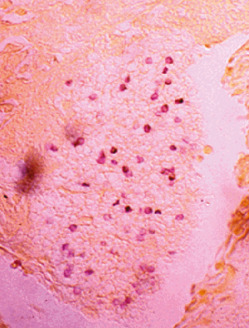
From Gabbe SG: Obstetrics, ed 6, Philadelphia, 2012, Saunders.

  • Infection can occur either as a result of activation of latent infection or by new acquisition.
  • Infection occurs via airborne transmission.
  • No transmission from vertebrate animals (mammals) to humans.
  • Increasing number of reports of nosocomial PCP transmission among immunocompromised patients (commonly solid organ transplant recipients).6

Diagnosis ⬆ ⬇

Differential Diagnosis

  • Infectious etiologies:
    1. Tuberculosis (especially in low- and middle-income countries)
    2. Bacterial pneumonia
    3. Viral pneumonia, including COVID-19 (SARS CoV-2)
    4. Atypical pneumonia, such as Mycoplasma pneumonia
    5. Cryptococcal pneumonia
    6. Histoplasma pneumonia (in endemic areas and advanced AIDS with CD4 cell count <50 cells/mm3)
    7. Coccidioides pneumonia (in endemic areas and advanced AIDS with CD4 cell count <50 cells/mm3)
    8. CMV pneumonitis (in CD4 cell count <50 cells/mm3)
    9. Mycobacterium avium complex (MAC) (in CD4 cell count <50 cells/mm3)
    10. Toxoplasmosis
    11. Legionella pneumonia
    12. Other fungal pneumonias, such as Aspergillus and Blastomycosis
    13. Strongyloidiasis
  • Noninfectious etiologies:
    1. Kaposi sarcoma
    2. Non-Hodgkin lymphoma
    3. Drug-induced lung injury
    4. Pulmonary edema
    5. Lymphocytic interstitial pneumonia
Workup

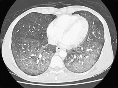
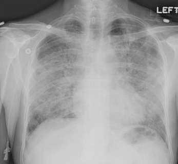
  • Chest x-ray (Fig. 2) or chest computed tomography (CT) (Fig. E3).
Figure 2 Chest Radiograph Showing Diffuse Interstitial Infiltrate in a Patient with Pneumocystis Jirovecii Pneumonia

From Firestein GS et al [eds]: Kelly’s textbook of rheumatology, ed 9, Philadelphia, 2013, Saunders.

Figure E3 Computed Tomography Scan of the Chest Demonstrating Ground-Glass Opacity in a Patient with Pneumocystis Jirovecii Pneumonia

From Firestein GS et al [eds]: Kelly’s textbook of rheumatology, ed 9, Philadelphia, 2013, Saunders.

  • Arterial blood gas for evaluation of severity of hypoxemia and risk stratification.
  • Because Pneumocystis cannot be cultured, diagnosis must be made through histopathologic or cytopathologic visualization of the organism from induced sputum, bronchoalveolar lavage (BAL) fluid, or tissue. Induced sputum is a common first diagnostic procedure. If negative, bronchoscopy with BAL is performed, which has sensitivity >90% to 97%,7 and rarely a transbronchial biopsy can be done to augment BAL results and to exclude other pathogens.
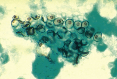
  • Several stains can be performed (Fig. E4), such as Giemsa, Diff-Quik, and Wright (all stain both the cystic and the trophic forms of the organism but not the cyst wall); Grocott-Gomori methenamine silver, Gram-Weigert, toluidine blue, and cresyl violet stain (stain the cyst wall), as well as immunofluorescent stains, with results depending on the lab experience and the quality of the specimen.

Figure E4 Cysts of Pneumocystis jirovecii in smear from bronchoalveolar lavage.

Methenamine silver stain.

Courtesy Dr. Russell K. Brynes/Centers for Disease Control and Prevention. In Ryan ET et al: Hunter’s tropical medicine and emerging infectious diseases, ed 10, 2019, Elsevier.

  • Polymerase chain reaction (PCR) can be used; however, this cannot distinguish between colonization and active disease.8
  • Serum 1,3-β-D glucan has a good negative predictive value (very sensitive, less specific); test characteristics are slightly better in people with HIV.9
  • Lactate dehydrogenase (LDH) is nonspecific; it is often elevated, but a normal LDH does not rule out PCP. Persistently elevated LDH may be predictive of mortality or lack of response to treatment.3,10
  • Metagenomic next-generation sequencing (mNGS) in blood or BAL fluid has demonstrated good performance for diagnosis of PCP.11
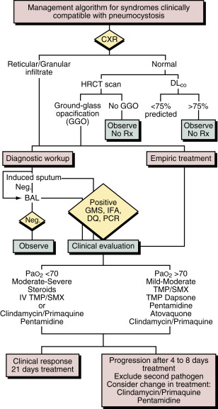
  • Fig. 5 describes an algorithm for the diagnostic evaluation and management of patients with suspected Pneumocystis pneumonia.
Figure 5 Algorithm for the Diagnostic Evaluation and Management of Patients with Suspected Pneumocystis Pneumonia

!!flowchart!!

BAL, Bronchoalveolar lavage; CXR, chest x-ray; DLCO, single-breath diffusing capacity for carbon monoxide; DQ, Diff-Quik (stain); GGO, ground-glass opacities; GMS, Gomori methenamine silver (stain); HRCT, high-resolution computed tomography; IFA, immunofluorescent antibody (stain); IV, intravenous; Neg., negative; PaO2, partial pressure of oxygen in arterial blood; PCR, polymerase chain reaction; Rx, treatment; TMP/SMX, trimethoprim-sulfamethoxazole.

From Bennett JE et al: Mandell, Douglas, and Bennett’s principles and practice of infectious diseases, ed 8, Philadelphia, 2015, Saunders.

Laboratory Tests

  • HIV antibody test and CD4 cell count (with CD4%) if cause of underlying immune deficiency state is unclear.
  • Arterial blood gas and calculation of the alveolar-arterial (A-a) oxygen gradient for determination of disease severity and need for adjunctive corticosteroids:
    1. Mild disease: A-a gradient <35 mm Hg and/or PaO2 >70 mm Hg
    2. Moderate disease: A-a gradient 35 to 45 mm Hg and PaO2 >60 mm Hg and <70 mm Hg
    3. Severe disease: A-a gradient >45 mm Hg and PaO2 <60 mm Hg
  • Elevated 1,3-β-D-glucan (>80 pg/ml).
  • Elevated lactate dehydrogenase.
Imaging Studies

  • Early in the disease course, the chest x-ray can be normal. In these cases, a high-resolution chest CT is recommended. This test is mostly useful in ruling out PCP as a chest CT without ground-glass opacities makes the diagnosis extremely unlikely.
  • Classically, PCP presents as bilateral, symmetric interstitial, reticular, or granular infiltrates in the perihilar region (“butterfly pattern”) on chest x-ray, and CT chest typically shows ground-glass opacities; however, variable radiographic presentations are possible (unilateral, asymmetric, nodular, miliary, etc.).8 Pneumatoceles, or cysts, which may cause pneumothorax, are more common in people with HIV.
Other Studies

If diagnostic uncertainty, PFTs can be used as adjunctive test. Low DLCO is highly sensitive but not specific for PCP infection. A normal DLCO argues against PCP.

Treatment ⬆ ⬇

Nonpharmacologic Therapy

  • Supplemental oxygen
  • Noninvasive or invasive ventilatory support if needed
  • Prompt chest tube placement if pneumothorax develops
Acute General Rx12

For confirmed or suspected PCP:

  • Preferred therapy: Trimethoprim-sulfamethoxazole (TMP-SMX; 15 to 20 mg/kg trimethoprim and 75 to 100 mg/kg sulfamethoxazole) divided and given q6 to 8h. Recent systematic reviews and meta-analyses have suggested that lower doses of TMP-SMX may be as effective and may be considered in older adults or those with renal dysfunction; however, official guidelines continue to recommend the higher dose of TMP-SMX at this time.13
    1. Mild PCP can be treated with PO formulation of TMP-SMX, usually two double-strength (DS) tablets PO tid.
    2. Moderate and severe disease should be treated with intravenous (IV) formulation of TMP-SMX at first but can be switched to PO after clinical improvement.
  • Alternative therapies: Use only if severe contraindication to TMP-SMX (e.g., Stevens-Johnson syndrome or toxic epidermal necrolysis [TEN]) and after development of treatment-limiting adverse reaction.
    1. Mild-moderate disease:
      1. TMP 15 mg/kg/day split into tid dosing plus dapsone 100 mg PO daily, OR
      2. Clindamycin 600 mg PO q8h plus primaquine 30 mg (base) PO daily, OR
      3. Atovaquone 750 mg PO bid (with food)
    2. Moderate-severe disease:
      1. Clindamycin (600 mg q6h or 900 q8h) IV and primaquine 30 mg (base) PO daily, OR
      2. Pentamidine (4 mg/kg) IV daily. May reduce dose to 3 mg/kg IV daily if toxicity develops. Careful monitoring is required, as IV pentamidine can cause nephrotoxicity, numerous electrolyte disturbances, and cardiac arrhythmias.
  • Adjunctive corticosteroids are indicated in moderate to severe PCP (PaO2 <70 mm Hg and A-a gradient >35 mm Hg):
    1. Prednisone 40 mg bid × 5 days, followed by 40 mg/day × 5 days, followed by 20 mg/day × 11 days (for total of 21 days)
    2. Alternative: IV methylprednisolone at 75% of prednisone dose
  • Therapy continued for 21 days.
  • Antiretroviral therapy in HIV positive individuals should be initiated as soon as possible, ideally within 2 wk of PCP diagnosis. Monitor for immune reconstitution inflammatory syndrome (IRIS).
  • Table 1 summarizes causes of deterioration in an HIV-infected patient receiving treatment for PCP.

Table 1 Causes of Deterioration in an HIV-Infected Person Receiving Treatment for PCP

EtiologyExplanation
Severe Progressive PCP
IatrogenicPulmonary edema due to IV fluid overload when giving TMP-SMX
IRIS following early initiation of ART
Side effects of therapyAnemia (e.g., caused by TMP-SMX), methemoglobinemia (e.g., caused by dapsone, primaquine)
Inadequate therapyIncorrect dosage or route of administration
Adjuvant glucocorticoids not given for treatment of moderate or severe PCP
Post bronchoscopySedation
Pneumothorax
PneumothoraxSpontaneous
Associated with intubation and positive pressure ventilation
Co-pathology in lungBacterial infection
Pulmonary Kaposi sarcoma
Intercurrent pulmonary embolism
Wrong diagnosisEmpiric diagnosis of PCP, and correct diagnosis is another pathology (e.g., bacterial pneumonia)

ART, Antiretroviral therapy; HIV, human immunodeficiency virus; IRIS, immune reconstitution inflammatory syndrome; IV, intravenous; PCP, Pneumocystis jirovecii pneumonia; TMP-SMX, trimethoprim-sulfamethoxazole.

Bennett JE et al: Mandell, Douglas, and Bennett’s principles and practice of infectious diseases, ed 8, Philadelphia, 2015, Saunders.

Prophylaxis

  • Primary prophylaxis:
    1. In people with HIV, primary prophylaxis is indicated when CD4 count <200 cells/mm3 or CD4% <14%. Acceptable regimens include one double-strength tablet PO daily, one single-strength tablet PO daily or one double-strength tablet PO three times a week. Can consider discontinuation of prophylaxis if CD4 cell count between 100 and 200/mm3 with undetectable HIV viral load for 3 to 6 mo following ART initiation12
    2. Commonly also applied to individuals on prolonged courses of steroids (e.g., >4 wk of >20 mg prednisone daily)
    3. Indicated in certain immunosuppressed adults with malignancy14
    4. Recommended in patients with rheumatologic, autoimmune, dermatologic diseases on biologic therapy, as well as in patients with solid organ transplant2,4,15
  • Secondary prophylaxis:
    1. After completion of therapy, secondary prophylaxis should be maintained in people with HIV with TMP-SMX (one single-strength tablet PO daily OR one double-strength tablet PO daily OR [alternative] one double-strength tablet PO three times weekly) until the CD4 cell count is >200 cells/mm3 and viral load is undetectable for at least 3 mo after ART initiation.12
    2. If PCP occurs at a CD4 cell count >200 cells/mm3 while on ART, continue PCP prophylaxis for life, regardless of how high the CD4 cell count rises as a result of ART.
  • Patients intolerant of TMP-SMX should be treated with dapsone (100 mg PO daily) or atovaquone (1500 mg PO daily).
  • Inhaled pentamidine (300 mg mo by standardized nebulizer) is less effective and is reserved for patients intolerant to other forms of prophylaxis. Inhaled pentamidine or dapsone without pyrimethamine should not be used for PCP prophylaxis in patients with HIV who are also seropositive for Toxoplasma gondii.12
Disposition

  • After completion of therapy, long-term ambulatory follow-up is mandatory to provide secondary prophylaxis of PCP and management of the underlying immunodeficiency syndrome.
  • Many patients have chronic respiratory complaints such as exertional dyspnea and may develop chronic obstructive pulmonary disease (COPD). These patients should be referred to a pulmonologist for evaluation and management.
Referral

  • To a pulmonologist for bronchoscopy if diagnosis cannot be confirmed by sputum examination
  • To an infectious disease specialist if case is severe or difficult to manage and to evaluate for underlying immune deficiency
  • To a pulmonologist for evaluation if persistent respiratory complaints such as exertional dyspnea after PCP treatment

Pearls & Considerations ⬆ ⬇

Comments

  • All patients, especially those with severe infection or intolerant of conventional therapy, should be followed by a physician experienced in the management of PCP and, if appropriate, in the long-term management of HIV infection or other underlying disease.
  • Patients with suspected PCP should be started on empiric PCP therapy (and adjunctive corticosteroids if indicated) without waiting for diagnostic procedures. Pneumocystis can be seen on microscopic examination days to weeks into PCP therapy.
  • Before administration of dapsone or primaquine, patients should be tested for G6PD deficiency.
  • Clinical and chest x-ray findings in patients with PCP may worsen in the first 5 days after treatment initiation. Wait to declare treatment failure until after at least 7 to 10 days of therapy.8
  • Mutations associated with resistance to sulfa drugs in other microorganisms have been documented in P. jirovecii, but their effect on clinical outcomes is uncertain.
  • Monitor for IRIS after ART initiation.
  • TMP-SMX can have a range of side effects, including neutropenia (absolute neutrophil count <500 cells/mm3), fever, hyperkalemia, acute kidney injury, rash. May be able to slowly reintroduce medication after resolution of most mild side effects. Most severe and deadly TMP-SMX side effect is Stevens-Johnson syndrome/TEN; avoid all sulfa drugs thereafter.
  • Severe and life-threatening hypoglycemia may occur 1 or 2 wk after starting IV pentamidine. Monitor closely and advise the patient of symptoms of hypoglycemia.
  • Coinfection with other pathogens can be seen.8
  • Consider indications for primary PCP prophylaxis in all immunosuppressed patients regardless of HIV status.
Related Content

Pneumocystis Pneumonia (Patient Information)

Acquired Immunodeficiency Syndrome (Related Key Topic)

Related Content ⬆

    1. Gold J.A.W. : Possible diagnostic delays and missed prevention opportunities in pneumocystis pneumonia patients without HIV: analysis of commercial insurance claims data-United States, 2011-2015Open Forum Infect Dis. ;7(7), 2020.doi:10.1093/ofid/ofaa255
    2. Ghembaza A. : Risk factors and prevention of Pneumocystis jirovecii pneumonia in patients with autoimmune and inflammatory diseasesChest. ;158(6):2323-2332, 2020.doi:10.1016/j.chest.2020.05.558
    3. Wang Y. : Risk factors of mortality from pneumocystis pneumonia in non-HIV patients: a meta-analysisFront Public Health. ;9, 2021.doi:10.3389/fpubh.2021.680108
    4. Fishman J.A. : Pneumocystis jiroveci in solid organ transplantation: guidelines from the American Society of Transplantation Infectious Diseases Community of PracticeClin Transplant. ;33(9), 2019.doi:10.1111/ctr.13587
    5. Roux A. : Pneumocystis jirovecii pneumonia in patients with or without AIDS, FranceEmerg Infect Dis. ;20(9):1490-1497, 2014.doi:10.3201/eid2009.131668
    6. Yiannakis E.P., Boswell T.C. : Systematic review of outbreaks of Pneumocystis jirovecii pneumonia: evidence that P. jirovecii is a transmissible organism and the implications for healthcare infection controlJ Hosp Infect. ;93(1):1-8, 2016.doi:10.1016/j.jhin.2016.01.018
    7. Huang L. : Suspected Pneumocystis carinii pneumonia with a negative induced sputum examination. Is early bronchoscopy useful?Am J Respir Crit Care Med. ;151(6):1866-1871, 1995.doi:10.1164/ajrccm.151.6.7767533
    8. White P. : Therapy and management of Pneumocystis jirovecii infectionJ Fungi. ;4(4), 2018.doi:10.3390/jof4040127
    9. Del Corpo O. : Diagnostic accuracy of serum (1-3)-β-D-glucan for Pneumocystis jirovecii pneumonia: a systematic review and meta-analysisClin Microbiol Infect. ;26(9):1137-1143, 2020.doi:10.1016/j.cmi.2020.05.024
    10. Zaman M.K., White D.A. : Serum lactate dehydrogenase levels and Pneumocystis carinii pneumonia. Diagnostic and prognostic significanceAm Rev Respir Dis. ;137(4):796-800, 1988.doi:10.1164/ajrccm/137.4.796
    11. Wang D. : Metagenomic next-generation sequencing is highly efficient in diagnosing Pneumocystis jirovecii pneumonia in the immunocompromised patientsFront Microbiol. ;13, 2022.doi:10.3389/fmicb.2022.913405
    12. Pneumocystis Pneumonia | NIH. Available at https://clinicalinfo.hiv.gov/en/guidelines/hiv-clinical-guidelines-adult-and-adolescent-opportunistic-infections/pneumocystis-0. Accessed August 11, 2022.
    13. Haseeb A. : Trimethoprim-Sulfamethoxazole (Bactrim) dose optimization in Pneumocystis jirovecii pneumonia (PCP) management: a systematic reviewInt J Environ Res Public Health. ;19(5), 2022.doi:10.3390/ijerph19052833
    14. Taplitz R.A. : Antimicrobial prophylaxis for adult patients with cancer-related immunosuppression: ASCO and IDSA clinical practice guideline updateJ Clin Oncol. ;36(30):3043-3054, 2018.doi:10.1200/JCO.18.00374
    15. Wolfe R.M. : Practice patterns of pneumocystis pneumonia prophylaxis in connective tissue diseases: a survey of infectious disease physiciansOpen Forum Infect Dis. ;6(9), 2019.doi:10.1093/ofid/ofz315