section name header

Basic Information

AUTHOR: Imrana Qawi, MD

Definition

Sarcoidosis is a chronic multisystem granulomatous disease of unknown cause characterized histologically by the presence of noncaseating granulomas and manifesting with a wide range of clinical disturbances.

Synonym

Boeck sarcoid

ICD-10CM CODES
D86Sarcoidosis
D86.0Sarcoidosis of lung
D86.1Sarcoidosis of lymph nodes
D86.2Sarcoidosis of lung with sarcoidosis of lymph nodes
D86.3Sarcoidosis of skin
D86.8Sarcoidosis of other and combined sites
D86.9Sarcoidosis, unspecified
Epidemiology & Demographics
Incidence (In U.S.)

Incidence is 11 in 100,000 in whites and 35 in 100,000 in blacks; presents most commonly in the winter and early spring. (The adjusted annual incidence among black Americans is roughly 3 times higher than among white Americans [35.5 cases/100,000, as compared with 10.9/100,000] and is likely more chronic and fatal in black Americans.)

Predominant Sex

Increased incidence in females.

Predominant Age

20 to 50 yr, second peak after 60 yr1

Genetics

Familial clustering has been described. Having a first-degree relative with sarcoidosis increases the risk for disease fivefold.2 There have been reports of association between sarcoidosis and gene products, specifically human leukocyte antigen (HLA) class II antigens, encoded by HLA-DRB1 and DQB1 alleles.3

Physical Findings & Clinical Presentation

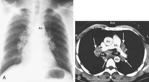
  • Clinical manifestations often vary with the stage of the disease and degree of organ involvement. Patients may be asymptomatic, but a chest radiograph may demonstrate findings consistent with sarcoidosis (see “Imaging Studies”). Nearly 50% of patients with sarcoidosis are diagnosed by incidental findings on chest radiograph. Lung involvement occurs in >90% of patients with sarcoidosis.
  • Frequent manifestations:
    1. Pulmonary manifestations: Dry, nonproductive cough; dyspnea; chest discomfort. Pleural effusion is an uncommon manifestation but can occur in stage II or III of pulmonary involvement.
    2. Constitutional symptoms: Fatigue, weight loss, anorexia, malaise, night sweats.
    3. Visual disturbances: Blurred vision, ocular discomfort, conjunctivitis, iritis, uveitis (65% of patients).
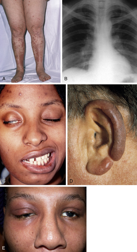
    4. Dermatologic manifestations (30% of patients): Erythema nodosum (10% of patients), macules, papules, subcutaneous nodules, hyperpigmentation, lupus pernio (indurated violaceous lesions on the nose, lips, ears, and cheeks that can erode into underlying cartilage and bone) (Fig. E1).
    5. Myocardial disturbances, arrhythmias, cardiomyopathy, various conduction abnormalities, and pericardial effusion. Cardiac sarcoidosis is much more common than clinically appreciated and is found in up to 25% of patients in the U.S.
    6. Splenomegaly, hepatomegaly, and rarely, can involve the pancreas.
    7. Rheumatologic manifestations: Arthralgias have been reported in up to 40% of patients. It typically affects the ankles but can also involve the knees, wrists, and small joints of the hands and feet.
    8. Löfgren syndrome, consisting of the triad of arthritis, erythema nodosum, and bilateral hilar adenopathy, occurs in 9% to 34% of patients. Fever is frequently present.
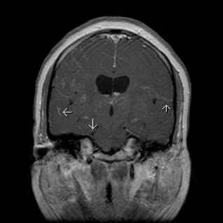
    9. Neurologic and other manifestations: Cranial nerve palsies, diabetes insipidus, meningeal involvement, parotid enlargement, hypothalamic and pituitary lesions, peripheral adenopathy. Neurosarcoidosis is detected in up to 25% of patients and can occur in the absence of apparent disease elsewhere. Cranial nerve dysfunction is the most common neurologic complication of sarcoidosis. Basilar granulomatous meningitis is the usual cause (Fig. E2), but the facial nerve may also be involved when parotitis is present.
    10. The presence of anterior uveitis, parotiditis, fevers, and facial nerve palsy is known as Heerfordt syndrome.
    11. Renal involvement in 7% to 22% in multisystem disease. Epididymis and the testis can be involved in male patients. Initial presentation can be of hydronephrosis due to external compression by retroperitoneal lymph nodes.
    12. Hypercalcemia is seen in about 10% to 13% of patients with multisystem involvement. Abnormal production of 1-alpha-hydroxylase and PTHrp is thought to contribute to the hypercalcemia in some patients with sarcoidosis. Hypercalciuria is also common.

Figure E1 Sarcoidosis.

A, Erythema nodosum. B, Bilateral hilar lymphadenopathy. C, Seventh nerve palsy. D, Lupus pernio. E, Lacrimal gland enlargement.

D, Courtesy MA Mir. From Mir MA: Atlas of clinical diagnosis, Philadelphia, 2003, Saunders.

Figure E2 Neurosarcoidosis

T1 Coronal Magnetic Resonance Imaging (MRI) with Contrast Shows Enhancing Leptomeninges (Arrows).

From Pina-Garza J, James KC. Fenichel’s clinical pediatric neurology, ed 8, Philadelphia. 2019, Elsevier.

Etiology & Pathogenesis

A cardinal feature of sarcoidosis is the presence of CD4+ T cells that interact with antigen-presenting cells to initiate the formation and maintenance of granulomas (Fig. E3). Multiple lines of evidence suggest that sarcoidosis may result from the interaction of multiple genes with environmental exposures or infection.

Figure E3 Simplified proposed pathogenetic sequence in sarcoidosis.

!!flowchart!!

Ab, Antibody; Ag, antigen.

From Weinberger SE: Principles of pulmonary medicine, ed 7, Philadelphia, 2019, Elsevier.

Genetic Predisposition to Sarcoidosis

Various HLA antigens have been implicated in diverse patient populations. Familial predisposition has been reported dating back to 1923, with a wide percentage of variability of affected relatives (0.4% to 21%) and heterogeneity based on genetic background. A more recent study was published by the ACCESS (A Case Controlled Etiologic Survey of Sarcoidosis) study group, which confirmed increased risk for family members with an odds ratio of 4.6 for all relatives. Absolute risk, however, for a family member to be affected was less than 1%. This study also showed a higher risk in white versus black American siblings and parents.

Immunopathogenesis of Sarcoidosis

Numerous chemokines and cytokines have been implicated in the development and/or resolution of the disease. In sarcoidosis, the alveolitis seen at disease presentation represents an increase in primarily lymphocytic cellularity, predominated by CD4 cells. Presence of increased neutrophils in the bronchoalveolar lavage of patients with sarcoidosis has been associated with persistence of disease, with spontaneous remission noted in 36% of patients with elevated neutrophil counts.

Diagnosis

Differential Diagnosis

  • Infection:
    1. Tuberculosis
    2. Fungal infection (e.g., Coccidioides, histoplasmosis, blastomycosis)
    3. Infectious mononucleosis
  • Malignancies:
    1. Lymphoma
    2. Metastatic cancer
    3. Lymphangitic carcinomatosis
  • Pneumoconioses including berylliosis and silicosis
  • Other interstitial lung diseases (e.g., hypersensitivity pneumonitis)
  • Vascular abnormalities (e.g., enlarged pulmonary arteries)
  • Parasitic infection
  • Other diseases such as amyloidosis
Workup

  • No pathognomonic diagnostic test exists for sarcoidosis, so the diagnosis remains one of exclusion. The presence of noncaseating granulomas does not establish the diagnosis, because conditions such as tuberculosis and malignancies, among others, can cause granulomas.
  • Workup is aimed at excluding critical organ involvement, determining extent and severity of disease, and excluding other disease. A complete neurologic and ophthalmologic examination is mandatory.
  • A complete occupational and environmental exposure history is recommended.
  • In patients with a history of beryllium exposure, beryllium lymphocyte proliferation testing should be obtained to assess for berylliosis.
  • Initial laboratory evaluation should include complete blood count, serum chemistries (alanine aminotransferase, aspartate aminotransferase, alkaline phosphatase, electrolytes, blood urea nitrogen, creatinine, serum calcium), urinalysis, 24-hour urinary excretion of calcium, C-reactive protein (CRP), erythrocyte sedimentation rate (ESR), and tuberculin skin test or QuantiFERON gold test for tuberculosis. Routine vitamin D screening is generally not recommended; if done, it should measure both 25-hydroxyvitamin D and 1,25-dihydroxyvitamin D because sarcoidosis increases conversion of the former to the latter.4
  • Chest radiograph and ECG should also be obtained in all patients suspected to have sarcoidosis.
  • Pulmonary function testing: Spirometry, diffusion capacity of carbon monoxide.
  • Biopsy (Fig. 4) in symptomatic patients should be done on accessible tissues suspected of sarcoid involvement (conjunctiva, skin, lymph nodes); bronchoscopy with transbronchial biopsy (85% diagnostic yield) is often performed in patients without any readily accessible site. Among patients with suspected Stage I/II pulmonary sarcoidosis undergoing tissue confirmation, the addition of endobronchial ultrasound (EBUS) transbronchial needle aspiration (TBNA) resulted in greater diagnostic yield. EBUS-TBNA of lymph nodes has a diagnostic yield of approximately 87%. Although in some studies, mediastinoscopy may have a higher diagnostic yield than EBUS-guided lymph node biopsy (98% and 87%, respectively), EBUS-TBNA is less invasive and has become the standard diagnostic modality for patients with mediastinal adenopathy with high clinical suspicion for sarcoidosis.
Figure 4 Diagnostic Approach to Selecting a Biopsy Site for Pathologic Confirmation of Granulomatous Inflammation Consistent with Sarcoidosis

This Approach Emphasizes Selection of a Relatively Noninvasive Biopsy Site When Possible, Biopsy of a Site Suspected to Be Clinically Involved Unless the Biopsy Would Be Highly Invasive, and Various Approaches When No Obvious Organ Involvement is Demonstrated or Only Organs Requiring Very Invasive Biopsies Demonstrate Potential Involvement. PET, Positron Emission Tomography.

!!flowchart!!

From Broaddus VC et al: Murray & Nadel’s textbook of respiratory medicine, ed 7, Philadelphia 2022, Elsevier.

Laboratory Tests

The following laboratory abnormalities are often present in sarcoidosis:

  • Hypergammaglobulinemia, anemia, leukopenia may be present.
  • Liver function test abnormalities are common, e.g., elevated alkaline phosphatase.
  • Hypercalcemia (11% of patients), hypercalciuria (40% of patients; attributable to increased gastrointestinal absorption, abnormal vitamin D metabolism, and increased calcitriol production by sarcoid granuloma).
  • Angiotensin-converting enzyme: Elevated in approximately 75% of patients with untreated sarcoidosis; nonspecific and poor sensitivity; generally not useful as a diagnostic tool or in following the course of the disease.
  • Serum adenosine deaminase (ADA); serum amyloid A (SAA) elevated but nonspecific.
Imaging Studies

  • Chest x-ray (Fig. 5): Pulmonary sarcoidosis is classified based on radiographic pattern. Adenopathy of the hilar and paratracheal nodes is a frequent finding. Parenchymal changes may also be present, depending on the stage of the disease (stage 0, normal radiograph; stage I, bilateral hilar adenopathy; stage II, stage I plus pulmonary infiltrate; stage III, pulmonary infiltrate without adenopathy; stage IV, advanced fibrosis with evidence of “honeycombing,” hilar retraction, bullae, cysts, and emphysema).
  • Computed tomography (CT) imaging: High-resolution CT may help detect early parenchymal abnormalities. A chest CT scan with contrast can better define mediastinal adenopathy.
  • For patients without apparent lung involvement, 18F-fluorodeoxyglucose positron emission tomography (FDG-PET) (Fig. E6) is useful in identifying sites for diagnostic biopsy.
  • FDG-PET and MRI with gadolinium are useful in patients with suspected cardiac and neurologic involvement.
  • Gallium-67 scan: Represents an older testing modality. It will localize in areas of granulomatous infiltrates; however, it is not specific and not necessary. The “panda” sign (localization in the lacrimal and salivary glands, giving a “panda” appearance to the face) is suggestive of sarcoidosis.

Figure 5 Sarcoid.

Marked lymphadenopathy (dotted lines) is seen in the region of both hila in the right paratracheal region (A). The transverse contrast-enhanced computed tomography (CT) scan of the upper chest (B) clearly shows the ascending and descending aorta (Ao) as well as the pulmonary artery (PA) and superior vena cava. The right and left mainstem bronchus area is also seen. The arrows indicate the extensive lymphadenopathy. LB, Left bronchus; RB, right bronchus.

From Mettler FA [ed]: Primary care radiology, Philadelphia, 2000, Saunders.

Figure E6 Positron Emission Tomography Scan Demonstrating Both Lung Parenchymal and Mediastinal Nodal Uptake

Also Demonstrated are Several Extrapulmonary Lymph Nodes and Other Areas with Increased Activity.

From Broaddus VC et al: Murray & Nadel’s textbook of respiratory medicine, ed 7, Philadelphia 2022, Elsevier.

Additional diagnostic tests:

  • Pulmonary function tests (spirometry, lung volumes, and diffusing capacity for carbon monoxide): May be normal; may reveal a restrictive ventilatory defect with reduced forced vital capacity and/or reduced DLCO; may reveal an obstructive pattern; or may reveal a combination.
  • Bronchoscopy: Flexible bronchoscopy and bronchoalveolar lavage (BAL) with transbronchial biopsy (showing noncaseating granulomas) are traditional methods for the minimally invasive diagnosis of sarcoidosis. With hilar adenopathy, EBUS-TBNA is the preferred method for lymph node sampling. BAL may show predominantly lymphocytosis, elevated ADA levels, and elevated CD4:CD8 ratio (4:1).
  • Mediastinoscopy: Now rarely used for lymph node sampling and diagnosis.
  • Routine eye examination is recommended, even for patients without ocular symptoms.
  • For patients with extracardiac sarcoidosis or suspected cardiac involvement, cardiac MRI is indicated.
  • Echocardiogram is indicated in patients with suspected pulmonary hypertension. If suggested by transthoracic echocardiogram, right heart catheterization should be done.

Treatment

TABLE 1 Indications for Use of Corticosteroids in Sarcoidosis

DisorderTreatment
IridocyclitisCorticosteroid eye drops; local subconjunctival deposit of cortisone
Posterior uveitisOral prednisone
Pulmonary involvementSteroids rarely recommended for stage I; typically used if infiltrate remains static or worsens over 3-mo period or the patient is symptomatic
Upper airway obstructionRare indication for intravenous steroids
Lupus pernioOral prednisone shrinks the disfiguring lesions
HypercalcemiaResponds well to corticosteroids
Cardiac involvementCorticosteroids usually recommended if patient has arrhythmias or conduction disturbances
Central nervous system involvementResponse is best in patients with acute symptoms
Lacrimal/salivary gland involvementCorticosteroids recommended for disordered function, not gland swelling
Bone cystsCorticosteroids recommended if symptomatic

From Andreoli TE (ed): Cecil essentials of medicine, ed 8, Philadelphia, 2010, Saunders.

Disposition

  • The majority of patients with sarcoidosis have spontaneous remission within 2 years and do not require treatment. Their course can be followed by periodic clinical evaluation, chest CT scans, and pulmonary function tests.
  • Blacks have increased rates of pulmonary involvement, a worse long-term prognosis, and more frequent relapses.
  • Up to one third of patients have unrelenting disease, leading to clinically significant organ impairment. Adverse prognostic factors in sarcoidosis include age of onset >40 years, cardiac involvement, neurosarcoidosis, progressive pulmonary fibrosis, chronic hypercalcemia, chronic uveitis, involvement of nasal mucosa, nephrocalcinosis, and presence of cystic bone lesions and lupus pernio.
  • Prognosis for Loeffler syndrome is good with remittance within 16 weeks in most patients. Symptoms can generally be controlled with NSAIDs. Low-dose glucocorticoids, hydroxychloroquine, and colchicine are also effective.
  • Pulmonary hypertension is a poor prognostic sign and warrants consideration of referral for lung transplant evaluation. Transplantation for sarcoidosis is performed in 3% to 5% of patients and posttransplant survival rate is approximately 70% at 5 yr. The main factors associated with worse survival are older age and extensive preoperative lung fibrosis.1
Referral

  • Patients with progressive pulmonary disease or development of pulmonary hypertension despite therapy should be referred for consideration of lung transplant candidacy.
  • Ophthalmologic examination is indicated in all patients with suspected sarcoidosis because ocular findings (iridocyclitis, uveitis, conjunctivitis, and keratopathy) are found in 25% of documented cases.
  • Dermatology consultation for possible biopsy is indicated for patients with possible skin manifestations.
  • Patients with suspected cardiac involvement should be referred to cardiology and those with CNS involvement referred to neurology.

Pearls & Considerations

Comments
  1. Serial spirometry and measurement of DLCO can be useful in following response to therapy and disease progression.
  2. Approximately 15% to 20% of patients with lung involvement advance to irreversible lung impairment (bronchiectasis, cavitation, progressive fibrosis, pneumothorax, and respiratory failure). Death from pulmonary failure occurs in 5% to 7% of patients with sarcoidosis.
  3. Newer treatment approaches are aimed at targeting mechanisms involving CD4 type 1 helper T cells.
  4. The diagnosis of sarcoidosis should be reconsidered in the presence of atypical manifestations or persistent/progressive disease despite appropriate therapy.
  5. Diagnostic biopsy may not be necessary in most patients presenting with asymptomatic bilateral lymphadenopathy (with no other evidence of malignancy), in those with Lofgren syndrome, or in those with Heerfordt syndrome.
Related Content

Sarcoidosis (Patient Information)

Related Content

    1. Maier L.A. : Executive summary of the NHLBI workshop report: leveraging current scientific advancements to understand sarcoidosis variability and improve outcomesAnn Am Thorac Soc. ;14(Suppl 6):S415-S420, 2017.doi:10.1513/AnnalsATS.201707-563OT
    2. Rossman M.D., Kreider M.E. : Lesson learned from ACCESS (A Case Controlled Etiologic Study of Sarcoidosis)Proc Am Thorac Soc. ;4(5):453-456, 2007.doi:10.1513/pats.200607-138MS
    3. Moller D.R. : Genetic, immunologic, and environmental basis of sarcoidosisAnn Am Thorac Soc. ;14(Suppl 6):S429-S436, 2017.doi:10.1513/AnnalsATS.201707-565OT
    4. Crouser E.D. : Diagnosis and detection of sarcoidosis. An official American Thoracic Society clinical practice guidelineAm J Respir Crit Care Med. ;201(8):e26-e51, 2020.