section name header

Basic Information ⬇

AUTHOR: Bharti Rathore, MD

Definition

Thrombotic thrombocytopenic purpura (TTP) is a rare autoimmune disorder characterized primarily by thrombocytopenia and microangiopathic hemolytic anemia and can be associated commonly with other hallmarks such as neurologic impairment, renal dysfunction, and fever. The laboratory hallmark of TTP is a severe deficiency of the ADAMTS13 factor (activity <10%).

Synonyms

Immune mediated thrombocytopenic purpura

iTTP

TTP

Upshaw-Schulman syndrome

ICD-10CM CODE
M31.1Thrombotic microangiopathy
Epidemiology & Demographics

  • About 90% of new TTP cases are seen in adults (mostly females between 18 and 50 yr).
  • The incidence of new TTP is 3 to 11 cases per million population per year. The prevalence is ∼10 cases per million population.
  • There is increased incidence in HIV/AIDS and during pregnancy.
Physical Findings & Clinical Presentation

  • The disease often begins as a flulike illness ultimately followed by development of characteristic clinical and laboratory abnormalities.
  • Most patients present with nonspecific constitutional symptoms (weakness, nausea, abdominal pain, vomiting).
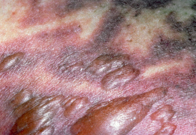
  • Purpura (Fig. E1 and Fig. E2).
  • Jaundice and pallor (from hemolysis).
  • Mucosal bleeding.
  • Fever.
  • Fluctuating levels of consciousness (caused by thrombotic occlusion of the cerebral vessels). However, one third of patients have no neurologic abnormalities.
  • Renal failure and neurologic events are usually end-stage features.
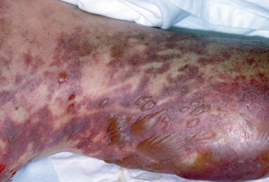
Figure E1 Thrombotic thrombocytopenic purpura.

From Micheletti RG et al: Andrews’ diseases of the skin, clinical atlas, ed 2, Philadelphia, 2023, Elsevier.

Figure E2 Thrombotic thrombocytopenic purpura.

From Micheletti RG et al: Andrews’ diseases of the skin, clinical atlas, ed 2, Philadelphia, 2023, Elsevier.

Etiology

  • Acquired TTP is an autoimmune disorder caused by autoantibody inhibition of ADAMTS13 activity.1
  • Hereditary TTP (also called Upshaw-Schulman syndrome) is caused by homozygous or compound heterozygous ADAMTS13 mutations2 (Table 1).
  • Many drugs, including clopidogrel, ticlopidine, penicillin, antineoplastic agents (gemcitabine, mitomycin C), calcineurin inhibitors (cyclosporine), oral contraceptives, and quinine, have been associated with TTP.
  • Other precipitating causes include infectious agents, pregnancy, malignancies, allogeneic stem cell transplantation, and neurologic disorders.
  • Acquired TTP has been reported in the setting of COVID-19 infection as well as after vaccination with traditional and mRNA COVID-19 vaccines.

TABLE 1 Etiology, Epidemiology, and Pathogenesis of TTP, HUS, and ADAMTS13-Related Parameters

Congenital TTPAcquired TTPHUS
EtiologyADAMTS13 mutationAntibody to ADAMTS13, endothelial cell activationEscherichia coli or other microorganisms
Epidemiology5-10 cases per year per million≤1 case per year per million1-5 cases per year per million, mainly in children
PathogenesisDefective cleavage of vWF multimers, massive secretion of ultra-large vWF multimers, increased platelet deposition under shear condition, occlusion of blood vessels in microcirculationIntoxication with Shiga-like toxin, damage of endothelial cells, enterohemorrhagic colitis, renal disorder
ADAMTS13 antigenVery low or absentLow or variableNormal or moderately decreased
ADAMTS13 activity≤5%-10%≤5%-10% or variable30%-100%
Inhibitor against ADAMTS13NoMostly yesNo

ADAMTS13, A disintegrin and metalloproteinase with a thrombospondin type 1 motif, member 13; HUS, hemolytic uremic syndrome; TTS, thrombotic thrombocytopenic purpura; vWF, von Willebrand factor.

From McPherson RA, Pincus MR: Henry’s clinical diagnosis and management by laboratory methods, ed 23, Philadelphia, 2017, Elsevier.

Diagnosis ⬆ ⬇

Differential Diagnosis

It is challenging to differentiate TTP from other thrombotic microangiopathies given the significant overlap in clinical presentation, but this distinction is critical in selecting an appropriate therapy for patients.

  • Disseminated intravascular coagulation (DIC)
  • Malignant hypertension
  • Vasculitis
  • Eclampsia or preeclampsia
  • Hemolytic-uremic syndrome (HUS) and atypical HUS
  • Gastroenteritis associated with serotoxin-producing serotype of Escherichia coli
Workup

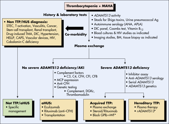
A comprehensive history, physical examination, and laboratory evaluation usually confirm the diagnosis (Fig. 3).

Figure 3 An approach to diagnosis and management of thrombotic microangiopathies.

!!flowchart!!

ADAMTS13, A disintegrin and metalloproteinase with thrombospondin type 1 motifs, member 13; Ag, antigen; aHUS, atypical hemolytic uremic syndrome; AKI, acute kidney injury; ANA, antinuclear antibody; APLA, antiphospholipid antibodies; BM, bone marrow; CAPS, catastrophic antiphospholipid syndrome; CFB, complement factor B; CFH, complement factor H; CFI, complement factor I; DGKE, diacyl glycerol kinase Ε; DIC, disseminated intravascular coagulation; GP, glycoprotein; HELLP, hemolysis, elevated liver enzymes, and low platelet count; HIV, human immunodeficiency virus; HUS, hemolytic uremic syndrome; MAHA, macroangiopathic hemolytic anemia; MCP, membrane cofactor protein; rADAMTS13, recombinant ADAMTS13; STEC, Shiga toxin-producing E. coli; TMA, thrombotic microangiopathy; TTP, thrombotic thrombocytopenic purpura; vWF, von Willebrand factor. Bullet points are diagnoses or therapies. ∗Indicates therapies under investigation.

From Hoffman R et al: Hematology: basic principles and practice, ed 7, Philadelphia, 2018, Elsevier.

Laboratory Tests

  • Severe anemia and thrombocytopenia (platelet count <50,000 or >50% reduction from previous counts)
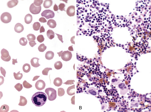
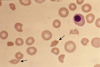
  • Peripheral blood smear (Fig. E4,Fig. E5) with numerous red blood cell (RBC) fragments (schistocytes)
  • Elevated blood urea nitrogen and creatinine
  • Hemolysis demonstrated by elevated reticulocyte count, indirect bilirubin, lactate dehydrogenase and decreased haptoglobin
  • Urinalysis: Hematuria (RBCs and RBC casts in urine sediment) and proteinuria
  • No laboratory evidence of DIC (normal fibrin degradation product, fibrinogen)
  • Hereditary TTP is diagnosed in cases of ADAMTS13 deficiency with concurrent absence of ADAMTS13 autoantibody inhibitor; the confirmation requires documentation of ADAMTS13 mutations2
  • Acquired TTP is diagnosed in cases with microangiopathic hemolytic anemia and thrombocytopenia without another apparent cause. An ADAMTS13 level <10% of normal activity is seen in acquired TTP, while levels >20% suggest other diagnoses

Figure E4 A, Peripheral Blood Smear of Thrombotic Thrombocytopenic Purpura

Numerous Red Cell Fragments (Schistocytes) are Present. No Platelets are Seen in This Field. B, The Bone Marrow Trephine Biopsy Section Demonstrates Increased Numbers of Megakaryocytes.

From Jaffe ES et al: Hematopathology, Philadelphia, 2011, Saunders.

Figure E5 Peripheral Blood Film Obtained from a 28-Yr-Old Woman Who Presented with Fever, Epistaxis, and Altered Mental Status

Note the absence of platelets and the presence of a nucleated erythrocyte and schistocytes (arrows) consistent with a microangiopathic process.

From Hoffman R et al: Hematology: basic principles and practice, ed 7, Philadelphia, 2018, Elsevier.

Treatment ⬆ ⬇

Acute General Rx

  • Discontinue any potential offending agents.
  • Initiate ADAMTS13 replacement by plasma infusion in patients with hereditary TTP.
  • TTP therapy consists of complementary approaches:
    1. Therapeutic plasma exchange (TPE)
    2. Immunosuppression
    3. Newer paradigms also include targeting von Willebrand factor-platelet interactions3
  • TPE reduces mortality rates from >90% to <20%. Daily TPE with replacement of 1.0 to 1.5 times the predicted patient plasma volume is the standard approach. TPE should be continued for a minimum of 2 days after the platelet count returns to normal (>150,000 cells/m3). There is no benefit to a tapering approach with TPE.
  • High-dose plasma infusion (25 ml/kg/day) may be useful when TPE cannot be promptly started and in patients with very severe or refractory disease between plasma exchange sessions. This approach can cause volume overload in patients with renal insufficiency.
  • Patients with hereditary TTP who experience severe plasma allergic reactions have been effectively treated with plasma-derived factor VIII concentrate that contains ADAMTS13.
  • Immunosuppression with the concurrent use of corticosteroids (prednisone 1 to 2 mg/kg/day) typically is initiated and tapered over a period of 3 to 4 wk after stable platelet counts are achieved.
  • Immunosuppression with the monoclonal anti-CD20 antibody rituximab is used in cases of suboptimally responsive TTP (inadequate platelet recovery after 3 to 5 days of TPE) and results in remissions in most patients.4 Frontline use of rituximab results in shorter hospitalization with fewer relapses but results in overtreatment in many cases.
  • Caplacizumab, an anti-von Willebrand factor humanized, bivalent, variable-domain-only immunoglobulin fragment, inhibits interaction between von Willebrand factor multimers and platelets. In a randomized trial, treatment with caplacizumab and TPE was associated with faster normalization of the platelet count and lower incidence of a composite of TTP-related death, recurrence of TTP, or a thromboembolic event during the treatment period than placebo.5 It has been FDA approved for use in combination with plasma exchange and immunosuppressive therapy for treatment of immune-mediated TTP.
  • Recombinant ADAMTS13 is being evaluated, and early data have demonstrated its efficacy in increasing ADAMTS13 activity in congenital TTP6 and potential efficacy as an adjunct to TPE.
  • Platelet transfusions are contraindicated except in severely thrombocytopenic patients with documented bleeding or those who are facing surgery or other invasive procedures in the setting of severe thrombocytopenia.
  • Use of antiplatelet agents (acetylsalicylic acid, dipyridamole) is controversial.
  • Splenectomy is performed in refractory cases.
  • Dialysis is rarely required.
Chronic Rx

  • Relapsing TTP may be initially retreated with TPE.
  • TTP unresponsive to standard therapy is usually effectively treated with rituximab and occasionally with chemotherapy agents (vincristine, cyclophosphamide, bortezomib).
  • Splenectomy done while patients are in remission has been used historically to decrease the frequency of relapses.
Disposition

  • Survival of patients with TTP currently exceeds 80% with TPE.
  • Relapse occurs in 20% to 40% of patients who have achieved initial remission.

Pearls & Considerations ⬆ ⬇

Comments

  • TTP should be considered in pregnant women with vague neurologic, gastrointestinal, or renal symptoms in either the obstetric triage or emergency department areas.
  • TTP is fatal in 90% of patients without therapy.
Related Content

Hemolytic-Uremic Syndrome (Related Key Topic)

Related Content ⬆

    1. Joly B.S. : Thrombotic thrombocytopenic purpuraBlood. ;129(21):2836-2846, 2017.
    2. Kremer Hovinga J.A. : Hereditary thrombotic thrombocytopenic purpuraN Engl J Med. ;38:1653-1662, 2019.
    3. Mazepa M.A. : How targeted therapy disrupts the treatment paradigm for acquired TTP: the risks, benefits, and unknownsBlood. ;134(5):415-420, 2019.
    4. Scully M. : A phase 2 study of the safety and efficacy of rituximab with plasma exchange in acute acquired thrombotic thrombocytopenic purpuraBlood. ;118(7):1746-1753, 2011.
    5. Scully M. : Caplacizumab treatment for acquired thrombotic thrombocytopenic purpuraN Engl J Med. ;380(4):335-346, 2019.
    6. Asmis LM et al: Recombinant ADAMTS13 for hereditary thrombotic thrombocytopenic purpura, N Engl J Med 387(25):2356-2361, 2022.