section name header

Basic Information

Definition

Enthesitis related arthritis (ERA) is a subtype of juvenile idiopathic arthritis (JIA) and is associated with the HLA B27 antigen. The International League of Associations for Rheumatology (ILAR) defines ERA using the following criteria: (1) Any patient with both arthritis and enthesitis; (2) the presence of arthritis or enthesitis with two of the following features: Sacroiliac joint tenderness, inflammatory spinal pain, or both; HLA B27 family history in 1st degree relative of medically confirmed HLA B27-associated disease; acute anterior uveitis; onset of arthritis in a boy after the age of 6 yr. Of note, patients with psoriasis or with psoriasis in a 1st degree relative are also excluded.

Synonyms

Juvenile enthesitis-related arthritis

Enthesitis related arthritis, juvenile

Enthesitis-related arthritis

ERA

Juvenile spondyloarthropathy

Enthesitis-related JIA

ICD 10-CM CODE
M08.8Other Juvenile Arthritis
Epidemiology & Demographics
Incidence

11 to 86 per 100,000 children.

Predominant Sex and Age

Most commonly observed in late childhood and early adolescence. The male:female ratio is 6:1.

Genetics

HLA B27 is positive in between 76 and 85% of patients with ERA.

Physical Findings & Clinical Presentation

  • The most common presenting symptoms of ERA include arthritis, enthesitis, and symptomatic uveitis.
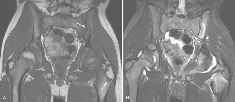
  • With the exception of the hip and sacroiliac joints, a majority of the observed arthritis is peripheral. The most common joints to be affected include the ankles, knees, and hips.
  • The observed arthritis is typically asymmetric and typically effects fewer than 4 joints.
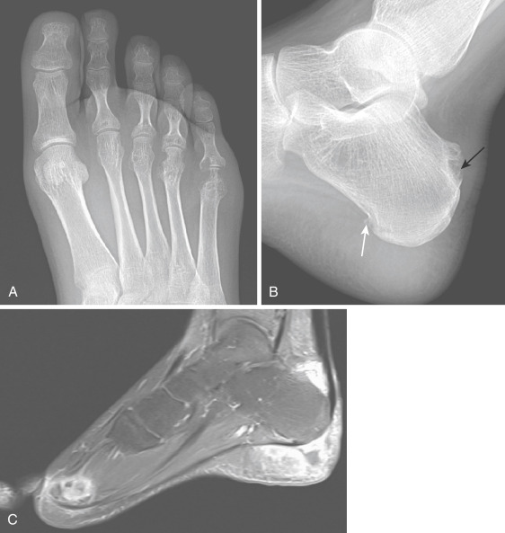
  • While pain and inflammation about the bones of the midfoot are less common, such symptoms are highly suggestive of the disease.
  • As the disease process progresses many patients go on to develop clinical and radiologic evidence of spinal and sacroiliac joint involvement.
  • Enthesitis is typically asymmetric and more frequently occurs in the lower extremities.
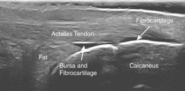
  • Enthesitis most frequently occurs at inferior pole of the patella, the plantar fascia insertion at the calcaneus, and finally the Achilles tendon insertion at the calcaneus. More chronic enthesitis may demonstrate evidence of erosion, calcifications, and heterotopic bone formation on radiographs.
  • The anterior uveitis observed with ERA is typically acute in onset and manifests with conjunctival injection, pain, and light sensitivity.
Etiology

The etiology of ERA is currently unknown; however, the strong association with HLA B27 suggests a genetic component of the disease.

Diagnosis

Differential Diagnosis

  • Other forms of JIA
  • Infectious arthritis
  • Mixed connective tissue disease
  • Leukemia
  • Reiter’s syndrome
  • Other systemic arthritides
Workup

  • A thorough history and physical exam are required to diagnose ERA.
  • A focused history should include a history of joint and/or enthesitis, back pain, ocular symptoms, and bowel symptoms. One should ensure that the patient does not have a personal or family history of psoriasis as this would exclude the diagnosis of ERA.
  • A focused physical exam should be performed to assess for the presence of spinal disease, sacroiliac pain and or instability, joint pain, and enthesitis.
Laboratory Tests

  • CBC, erythrocyte sedimentation rate, C-reactive protein, HLA B27
  • Antinuclear antibodies, rheumatoid factor, and anticyclic citrullinated peptide antibodies are all typically absent; however, they are used to exclude other differential diagnoses.
Imaging Studies

  • X-ray (Fig. E1) can be used to detect any evidence of arthritic changes (Box 1).
  • MRI (Fig. E2) or ultrasound (Fig. E3) may be used to detect and/or confirm sacroiliitis or enthesitis, which can be frequently missed on physical exam in the early stages of the disease.

BOX E1 Radiographic Features of Enthesitis-Related Arthritis and Spondyloarthropathies

Peripheral Joints

Asymmetric involvement of large lower limb joints

Involvement of interphalangeal joint of the hallux

New bone at the margins of erosions

Affected joints-show swelling, effusion, epiphyseal overgrowth, erosions, osteopenia, cartilage space narrowing, and, rarely, fusion

Dactylitis-swelling and periosteal new bone of fingers or toes

Periosteal new bone-e.g., metatarsals, proximal femur

Entheses

Especially tibial tubercle and posterior aspect of calcaneus

Swelling, erosion, new bone formation

Sacroiliitis

Radiographic changes generally delayed until late teens

Asymmetric involvement may occur early, then become symmetric

Erosions occur first on the iliac side of sacroiliac joint

Pseudowidening occurs from erosion

Sclerosis and finally ankylosis develop

From Petty RE et al: Textbook of pediatric rheumatology, ed 8, Philadelphia, 2021, Elsevier.

Figure E1 A 17-yr-old girl with enthesitis-related arthritis.

A, The forefoot x-ray examination shows joint space narrowing and extensive bone erosions involving multiple metatarsophalangeal joints, especially the fifth metatarsophalangeal joint. B, A spur is noted in the hindfoot x-ray examination at the insertion of the plantar fascia (white arrow) and erosive changes at the calcaneal tendon insertion (black arrow). C, Sagittal contrast-enhanced fat-suppressed T1-weighted magnetic resonance imaging shows intense enhancement adjacent to the calcaneal tendon, heel, and metatarsophalangeal joint.

From Petty RE et al: Textbook of pediatric rheumatology, ed 8, Philadelphia, 2021, Elsevier.

Figure E2 An 11-yr-old boy with enthesitis-related arthritis.

A, Magnetic resonance imaging shows bilateral hip joint effusions and abnormal bone signal adjacent to the left sacroiliac joint and left greater trochanter (arrows). Whereas the coronal T1-weighted image (A) shows low signal in these regions (arrows), the coronal short tau inversion recovery image (B) shows increased signal intensity.

From Petty RE et al: Textbook of pediatric rheumatology, ed 8, Philadelphia, 2021, Elsevier.

Figure E3 A Longitudinal Scan of the Posterior Heel with the Achilles Tendon Insertion Shows the Various Structures of the Enthesis, Including Fibrocartilage, Retrocalcaneal Fat Pad and Bursa, and the Tendon with its Enthesis

From Petty RE et al: Textbook of pediatric rheumatology, ed 8, Philadelphia, 2021, Elsevier.

Treatment

Nonpharmacologic Therapy

Physical therapy, heat therapy, and cold therapy can be used to address the musculoskeletal pain associated with the disease.

Acute General Rx

Acute treatment typically includes monotherapy or combination therapy with NSAIDs and methotrexate/sulfasalazine

Chronic Rx

If patients are not able to achieve control, of their symptoms with the above regimen, antitumor necrosis factor (anti-TNF) medications such as etanercept and adalimumab

Referral

ERA should be managed by a multidisciplinary team including a referral to physical therapy and a certified rheumatologist.

Related Content

Juvenile Idiopathic Arthritis (Related Key topic)

Related Content

  1. Ferjani H.L. : Enthesitis-related arthritis and spondylarthritis: the same disease or disparate entities?Expert Rev Clin Immunol. :1-7, 2021.doi:10.1080/1744666X.2022.2010547