section name header

Basic Information

AUTHOR: Fred F. Ferri, MD

Definition

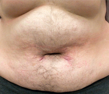
Hirsutism is the development of stiff, pigmented (terminal) facial and body hair (male distribution) in women (Fig. E1) as a result of excess androgen production.

Figure E1 Hirsutism.

From Lebwohl MG et al: Treatment of skin disease, comprehensive therapeutic strategies, ed 6, Philadelphia, 2022, Elsevier.

Synonym

Excessive hair growth

ICD-10CM CODE
L68.0Hirsutism
Epidemiology & Demographics

  • Overall prevalence unknown, estimated 5% to 10% in reproductive age women.
  • Race and genetics should be considered. Some distinct ethnic populations have minimal body hair and others (Mediterranean, Middle Eastern, South Asian) have moderate to large amounts of body hair while serum androgen levels are similar.
  • Social norms and culture also determine how much body hair is cosmetically acceptable.
  • Half of all cases of mild hirsutism do not have hyperandrogenemia. “Patient-important hirsutism” refers to hirsutism causing woman sufficient distress to seek care.
  • Incidence and presentation of hirsutism is dependent on underlying cause of androgen excess (see “Differential Diagnosis”).
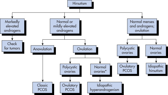
  • Most women with hirsutism have polycystic ovary syndrome (PCOS). PCOS accounts for 95% of cases of hirsutism.
Physical Findings & Clinical Presentation

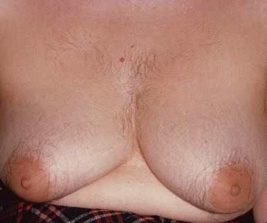
  • Timing of symptoms: Abrupt onset, short duration, rapid progression, progressive worsening, more severe signs of virilization (Fig. E2), or later age of onset suggest androgen-producing tumor, late-onset congenital adrenal hyperplasia, or Cushing syndrome. Weight increases may produce increased androgen production.
  • Menstrual history: Menarche, cycle regularity and symptoms of ovulation, fertility, and contraception use. Anovulatory cycles are the most common underlying cause of androgen excess.
  • Medication use history: Some drugs cause hirsutism or produce androgenic effects (danazol, phenytoin, valproic acid, androgenic progestins [e.g., norgestrel], cyclosporin, minoxidil, metoclopramide, phenothiazines, methyldopa, diazoxide, and penicillamine).
  • Family history: Known or suspected family history of hirsutism, congenital adrenal hyperplasia, insulin resistance, PCOS, infertility, obesity, menstrual irregularity may be found.
  • Physical exam reveals deepening voice, body habitus, increased muscle mass, galactorrhea; abdominal and pelvic exam.
  • Associated cutaneous manifestations are acne, acanthosis nigricans, striae, hair distribution, location and quantity, frontotemporal balding, muscle mass, clitoromegaly.
  • Ferriman-Gallwey scale, a simple, pictorial system of scoring nine body areas, is the most common tool used to quantify hirsutism. It may be unreliable in non-Caucasian women of other ethnicities.

Figure E2 Hirsutism.

From James WD et al: Andrews’ diseases of the skin, ed 12, Philadelphia, 2016, Saunders.

Etiology

  • Presence of hirsutism indicates androgen excess. Total testosterone may be normal, but free testosterone is elevated.
  • Androgens induce vellus hair follicles (soft, unpigmented hair) in sex-specific areas (upper lip, chin, midsternum, upper abdomen, back, buttocks) to develop into thicker, more heavily pigmented terminal hairs.
  • Anovulatory ovaries are usual source of excess androgens through thecal cell steroidogenesis and conversion of androstenedione to testosterone. The most common cause of hirsutism is polycystic ovary syndrome, which accounts for three out of every four cases.
  • Conditions that decrease hepatic production of sex hormone binding globulin (SHBG) decrease protein-bound testosterone and increase free testosterone fraction (e.g., low estrogen, high androgen, and hyperinsulinemic states).
  • Late-onset, congenital adrenal hyperplasia enzyme deficiency (most commonly 21-hydroxylase deficiency) produces excess 17 hydroxyprogesterone (17-OHP) and overproduction of androstenedione.
  • Rare ovarian tumors primarily derived from Sertoli-Leydig cells, granulosa theca cells, or hilus cells produce excess androgens.
  • Rare adrenal tumors produce excess androgens.
  • Rare pituitary or hypothalamic tumors produce excess prolactin and can lead to anovulation.
  • Box E1 summarizes causes of androgen excess in women of reproductive age.

BOX E1 Causes of Androgen Excess in Women of Reproductive Age

Ovarian

Polycystic ovary syndrome (PCOS)

Hyperthecosis (a severe PCOS variant)

Ovarian tumor (e.g., Sertoli-Leydig cell tumor)

Adrenal

Nonclassic adrenal hyperplasia

Cushing syndrome

Glucocorticoid resistance

Adrenal tumor (e.g., adenoma, carcinoma)

Specific Conditions of Pregnancy

Luteoma of pregnancy

Hyperreaction luteinalis

Aromatase deficiency in fetus

Other

Hyperprolactinemia, hypothyroidism

Medications (danazol, testosterone, anabolizing agents)

Idiopathic hirsutism (normal serum testosterone in an ovulatory woman)

Idiopathic hyperandrogenism (patients who do not fall into any of the other categories listed)

From Melmed S et al: Williams textbook of endocrinology, ed 12, Philadelphia, 2011, Saunders.

Diagnosis

Differential Diagnosis (Table E1

  • Androgen-independent vellus hair: Soft, unpigmented hair that covers entire body
  • Hypertrichosis: Diffusely increased total body hair (vellus or lanugo-type) not restricted to androgen-dependent areas often an adverse response to a medication or systemic illness (e.g., anorexia nervosa, porphyria, malnutrition, hypothyroidism)
  • PCOS 75%
  • Idiopathic 5% to 15%
  • Congenital adrenal hyperplasia 1% to 8%
  • Insulin resistance syndrome 3% to 4%
  • Cushing syndrome <1%
  • Drug induced <1%
  • Ovarian tumor <1%
  • Adrenal tumor <1%
  • Hyperthecosis <1%
  • Hyperprolactinemia <1%

TABLE E1 Differential Diagnosis of Hirsutism and Virilization

SourceDiagnosis
NonspecificExogenous, iatrogenic
Abnormal gonadal or sexual development
PregnancyAndrogen excess in pregnancy, luteoma or hyperreactio luteinalis
PeripheryIdiopathic hirsutism
OvaryPolycystic ovary syndrome
Functional or idiopathic hyperandrogenism
Stromal hyperthecosis
Ovarian tumors
Adrenal glandAdrenal tumors
Cushing syndrome
Adult-onset congenital adrenal hyperplasia

PCOS, Polycystic ovary syndrome.

Idiopathic hirsutism and polycystic ovary syndrome do not present with virilization.

The hyperandrogenism in PCOS can also be of adrenal origin, at least in part.

Functional hyperandrogenism may well be a type of PCOS, but without clearly defined polycystic ovaries on ultrasound, and can also have an adrenal source of hyperandrogenism.

From Gershenson DM et al: Comprehensive gynecology, ed 8, Philadelphia, 2022, Elsevier.

Workup

  • Hirsutism is a clinical diagnosis. Fig. E3 is an algorithm for the diagnosis of hirsutism.
  • Management of hirsutism is largely independent of the etiology.
  • Workup in selected hirsute women is directed to determine underlying cause of androgen excess.
  • See specific conditions for more detailed workup of individual diagnoses.

Figure E3 An algorithm for the diagnosis of hirsutism.

Note that women with ovulatory polycystic ovary disease (PCOS) need only have hirsutism and can have “normal” androgen levels; also the ultrasound diagnosis of polycystic ovaries is not always accurate.

From Stanczyk CE, Lobo RA: Evaluation of hormonal status. In: Strauss JF, Barbieri RL (eds): Yen and Jaffe’s Reproductive Endocrinology, ed 8, Philadelphia, 2019, Elsevier, p. 899.

Laboratory Tests

Establishing laboratory evidence of excess androgens in women with moderate or severe hirsutism, sudden onset, rapid progression, or associated menstrual dysfunction, central obesity, clitoromegaly, or acanthosis nigricans is an approach consistent with guidelines from the Endocrine Society, the American College of Obstetricians and Gynecologists, the Androgen Excess and Polycystic Ovary Syndrome Society, and the American Association of Clinical Endocrinologists.

  • Total plasma testosterone (normal range 20-60 ng/dl [0.69-2.1 nmol/L]) or free testosterone: Early morning on day 4 to 10 of menstrual cycle to screen for testosterone-secreting tumors. If moderately or markedly elevated (total testosterone >150 ng/dl [5.2 nmol/L], free testosterone >2 ng/dl [0.07 nmol/L]) may image adrenals and ovaries for androgen-secreting tumors.

Other laboratory test considerations if appropriate:

  • Prolactin: Moderately elevated values should prompt imaging of pituitary-hypothalamic region.
  • 17-OHP (17 α-hydroxyprogesterone): Screen for adrenal enzyme deficiencies. Morning value >200 ng/dl in early follicular phase suggests nonclassic (late onset) congenital adrenal hyperplasia due to 21-hydroxylase deficiency and may be confirmed with high-dose (250 mcg) ACTH stimulation test.
  • Thyroid-stimulating hormone (TSH): Rule out hypothyroidism.
  • Dehydroepiandrosterone sulfate (DHEA-S): Screen for adrenal androgen production as almost entirely produced by adrenals. Levels >700 mcg/dl (13.6 nmol/L) raise suspicion for adrenal androgen-secreting tumor.

Additional laboratory test considerations if appropriate:

  • Follicle-stimulating hormone (FSH): Rule out hypoestrogenic state (perimenopausal)
  • Luteinizing hormone (LH): Typically elevated in PCOS with low or normal FSH
  • 24-h urinary free cortisol: Rule out Cushing syndrome and overproduction of cortisol
  • Overnight single-dose dexamethasone suppression test: Rule out Cushing syndrome and adrenal hyperfunction
  • Fasting blood sugar (FBS), 2-h 75-g oral glucose tolerance test, fasting insulin levels: Rule out insulin resistance syndrome
  • Table E2 summarizes laboratory tests for the differential diagnosis of androgen excess

TABLE E2 Laboratory Tests for the Differential Diagnosis of Androgen Excess

Initial Testing
Total testosterone
Prolactin
Thyroid-stimulating hormone
Further Testing Based on Clinical Presentation
17-Hydroxyprogesterone (8:00 A.M.)
17-Hydroxyprogesterone 60 min after IV ACTH
Cortisol (8:00 A.M.) after 1 mg dexamethasone at midnight
DHEAS
Androstenedione
Imaging of ovaries (transvaginal ultrasonography)
Imaging of adrenals (abdominal ultrasonography, CT, MRI)
Nuclear imaging after IV administration of radiolabeled cholesterol

ACTH, Adrenocorticotropic hormone; CT, computed tomography; DHEAS, dehydroepiandrosterone sulfate; IV, intravenous; MRI, magnetic resonance imaging.

From Melmed S et al: Williams textbook of endocrinology, ed 12, Philadelphia, 2011, Saunders.

Imaging Studies

Imaging study considerations if appropriate:

  • Pelvic ultrasound (high resolution, transvaginal): Rule out ovarian tumor if total testosterone is elevated.
  • Abdominal computed tomography (CT)/MRI: Rule out adrenal tumor if elevated DHEAS.
  • Pituitary-hypothalamic region CT/MRI: Rule out pituitary tumor if prolactin elevated.
  • Laparoscopy/laparotomy: Rule out small ovarian tumor in cases of elevated testosterone levels without radiologic evidence of adrenal or ovarian pathology.

Treatment

Nonpharmacologic Therapy

  • Weight reduction: Can reduce androgen production indirectly by reducing insulin-stimulated theca cell androgen production and improve menstrual function, and slow hair growth in obese women
  • Cosmetic: Temporary:
    1. Shaving: Does not stimulate hair growth; lasts days, leaves stubble
    2. Epilation: Electronic plucking
    3. Bleaching: Removes hair pigment. May cause skin irritation
    4. Mechanical waxing/plucking
    5. Depilatories: Gels, lotions, or creams that chemically disrupt sulfide bonds of hair causing dissolution of hair shaft. No stubble
    6. Photoepilation (laser and intense pulsed light [IPL]): Hair follicles destroyed by wavelengths of light absorbed by melanin. Good for pigmented hair; laser treatment is more effective than shaving, waxing, and electrolysis. It lasts 3 to 6 mo as vellus follicles remain and can be converted to terminal pigmented hair under excess androgens
  • Cosmetic: Permanent:
    1. Electrolysis: Destroys individual hair follicles. May be expensive and time consuming
Acute General Rx

See “Pharmacologic Therapy.”

Chronic Rx

See “Pharmacologic Therapy.”

Pharmacologic Therapy

  • Usually second-line treatment following nonpharmacologic, physical methods of hair control, and in consideration of patient’s comorbidities and risk factors, patient preferences, area of excess hair amenable to treatment, and access and affordability of treatments
  • Pharmacologic treatments categorized as topical, oral contraceptive pills (OCPs), antiandrogens (potential adverse effects on a developing male fetus, so use with reliable contraception), other treatments directed at specific underlying etiology
  • Topical: Eflornithine topical cream 13.9%: Unclear mechanism of action; may inhibit ornithine decarboxylase, retarding hair growth. Temporary cosmetic treatment for facial hair. Applied directly to unwanted facial hair bid with at least 8-h spaced applications. Does not remove hair, rather slows growth. Slow response over 4 to 8 wk. Hair growth returns upon discontinuation of treatment
  • OCPs: Suppress ovarian steroidogenesis and LH through low-dose estrogen and low androgenic progestational agents. Slow response to treatment. Suppresses new hair growth. Established hair unaffected. Low-dose OCPs with low androgenic progestational agents, for example, desogestrel, drospirenone, norgestimate. Avoid norgestrel and levonorgestrel (higher androgenic progestational agents)
  • Antiandrogens: Spironolactone: When OCPs unacceptable or may be added for disappointing results after 6 mo of OCP treatment:
    1. Aldosterone-antagonist diuretic inhibits adrenal and ovarian biosynthesis of androgens. May result in ovulation, so consider contraception needs
    2. Slow response usually 6 mo or more
    3. 200 mg PO qd, then decrease to 25 to 50 mg qd maintenance
    4. May cause hyperkalemia
    5. Anovulatory, unopposed estrogen states require progestin management
Referral

  • To endocrinologist if difficulty in determining diagnosis, achieving therapeutic goals, or resistant to first-line therapies. Prepubertal and postmenopausal hirsutism is suspicious for neoplastic or secondary endocrine causes and should be referred for further evaluation
  • Consider referral or consultation for following therapies:
    1. Finasteride: Antiandrogen, in hair follicle blocks 5α-reductase conversion of testosterone to intranuclearly active 5α-dihydrotestosterone (DHT):
      1. Use only with reliable contraception because DHT necessary for normal male fetus urogenital development
      2. Not FDA approved for treatment of hirsutism
      3. 1 to 5 mg PO qd
    2. Flutamide: Inhibits androgen uptake and receptor binding:
      1. Not recommended by Endocrine Society Clinical Practice Guidelines and not FDA approved for treatment of hirsutism, but used by some European endocrinologists
      2. Use only with reliable contraception
      3. Reserved for women with severe, resistant hirsutism because of risk of hepatic dysfunction
    3. Cyproterone acetate: Antiandrogen that competes with DHT for binding androgen receptors. Used as progestin component of OCPs outside the U.S.
  • Other treatments directed at specific underlying etiology:
    1. Metformin/thiazolidinediones: Therapy reserved for documented insulin-resistant states
    2. GnRH agonists: Recommended only in women with severe hyperandrogenemia (e.g., ovarian hyperthecosis) with suboptimal response to combination low-dose estrogen/progestin pills and antiandrogen treatment. Inhibits gonadotropin and consequently ovarian androgen and estrogen secretion
    3. Dexamethasone: Adrenal glucocorticoid suppression is reserved for diagnosis of adrenal enzyme deficiency
    4. Total abdominal hysterectomy/bilateral salpingo-oophorectomy reserved for recalcitrant hirsutism in older female with hyperthecosis and undesired fertility

Pearls & Considerations

Comments

  • Hirsutism is both an endocrine and cosmetic problem for patients.
  • Ovulation induction therapy is indicated in women desiring pregnancy.
  • Delay checking serum androgens until oral contraceptives have been discontinued for 2 to 3 mo.
  • Evaluation of incidental adrenal mass is always warranted.
Related Content

Hirsutism (Patient Information)

Polycystic Ovary Syndrome (Related Key Topic)