section name header

Basic Information

AUTHOR: Fred F. Ferri, MD

Definition

Hypopituitarism (from the Latin pituita, meaning “phlegm”) is the deficiency of one or more of the hormones of the anterior or posterior pituitary gland resulting from diseases of the hypothalamus or pituitary gland. Panhypopituitarism indicates the loss of all the pituitary hormones but is often used in clinical practice to describe patients deficient in growth hormone (GH), gonadotropins, corticotropin, or thyrotropin in whom posterior pituitary function remains intact.

Synonyms

Panhypopituitarism

Pituitary insufficiency

ICD-10CM CODES
E23.0Hypopituitarism
E23.1Drug-induced hypopituitarism
E89.3Postprocedural hypopituitarism
Epidemiology & Demographics

Incidence of 4.2 cases/100,000 persons

Physical Findings & Clinical Presentation

Symptoms depend on type of onset, number and severity of hormone deficiencies, their target organs, and age of onset (Table 1). Physiologic regulators of pituitary hormone secretion are summarized in Table 2.

  • Mass effect of a pituitary tumor can cause headaches and visual disturbances (typically as bitemporal hemianopsia).
  • Rhinorrhea.
  • Corticotropin deficiency:
    1. Fatigue and weakness, no appetite, abdominal pain, nausea, vomiting, failure to thrive in children, and hyponatremia. If the onset is abrupt, hypotension and shock.
  • Thyrotropin deficiency:
    1. Fatigue and weakness, weight gain, cold intolerance, anemia, constipation
    2. Bradycardia, hung-up reflexes, pretibial edema, change in voice, and hair loss
  • Gonadotropin deficiency:
    1. Loss of libido, erectile dysfunction, amenorrhea, hot flashes, dyspareunia, infertility, gynecomastia, decreased muscle mass, and anemia
  • GH deficiency:
    1. Growth retardation in children
    2. Easy fatigue, hypoglycemia
    3. Lean mass is reduced and fat mass is increased, leading to obesity
    4. Decreased bone mineral density, increased low-density lipoprotein cholesterol, obesity, increased inflammatory cardiovascular markers (interleukin-6 and C-reactive protein)
  • Hyperprolactinemia:
    1. Galactorrhea, hypogonadism, inability to lactate after delivery
    2. Posterior pituitary (vasopressin; antidiuretic hormone [ADH] deficiency): Diabetes insipidus with polyuria, polydipsia, nocturia, hypotension, and dehydration

TABLE 1 Clinical Effects of Pituitary Hormone Deficiencies

In panhypopituitarism, all are combined, although diabetes insipidus (ADH deficiency) is often absent
HormoneContextSymptoms and Signs
ACTHAcuteFatigue, weakness, hypotension, weakness, vomiting
ChronicAs in Addison disease, except lack of pigmentation, electrolyte disturbance and hypovolemia
Hypoglycemia, weight loss
TSHChildrenGrowth retardation
AdultsAs for primary hypothyroidism, thyroid atrophy
GHChildrenGrowth retardation, increased adiposity
AdultsExercise capacity, lean mass, fat mass, cardiovascular risk
GonadotropinsChildrenFailure of sexual maturation, lack of pubertal growth spurt
MenAs for primary hypogonadism, testicular atrophy, infertility
WomenSecondary amenorrhea, infertility, osteoporosis
ProlactinWomenFailure of lactation
Vasopressin (ADH)Polyuria, dilute urine, thirst, nocturia, hypernatremia

ACTH, Adrenocorticotropic hormone; ADH, antidiuretic hormone; GH, growth hormone, TSH, thyroid-stimulating hormone.

From Talley NJ et al: Essentials of internal medicine, ed 4, Chatswood, NSW, 2021, Elsevier Australia.

TABLE E2 Physiologic Regulators of Pituitary Hormone Secretion

HormoneStimulatorInhibitor
Adrenocorticotropic hormone (ACTH)Corticotrophin-releasing hormone (CRH)
Thyroid-stimulating hormone (TSH)Thyrotropin-releasing hormone (TRH)
Luteinizing hormone/follicle-stimulating hormone [LH/FSH)Gonadotropin-releasing hormone (GnRH)Prolactin
Growth hormone (GH)Growth-hormone-releasing hormone (GHRH)
Ghrelin
Somatostatin
Prolactin (PRL)TRHDopamine
Etiology

It can be congenital or acquired:

  • Congenital: Mutations in transcription factors produce multiple hormonal deficiencies. Mutations in genes produce single hormonal deficiency.
  • Acquired (Table 3): The result of destruction of pituitary cells caused by:
    1. Pituitary apoplexy: Hemorrhage or infarction of the pituitary gland. Predisposing factors include diabetes mellitus, anticoagulation therapy, head trauma, and radiation therapy. Sheehan syndrome: Postpartum necrosis, a rare complication after pregnancy.
    2. Infiltrative disease, including sarcoidosis, hemochromatosis, histiocytosis X, Wegener granulomatosis, lymphocytic hypophysitis, and infection of the pituitary (tuberculosis, mycosis, syphilis).
    3. Primary empty sella syndrome: Flattening of the pituitary gland caused by extension of the subarachnoid space and filling of cerebrospinal fluid into the sella turcica.
    4. Pituitary tumors: Classified by size (microadenomas, <10 mm; macroadenomas, >10 mm) and function. Prolactin-secreting tumors and nonfunctioning tumors account for the majority of pituitary adenomas.
    5. Suprasellar tumors: Craniopharyngiomas are the most common.

TABLE 3 Causes of Acquired Pituitary Insufficiency

Traumatic
  • Surgical resection
  • Radiation damage
  • Traumatic brain injury
Infiltrative/Inflammatory
  • Primary hypophysitis
  • Lymphocytic
  • Granulomatous
  • Xanthomatous
  • Secondary hypophysitis
  • Sarcoidosis
  • Langerhans cell histiocytosis
  • Infections
  • Granulomatosis with polyangiitis
  • Takayasu disease
  • Hemochromatosis
Infections
  • Tuberculosis
  • Pneumocystis jirovecii infection
  • Fungal (histoplasmosis, aspergillosis)
  • Parasites (toxoplasmosis)
  • Viral (cytomegalovirus)
Vascular
  • Pregnancy related
  • Aneurysm
  • Apoplexy
  • Diabetes
  • Hypotension
  • Arteritis
  • Sickle cell disease
Neoplastic
  • Pituitary adenoma
  • Parasellar mass
  • Rathke cyst
  • Dermoid cyst
  • Meningioma
  • Germinoma
  • Ependymoma
  • Glioma
  • Craniopharyngioma
  • Hypothalamic hamartoma, gangliocytoma
  • Pituitary metastatic deposits
  • Hematologic malignancy
  • Leukemia
  • Lymphoma
Functional
  • Nutritional
  • Caloric restriction
  • Malnutrition
  • Excessive exercise
  • Critical illness
  • Acute illness
  • Chronic renal failure
  • Chronic liver failure
  • Hormonal
  • Hyperprolactinemia
  • Hypothyroidism
  • Drugs
  • Anabolic steroids
  • Glucocorticoid excess
  • GnRH agonists
  • Estrogen
  • Dopamine
  • Somatostatin analog
  • Thyroid hormone excess

GnRH, Gonadotropin-releasing hormone.

Modified from Kaiser U, Ho KKY: Pituitary physiology and diagnostic evaluation. In Melmed S et al (eds): Williams textbook of endocrinology, ed 13, Philadelphia, 2016, Elsevier; and Kliegman RM: Nelson textbook of pediatrics, ed 21, Philadelphia, 2020, Elsevier, Table 8.5, p. 193.
Diagnosis

The diagnosis of hypopituitarism is suspected by clinical history and physical findings and is established by blood tests to confirm the presence of hormone deficiency.

Differential Diagnosis

The differential diagnosis is as outlined under “Etiology.”

Workup

Includes baseline determination of each anterior pituitary hormone followed by dynamic provocative stimulation tests, radiograph imaging, and formal visual field testing. Table 4 summarizes testing for assessment of anterior pituitary function.

TABLE 4 Assessment of Anterior Pituitary Function

TestDoseNormal ResponseSide Effects
ACTH
Insulin tolerance0.1-0.15 U/kg IVPeak cortisol response >18 μg/dl, or increase by 7 μg/dlSweating, palpitation, tremor
MetyraponeOral administration of 30 mg/kg at 11 P.M.Peak 11-DOC 7 μg/dl
Peak cortisol 7 μg/dl
Peak ACTH >75 pg/ml
Nausea, insomnia, adrenal crisis
CRH stimulation100 μg IVPeak ACTH two-fold to four-fold
Peak cortisol 20 μg/dl or 7 μg/dl
Flushing
ACTH stimulation250 μg IV or IM or 1 μg IVPeak cortisol 20 μg/dlRare
TSH
Serum T4 (free T4)
Total T3
TSH-third generation
TRH stimulation
200-500 μg IVPeak TSH 2.5-fold or 5-6 mU/L (females), 2-3 mU/L (males)Flushing, nausea, urge to micturate
PRL
Serum PRL
TRH stimulation
200-500 μg IVPRL 2.5-foldFlushing, nausea, urge to micturate
LH/FSH
Serum LH and FSH
Serum testosterone
GnRH stimulation
100 μg IVElevated in menopause and in men with primary testicular failure
300-900 ng/ml (age-adjusted normal ranges)
LH two-fold to three-fold, or by 10 IU/LFSH 1.5-2-fold, or by 2 IU/L
Rare
GH
Insulin tolerance0.1-0.15 U/kgGH peak >5 μg/LSweating, palpitation, tremor
Glucagon1-1.5 mg IMGH peak >3 μg/LNausea, headaches
L-Arginine plus GHRH0.5 g/kg (max 30 g)
IV over 30 min
Peak GH >9 μg/LNausea
GHRH1 μg/kgFlushing

ACTH, Adrenocorticotropic hormone; CRH, corticotropin-releasing hormone; 11-DOC, 11-deoxycorticosterone; FSH, follicle-stimulating hormone; GH, growth hormone; GHRH, growth hormone-releasing hormone; GnRH, gonadotropin-releasing hormone; IM, intramuscular; IV, intravenous; LH, luteinizing hormone; PRL, prolactin; T3, triiodothyronine; T4, thyroxine; TSH, thyroid-stimulating hormone; TRH, thyrotropin-releasing hormone.

From Melmed S et al: Williams textbook of endocrinology, ed 14, Philadelphia, 2019, Elsevier.

Laboratory Tests

  • Corticotropin deficiency:
    1. The presence of a 9:00 A.M. cortisol level >20 mcg/dl or <4 mcg/dl usually confirms sufficiency or deficiency, respectively.
    2. Corticotropin stimulation test using 250 mcg of corticotropin given intravenous (IV) and measuring serum cortisol before and 30 and 60 min after administration. A normal response is an increase in serum cortisol level >20 mcg/dl.
    3. With pituitary disease these test results may be indeterminate, and more dynamic testing such as an insulin-tolerance or metyrapone test may be necessary.
  • Thyrotropin deficiency:
    1. Thyroid-stimulating hormone (TSH) and free T4 measurements
    2. Primary hypothyroidism shows elevated TSH with low free T4. Secondary hypothyroidism shows normal or low TSH with low free T4 and low T3 resin uptake.
  • Gonadotropin deficiency:
    1. Follicle-stimulating hormone (FSH), luteinizing hormone (LH), estrogen, and testosterone measurements.
    2. In men, hypogonadotropic hypogonadism is seen with low testosterone levels and normal or low FSH and LH levels (ideally measured at 9:00 A.M. because of diurnal rhythm). Check free testosterone if patient is obese.
    3. In premenopausal women with amenorrhea, low estrogen with normal or low FSH and LH levels is typically seen.
  • GH deficiency:
    1. Insulin-induced hypoglycemia stimulation test using 0.1 to 0.15 unit/kg regular insulin given IV and measuring GH 30, 60, and 120 min after administration. A normal response is a GH level >3 mcg/dl. This test is contraindicated in seizure disorder or ischemic heart disease.
    2. Combination of GH-releasing hormone plus arginine is an alternative test, with a diagnostic threshold of 9 mcg/L.
    3. Because the relation between serum insulin-like growth factor (IGF)-1 and GH levels blurs with age, a normal serum IGF-1 does not exclude the diagnosis in older adults.
  • Hyperprolactinemia: Prolactin levels may be elevated in prolactin-secreting pituitary adenomas.
  • Vasopressin deficiency:
    1. Urinalysis shows low specific gravity.
    2. Urine osmolality is low.
    3. Serum osmolality is high.
    4. Fluid deprivation test over 18 h with inability to concentrate the urine.
    5. Serum vasopressin level is low.
    6. Electrolytes may show hyponatremia and exclude hyperglycemia.
Imaging Studies

  • Imaging is the first step in identifying an underlying cause.
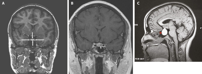
  • MRI (Fig. E1) is more sensitive than computed tomography (CT) in visualizing the pituitary fossa, sella turcica, optic chiasm, pituitary stalk, and cavernous sinuses. It is also more sensitive in detecting pituitary microadenomas. CT with contrast can be used if MRI is not available.
  • Surveillance scan at baseline and 12 mo thereafter depending on protocol and clinical symptoms.

Figure E1 Magnetic Resonance Images of Normal Pituitary (A), Microadenoma (B), and Macroadenoma (C)

From (A) MacMaster FP et al: Effect of antipsychotics on pituitary gland volume in treatment-naïve first-episode schizophrenia: a pilot study, Schizphr Res 92(1-3):207-210, 2007. (B) Torigian DA et al: The role of CT, MR imaging and ultrasonography in endocrinology, PET Clin 2007;2(3):395-408, 2007. (C) Klatt EC: Robbins & Cotran atlas of pathology, ed 4, Philadelphia, 2021, Elsevier.

Treatment

Three-fold: Removing underlying cause (surgery or radiation), treating hormonal deficiencies, and addressing any other repercussions from deficiency. Table 5 summarizes replacement therapy for adult hypopituitarism.

TABLE 5 Replacement Therapy for Adult Hypopituitarisma

Deficient HormoneTreatment
ACTHHydrocortisone, 10-20 mg/day in divided doses
Cortisone acetate, 15-25 mg/day in divided doses
TSHl-Thyroxine, 0.05-0.2 mg/day according to T4 levels
FSH/LHMales:
Testosterone enanthate, 200 mg IM every 2-3 wk
Testosterone undecanoate, 1000 mg IM every 3-6 mo
Testosterone skin patch, 2.5-5.0 mg/day-can increase dose up to 7.5 mg/day
Testosterone gel, 5-10 g/day (delivering 50-100 mg/day)
For fertility: hCG three times weekly, or hCG + FSH or menopausal gonadotropin or GnRH
Females (nonoral route is recommended):
Estradiol skin patch, 4-8 mg, twice weekly
Estradiol gel
Conjugated estrogen, 0.65 mg/day
Micronized estradiol, 1 mg/day
Estradiol valerate, 1-2 mg
Piperazine estrone sulfate, 1.25 mg
All of the estrogens are administered with progesterone or progestin sequentially or in combination if uterus present
For fertility: Menopausal gonadotropin and hCG or GnRH
GHAdults: Somatotropin, 0.2-1.0 mg/day SC Children: Somatotropin, 0.02-0.05 mg/kg/day
VasopressinIntranasal desmopressin, via rhinal tube, 5-20 μg twice daily
Oral DDAVP, 300-600 μg/day, usually in divided doses

ACTH, Adrenocorticotropic hormone; DDAVP, desmopressin acetate; FSH, follicle-stimulating hormone; GH, growth hormone; GnRH, gonadotropin-releasing hormone; hCG, human chorionic gonadotropin; IM, intramuscularly; LH, luteinizing hormone; SC, subcutaneously; T4, thyroxine; TSH, thyroid-stimulating hormone.

a Doses shown should be individualized and reassessed during stress, surgery, or pregnancy.

From Melmed S et al: Williams textbook of endocrinology, ed 14, Philadelphia, 2019, Elsevier.

Nonpharmacologic Therapy

  • IV fluid resuscitation, correction of electrolyte and metabolic abnormalities with potassium bicarbonate, and oxygen therapy.
  • Transsphenoidal surgery for tumors causing specific symptoms.
  • Radiation or stereotactic radiosurgery (“gamma knife”) for medically unresponsive, surgically unresectable tumors and tumors for which other modalities are contraindicated. It is both safe and effective for recurrent or residual pituitary adenomas.
Acute General Rx

Acute situations such as adrenal crisis or myxedema coma can occur in untreated hypopituitarism and should be treated accordingly with IV corticosteroids (e.g., hydrocortisone 100- to 250-mg bolus followed by hydrocortisone 100 mg IV q6h for 24 h) and levothyroxine (e.g., 5-8 mcg/kg IV over 15 min, then 100 mcg IV q24h).

Chronic Rx

Treatment is lifelong:

  • Adrenocorticotropic hormone (ACTH) deficiency: Hydrocortisone 10 mg orally (PO) every morning and 5 mg PO every evening or prednisone 5 mg PO every morning and 2.5 mg PO every evening. Dexamethasone or prednisone is often preferred because of longer duration of action.
  • LH and FSH deficiency:
    1. In men, testosterone enanthate or propionate 200 to 300 mg intramuscular (IM) every 2 to 3 wk, or transdermal testosterone scrotal patches can be tried.
    2. In women who are not interested in fertility, conjugated estrogen 0.3 to 1.25 mg/day and held the last 5 to 7 days of each mo with the addition of medroxyprogesterone 10 mg/day given during days 15 to 25 of the normal menstrual cycle. In those who have secondary hypogonadism and wish to become pregnant, pulsatile gonadotropic-releasing hormone may be of benefit.
  • TSH deficiency: Levothyroxine 0.05 to 0.2 mg/day. Only free thyroxine should be used to monitor appropriate dosing to maintain thyroxine level in the mid to upper half of normal range.
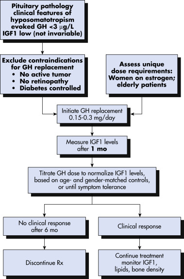
  • GH deficiency (Fig. 2):
    1. GH replacement in children is universally accepted.
    2. GH replacement in adults is not generally recommended and requires careful consideration of each individual case. It may have effects on quality of life, body composition, bone density, and cardiovascular risk factors.
    3. Side effects of replacement include peripheral edema, arthralgia, and headaches.
    4. Usual GH dose is between 0.2 and 0.4 mg, determined by the age and sex of a patient and increments of 0.1 mg every 2 to 4 wk until serum IGF-1 is in the upper part of the normal range. Young adults and women taking estrogen require a higher dose.
  • ADH deficiency:
    1. Desmopressin (DDAVP) 10 to 20 mcg by intranasal spray or 0.05 to 0.1 mg PO bid is used in patients with diabetes insipidus.
    2. Vasopressin: 5 to 10 U given IM or subcutaneous q6h.

Figure 2 Management of somatotropin deficiency in adults.

!!flowchart!!

Patients older than 60 yr require lower maintenance doses. Women receiving oral estrogen require higher doses than those receiving transdermal estrogen preparations. GH, Growth hormone; IGF1, insulin-like growth factor 1; Rx, treatment.

From Melmed S et al: Williams textbook of endocrinology, ed 14, Philadelphia, 2019, Elsevier.

Disposition

  • Hormone replacement therapy is adjusted according to serum hormone monitoring.
  • If untreated can lead to adrenal crisis, severe hyponatremia and hypothyroidism, metabolic abnormalities, and death.
  • Complications: Visual deficit, adrenal crisis, susceptibility to infection and other stressors.
  • Prognosis: Stable patients have a favorable prognosis with replacement hormone therapy. Patients with acute decompensation are in critical condition with a high mortality rate.
Referral

Consultation with an endocrinologist and neurosurgeon for surgical treatment

Pearls & Considerations

Comments

  • Mineralocorticoid replacement is not necessary in secondary adrenal insufficiency because the renin-angiotensin-aldosterone system is unaffected by pituitary failure.
  • Patients with adult-acquired GH deficiency must meet at least two criteria before replacement therapy: A poor GH response to at least two standard stimuli and hypopituitarism from pituitary or hypothalamic damage. The criteria are different in children in whom GH is required for normal growth.
  • Prevention of acute decompensation can be accomplished by reminding patients to increase the dose of hydrocortisone in response to stress.
  • Medical therapy should precede surgical therapy.
Related Content

Hypopituitarism (Patient Information)