section name header

Basic Information

AUTHOR: Fred F. Ferri, MD

Definition

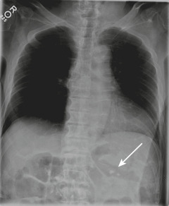
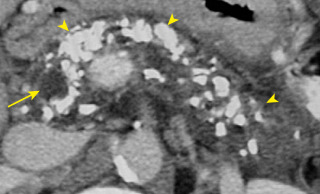
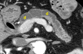
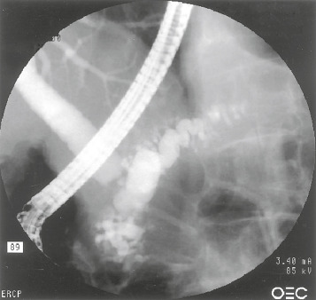
Chronic pancreatitis is a recurrent or persistent inflammatory process of the pancreas characterized by chronic pain and by pancreatic exocrine and/or endocrine insufficiency. It is classified anatomically as either large-duct disease or small-duct (minimal change) disease.

ICD-10CM CODES
K86.1Other chronic pancreatitis
K86.0Alcohol-induced chronic pancreatitis
Epidemiology & Demographics

  • Chronic pancreatitis occurs in approximately 5 to 10 per 100,000 persons in industrialized countries and is usually associated with alcohol use, smoking, and certain gene mutations. It typically begins with recurrent painful bouts of pancreatitis followed by the insidious onset of chronic debilitating pain during the next 3 to 5 years after an initial episode.1
  • Average age at diagnosis is 35 to 55 yr; male:female ratio is 5:1.
  • Annual incidence in the U.S. is 5 to 8 per 100,000 adults.
  • Prevalence in the U.S. is 42 to 73 per 100,000 adults.2
Physical Findings & Clinical Presentation

  • Persistent or recurrent epigastric and left upper quadrant pain (70% of patients) that may radiate to the back
  • Tenderness over the pancreas, muscle guarding
  • Significant weight loss
  • Bulky, foul-smelling stools, greasy in appearance
  • Epigastric mass (10% of patients)
  • Jaundice (5% to 10% of patients)
Etiology

  • Chronic alcoholism (most common cause)
  • Obstruction (ampullary stenosis, tumor, trauma [with pancreatic duct stricture], pancreas divisum, annular pancreas)
  • Tobacco
  • Recurrent pancreatitis
  • Vascular disease/ischemia
  • Hypertriglyceridemia
  • Chronic kidney disease
  • Hereditary pancreatitis
  • Severe malnutrition
  • Idiopathic
  • Untreated hyperparathyroidism (hypercalcemia)
  • Mutations of the cystic fibrosis transmembrane conductance regulator (CFTR) gene and the TF genotype
  • Other genetic mutations (cationic trypsinogen gene, chymotrypsinogen C gene, calcium-sensing receptor gene, claudin-2 gene, serine protease inhibitor, Kazal type 1 gene)
  • Autoimmune pancreatitis (AIP): (5% of chronic pancreatitis cases): Presents clinically with jaundice (63% of patients) and abdominal pain (35%). Computed tomography (CT) may reveal diffusely enlarged pancreas, enhanced peripheral rim of hypoattenuation “halo,” and low-attenuation mass in head of pancreas. Laboratory values reveal elevated serum immunoglobulin (Ig) G4, elevated serum Ig or gamma-globulin level, presence of antilactoferrin antibody (ALA), anticarbonic anhydrase (ACA) II level, antismooth-muscle antibody (ASMA), or antinuclear antibody (ANA)
  • Sclerosing pancreatitis: A form of chronic pancreatitis characterized by infrequent attacks of abdominal pain, irregular narrowing of the pancreatic duct, and swelling of the pancreatic parenchyma; patients have high levels of serum immunoglobulins (IgG4); chronic sclerosing pancreatitis is also known as autoimmune pancreatitis

Diagnosis

Differential Diagnosis

  • Pancreatic cancer
  • Peptic ulcer disease
  • Cholelithiasis with biliary obstruction
  • Malabsorption from other etiologies
  • Recurrent acute pancreatitis
  • Renal insufficiency
  • Intestinal ischemia or infarction
  • Other: Crohn disease, gastroparesis, inflammatory bowel disease
Workup

Medical history with focus on alcohol use, laboratory tests, diagnostic imaging. Table 1 summarizes available diagnostic tests for chronic pancreatitis.

TABLE 1 Available Diagnostic Tests for Chronic Pancreatitis

Tests of Pancreatic StructureTests of Pancreatic Function
EUSDirect hormonal stimulation (with pancreatic stimulation by secretin or CCK or both):
Using oroduodenal tube
Using endoscopy
MRI with MRCP, with or without secretin stimulationFecal elastase
CTSerum trypsinogen (trypsin)
ERCPFecal chymotrypsin
Abdominal USFecal fat
Plain abdominal filmBlood glucose level

Tests are listed in estimated order of decreasing sensitivity for each category.

CCK, Cholecystokinin; CT, computed tomography; ERCP, endoscopic retrograde cholangiopancreatography; EUS, endoscopic ultrasonography; MRCP, magnetic resonance cholangiopancreatography; MRI, magnetic resonance imaging.

See text for explanations.

From Feldman M et al: Sleisenger and Fordtran’s gastrointestinal and liver disease, ed 10, Philadelphia, 2016, Elsevier.

Laboratory Tests

  • Serum amylase and lipase may be elevated (normal amylase levels, however, do not exclude the diagnosis).
  • Hyperglycemia, glycosuria, hyperbilirubinemia, and elevated serum alkaline phosphatase may also be present.
  • Fecal elastase level: Simple, inexpensive and widely available. Levels below 50 mcg per gram of stool are indicative of steatorrhea. Levels between 51 and 200 mcg are below normal but can be due to several other disorders (IBD, renal failure, diabetes, watery diarrhea) and are not specific for steatorrhea.
  • 72-h fecal fat determination over period of 48 or 72 h reveals excess fecal fat. This test is rarely performed and is generally available only in academic centers and major reference laboratories. It can be useful if the fecal elastase level and vitamin A or E levels are low but the patient does not exhibit the classic symptoms of steatorrhea.
  • Secretin stimulation test is the best test for diagnosing pancreatic exocrine insufficiency. Pancreatic secretory function tests are summarized in Table 2.
  • Lipid panel: Significantly elevated triglycerides can cause pancreatitis.
  • Serum calcium: Hyperparathyroidism is a rare cause of chronic pancreatitis.
  • Elevated levels of serum IgG4 are found in sclerosing pancreatitis and AIP.
  • Elevated serum Ig or gamma-globulin level, presence of ALA, ACA II level, ASMA, or ANA in AIP.
  • Decreased levels of serum fat-soluble vitamins (A and E) and other micronutrients (zinc, magnesium, and vitamin B12).

TABLE 2 Pancreatic Secretory Function Tests

TestDescriptionAdvantagesDisadvantagesClinical Indications
Direct
SecretinMeasurements of volume and HCO3– secretion into the duodenum after IV secretinProvide the most sensitive and specific measurements of exocrine pancreatic functionRequire duodenal intubation and IV administration of hormones; not widely availableDetection of mild, moderate, or severe exocrine pancreatic dysfunction
CCKMeasurements of duodenal outputs of amylase, trypsin, chymotrypsin, and/or lipase after IV CCK
Secretin and CCKMeasurements of volume, HCO3–, and enzymes after IV secretin and CCK
Indirect (Requiring Duodenal Intubation)
Lundh test mealMeasurement of duodenal trypsin concentration after oral ingestion of a test mealDoes not require IV administration of hormonesRequires duodenal intubation, a test meal, and normal anatomy, including small intestinal mucosa; not widely availableDetection of moderate or severe exocrine pancreatic dysfunction when a direct test cannot be done (e.g., due to limited availability)
Indirect (Tubeless)
Fecal fatMeasurement of fat in the stool after ingesting meals with a known amount of fatProvides a quantitative measurement of steatorrheaRequires sufficient dietary fat intake and collection of stool; only detects severe pancreatic dysfunctionDetection of severe exocrine pancreatic dysfunction and steatorrhea
Fecal chymotrypsinMeasurement of chymotrypsin or elastase 1 in the stoolDo not require IVs, tubes, or administration of oral substratesInsensitive for detecting mild or moderate dysfunctionDetection of severe exocrine pancreatic dysfunction
Fecal elastase 1
NBT-PABAOral ingestion of NBT-PABA or fluorescein dilaurate with a meal, followed by measurements of PABA or fluorescein in serum or urineProvide simple measurements for severe pancreatic dysfunctionDo not detect mild or moderate dysfunction; results may be abnormal in patients with small intestinal mucosal diseaseDetection of severe exocrine pancreatic dysfunction
Fluorescein dilaurate

CCK, Cholecystokinin; IV, intravenous; NBT-PABA, N-benzoyl-L-tyrosyl-p-aminobenzoic acid.

From Feldman M et al: Sleisenger and Fordtran’s gastrointestinal and liver disease, ed 10, Philadelphia, 2016, Elsevier.

Imaging Studies

  • Plain abdominal x-rays may reveal pancreatic calcifications (Fig. 1) in 25% of patients (95% specific for chronic pancreatitis).
  • Ultrasound of abdomen may reveal duct dilation, pseudocyst, calcification, and presence of ascites.
  • Contrast-enhanced CT scan of abdomen is the initial modality of choice. It is useful to detect calcifications (Fig. E2), evaluate for ductal dilation (Fig. E3), AIP, and rule out pancreatic cancer. CT in AIP reveals narrowed main pancreatic duct and a homogenous “sausage-shaped” pancreas.
  • Magnetic resonance cholangiopancreatography (MRCP) and endoscopic ultrasonography (EUS) have similar specificity and sensitivity to CT.
  • EUS (Fig. E4) has a sensitivity of 97% and a specificity of 60% for chronic pancreatitis and a very low complication rate; however, it is invasive and observer dependent. The diagnosis of chronic pancreatitis on EUS is summarized in Table E3. Fine-needle aspiration biopsy combined with EUS is also the preferred modality for evaluation of cystic or mass lesions to determine malignancy.
  • MRCP is more expensive, takes longer, can miss calcification,3 and is unsuitable for patients with claustrophobia.
  • Endoscopic retrograde cholangiopancreatography (ERCP) (Fig. E5) had been traditionally used to evaluate for the presence of dilated ducts, strictures, pseudocysts, and intraductal stones; however, it is no longer recommended due to complications and availability of noninvasive imaging.
Figure 1 A 51-Yr-Old Man with Chronic Pancreatitis

A Plain Radiograph Demonstrates Multiple Punctate Foci of Calcification Overlying the Expected Location of the Pancreas (Arrow), a Finding Consistent with Chronic Pancreatitis.

From Soto JA: Emergency radiology, the requisites, ed 2, 2017, Elsevier.

Figure E2 Chronic Pancreatitis: Calcifications

Numerous Coarse Calcifications are Seen Throughout the Pancreas (Arrowheads) in This Patient with Recurrent Alcoholic Pancreatitis. The Common Bile Duct (Arrow) is Mildly Dilated Because of a Benign Stricture in the Pancreatic Head.

From Webb WR et al: Fundamentals of body CT, ed 4, Philadelphia, 2015, Saunders.

Figure E3 Chronic Pancreatitis: Dilated Pancreatic Duct

The Pancreatic Duct (D) Shows Marked Beaded Dilation. The Pancreatic Parenchyma is Severely Atrophied.

From Webb WR et al: Fundamentals of body CT, ed 4, Philadelphia, 2015, Saunders.

Figure E4 Endoscopic Ultrasound in a Patient with Chronic Pancreatitis, Demonstrating a Dilated Pancreatic Duct (Marks on Margin of Main Duct)

From Goldman L, Schafer AI: Goldman’s Cecil medicine, ed 24, Philadelphia, 2012, Saunders.

Figure E5 Film from an Endoscopic Retrograde Cholangiopancreatography (ERCP) Showing a Markedly Dilated Pancreatic Duct with Alternating Strictures and Dilation

This “chain of Lakes” Appearance is Diagnostic of Chronic Pancreatitis.

From Feldman M et al: Sleisenger and Fordtran’s gastrointestinal and liver disease, ed 10, Philadelphia, 2016, Elsevier.

TABLE E3 Diagnosis of Chronic Pancreatitis on Endoscopic Ultrasound (EUS)

Standard EUS Grading SystemRosemont Criteria for EUS Diagnosis
Parenchymal abnormalitiesHyperechoic foci
Hyperechoic strands
Lobularity of contour
Cysts
Major featuresHyperechoic foci with shadowing (major A)
Main pancreatic duct calculi (major A)
Lobularity with honeycombing (major B)
Ductal abnormalitiesMain duct dilation
Main duct irregularity
Hyperechoic ductal walls
Visible side branches
Calcification
Minor featuresLobularity without honeycombing
Hyperechoic foci without shadowing
Stranding
Cysts
Irregular main pancreatic duct contour
Main pancreatic duct dilation
Hyperechoic duct margin
Dilated side branches
In the standard EUS grading system, each finding counts equally, and the score is the total number of findings. In the Rosemont system, the diagnostic strata are as follows:
Most consistent with chronic pancreatitisOne major A feature and 3 minor features or One major A feature and major B feature or Two major A features
Suggestive of chronic pancreatitisOne major A feature and <3 minor features or One major B feature and 3 minor features or5 minor features
Indeterminate for chronic pancreatitisThree to four minor features or One major B feature with <3 minor features
Normal2 minor features

From Feldman M et al: Sleisenger and Fordtran’s gastrointestinal and liver disease, ed 10, Philadelphia, 2016, Elsevier.

Treatment

Nonpharmacologic Therapy

  • Avoidance of alcohol and tobacco
  • Frequent, small-volume, low-fat meals
Acute General Rx

  • Avoidance of narcotics if possible (simple analgesics or NSAIDs can be used). Fig. 6 describes an approach to the patient with painful chronic pancreatitis. Management of chronic pancreatitis focuses on treatment of symptoms.
  • Treatment of steatorrhea with pancreatic supplements (e.g., Pancrease, Creon, pancrelipase titrated prn based on the amount of steatorrhea and patient’s weight loss). Enzyme products for the treatment of chronic pancreatitis are summarized in Table 4. All non-enteric-coated enzymes should be used with acid-suppressing medications. Proton pump inhibitors and H2 blockers reduce inactivation of the enzymes from gastric acid.
  • Antioxidants (vitamin A, selenium, vitamin E) may be helpful for pain control in chronic pancreatitis.
  • Percutaneous or via EUS celiac plexus blockade with corticosteroids or neurolysis with ethanol may provide temporary pain relief.
  • Treatment of complications (e.g., type 1 diabetes mellitus).
  • Autoimmune pancreatitis (AIP): Glucocorticoid therapy in patients with AIP and sclerosing pancreatitis can induce clinical remission and significantly decrease serum concentrations of IgG4, immune complexes, and the IgG4 subclass of immune complexes. Starting dose of oral prednisolone is 0.6 to 1.0 mg/kg/day tapered over 3 mo. Recurrent AIP is treated with glucocorticoids and immunomodulators (azathioprine, mycophenolate, mercaptopurine) or rituximab.

TABLE 4 Enzyme Products for the Treatment of Chronic Pancreatitis

ProductFormulationLipase Content per Pill or Capsule (USP units)
CreonEnteric-coated capsule3000; 6000; 12,000; 24,000; 36,000
ZenpepEnteric-coated capsule3000; 5000; 10,000; 15,000; 20,000; 25,000
PancreazeEnteric-coated capsule4200; 10,500; 16,800; 21,000
UltresaEnteric-coated capsule13,800; 20,700; 23,000
PertzyeEnteric-coated with bicarbonate8000; 16,000
ViokaseNon-enteric-coated tablet10,440; 20,880

The total dose of lipase per meal should be titrated based on response but usually requires at least 60,000 and usually 90,000 USP units (30,000 international units) of lipase per meal and one half that amount with snacks. The dose should be split equally during the meal and immediately after the meal.

Non-enteric-coated agents require cotreatment with an histamine 2 receptor antagonist (H2RA) or proton pump inhibitor (PPI) to avoid denaturation of the enzymes by gastric acid.

From Feldman M et al: Sleisenger and Fordtran’s gastrointestinal and liver disease, ed 10, Philadelphia, 2016, Elsevier.

Figure 6 Approach to the Patient with Painful Chronic Pancreatitis

Etoh, Alcohol; Eus, Endoscopic Ultrasound; Mrcp, Magnetic Resonance Cholangiopancreatography.

!!flowchart!!

From Goldman L, Ausiello D [eds]: Cecil textbook of medicine, ed 24, Philadelphia, 2012, Saunders.

Chronic Rx

  • Surgical intervention (e.g., distal pancreatectomy or pancreaticoduodenectomy) may be necessary in painful chronic pancreatitis to eliminate biliary tract disease and improve flow of bile into the duodenum by eliminating obstruction of pancreatic duct.3
  • ERCP with endoscopic sphincterectomy and stone extraction and for treatment of pancreatic duct strictures is less invasive and more widely available than surgery and may be preferred as first-line treatment in painful chronic pancreatitis.
  • Percutaneous or EUS-guided celiac plexus blockade using glucocorticoids is effective in providing short-term pain relief in nearly half of patients.
Disposition

  • Long-term survival is poor (50% of patients die within 10 yr from chronic pancreatitis or malignancy).
  • Prognosis is best in patients with recurrent acute pancreatitis resulting from cholelithiasis, hyperparathyroidism, or stenosis of the sphincter of Oddi.
Referral

Gastrointestinal referral for ERCP, surgical referral in selected patients

Related Content

Chronic Pancreatitis (Patient Information)

Malabsorption (Related Key Topic)

Pancreatitis, Acute (Related Key Topic)

Related Content

    1. Vege S.S., Chari S.T. : Chronic pancreatitisN Engl J Med. ;386:869-878, 2022.
    2. Singh V.K. : Diagnosis and management of chronic pancreatitis: a reviewJAMA. ;322:2422-2434, 2019.
    3. Beyer G. : Chronic pancreatitisLancet. ;396:499-512, 2020.
    4. Hines O.J. : Tracing the evidence to address painful chronic pancreatitis with surgeryJAMA. ;323:219-220, 2020.
    5. Issa Y. : Effect of early surgery vs endoscopy-first approach on pain in patients with chronic pancreatitis: the ESCAPE randomized clinical trialJAMA. ;323(3):237-247, 2020.