AUTHOR: Bharti Rathore, MD
Thyroid carcinoma is a primary neoplasm of the thyroid and consists of four major subtypes: Papillary, follicular, anaplastic, and medullary. A classification of thyroid neoplasms is described in Table E1.
TABLE E1 Classification of Thyroid Neoplasms
From Melmed S et al: Williams textbook of endocrinology, ed 14, Philadelphia, 2019, Elsevier.
Papillary carcinoma of thyroid
Follicular carcinoma of thyroid
Anaplastic carcinoma of thyroid
Medullary carcinoma of thyroid
|
TABLE 2 Inherited Syndromes Associated With Thyroid Cancer
Multiple endocrine neoplasia (MEN) 2A and 2B | |||
Isolated familial medullary thyroid cancer | |||
Gardner syndrome | |||
Familial adenomatous polyposis | |||
Carney complex | |||
Cowden syndrome | |||
Familial nonmedullary thyroid cancer |
From Cameron JL, Cameron AM: Current surgical therapy, ed 10, Philadelphia, 2011, Saunders.
Figure E1 Thyroid cancer pathways.
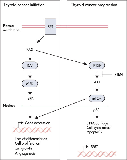
Diagram shows the key molecular signaling pathways involved in thyroid cancer. The box on the left shows the mitogen-activated protein kinase pathway, which is activated by mutation in most thyroid cancers. These events are believed to initiate thyroid cancer development and lead to altered gene expression, which promotes cell proliferation, cell growth, angiogenesis, and loss of differentiation. The box on the right shows pathways altered in advanced thyroid cancers, which are believed to promote tumor progression. This includes the PI3K-mTOR pathway, the p53 tumor suppressor, and alterations in the promoter for TERT. Blue boxes represent factors for which targeted treatments are available that have been approved by the U.S. Food and Drug Administration. mTOR, Mammalian target of rapamycin; PI3K, phosphatidylinositol-3-kinase; TERT, telomerase reverse transcriptase.
From Cabanillas ME et al: Thyroid cancer, Lancet 388[10061]:2783-2795, 2016.
The workup of thyroid carcinoma includes laboratory evaluation and diagnostic imaging. Key features of thyroid malignancies are summarized in Table 3. Diagnosis is confirmed with fine-needle aspiration or surgical biopsy. At diagnosis, the vast majority of thyroid cancers are well differentiated, with excellent prognosis. The characteristics of thyroid carcinoma vary with the type:
TABLE 3 Thyroid Malignancies-Key Features
Description | Pattern of Spread | |
---|---|---|
Papillary carcinoma (70%-80%) | Low-grade tumors with a good prognosis (histologically multicentric) ▸ Tumors concentrate radio-iodine | Early lymph node spread (metastatic lymph nodes may be normal in size, cystic, calcified, hemorrhagic, or contain colloid) ▸ Distant metastases are rare (and usually to the lungs) |
Follicular carcinoma (10%-20%) | Slow growing ▸ Tumors concentrate radio-iodine | It rarely metastasizes to the regional lymph nodes ▸ The tendency is to spread via the bloodstream and disseminate to the lungs, bones, or liver |
Anaplastic carcinoma (1%-2%) | Undifferentiated malignant tumors that do not concentrate radio-iodine ▸ There is a poor prognosis ▸ They tend to occur in older patients ▸ Punctate calcification and necrosis frequently are present | Lymphatic metastases occur in the majority of patients |
Medullary carcinoma (5%-10%) | This originates from the parafollicular C cells ▸ It does not concentrate radio-iodine ▸ It may be sporadic or familial (and associated with the MEN type II syndrome or other endocrine neoplasms) ▸ It is usually a unilateral, solitary lesion ▸ Calcification is seen in 10% ▸ 123I-MIBG and somatostatin analogs (e.g., octreotide) can be used for evaluation ▸ Circulating calcitonin levels are usually elevated | It may invade locally, spread to the regional nodes, or demonstrate hematogenous spread to the lungs, bones, or liver |
Lymphoma (10%) | It is usually a non-Hodgkin lymphoma ▸ It occurs in one third of patients with Hashimoto thyroiditis (a MALT-type lymphoma) ▸ It presents as a rapidly enlarging, solitary nodule (80%) or as multiple nodules (imaging cannot distinguish between a lymphoma and thyroiditis) ▸ Necrosis and calcification are uncommon | It can involve the nodes with spread to the GI tract |
Metastases (1%) | The most common primary is renal cell carcinoma |
From Grant LA: Grainger & Allisons diagnostic radiology essentials, ed 2, Philadelphia, 2019, Elsevier.
TABLE 4 The Bethesda System for Thyroid Cytopathology
Category | Risk of Malignancy (%) | Recommended Management |
---|---|---|
Nondiagnostic or unsatisfactory | 1-4 | Repeat FNA with ultrasound guidance |
Benign | 0-3 | Clinical follow-up |
Atypia of undetermined significance (AUS) or follicular lesion of undetermined significance (FLUS) | 5-15 | Repeat FNA∗ |
Follicular neoplasm or suspicious for follicular neoplasm | 15-30 | Lobectomy |
Suspicious for malignancy | 60-75 | Lobectomy with or without frozen section or total thyroidectomy |
Malignant | 97-99 | Total thyroidectomy |
FNA, Fine-needle aspiration.
∗Lobectomy also can be considered depending on clinical or sonographic characteristics.
From Niederhuber JE: Abeloffs clinical oncology, ed 6, Philadelphia, 2020, Elsevier.
The serum thyroglobulin level was 45 ng/ml during levothyroxine suppressive treatment. A, A total-body scan was performed 3 days after administration of 100 mCi (3.7 GBq). There is no visible uptake in the neck or thorax. Notice the accumulation of radioiodine in the stomach, colon, and bladder. B, Positron emission tomography scan using [18F]-fluorodeoxyglucose (18FDG) with maximal intensity projection demonstrates significant uptake in the upper mediastinum. C, In fusion images of 18FDG-PET and CT scans, axial and coronal slices localized the FDG uptake in the right paratracheal mediastinum, corresponding to a lymph node metastasis that subsequently was excised. Serum thyroglobulin became undetectable during thyroid hormone treatment.
From Melmed S et al [eds]: Williams textbook of endocrinology, ed 14, Philadelphia, 2019, Saunders.
Figure E3 Papillary adenocarcinoma.
A 74-yr-old woman presented with a thyroid nodule. A, An 123I thyroid scan (anterior view) demonstrates a cold nodule (arrows) in the upper portion of the left thyroid lobe. The hot spot below the thyroid is a suprasternal marker. B, An axial computed tomography scan demonstrates an irregular density in the left thyroid lobe at the level of the lesion seen on the radionuclide scan. The lesion contains a single area of calcification (arrow) and is not demarcated sharply from normal thyroid tissue. At operation, there proved to be extracapsular extension.
From Skarin AT: Atlas of diagnostic oncology, ed 3, St Louis, 2003, Mosby.
TABLE E5 The Tumor-Node-Metastasis (TNM) Scoring System
Definition of TNM | ||
---|---|---|
Category | 2010 Version (AJCC 7th Edition) | 2017 Version (AJCC 8th Edition) |
Primary Tumor (T) | ||
T0 | No evidence of primary tumor | No evidence of primary tumor |
T1 | Tumor ≤2 cm limited to the thyroid | Tumor ≤2 cm limited to the thyroid |
T1a: ≤1 cm | T1a: ≤1 cm | |
T1b: >1-2 cm | T1b: >1-2 cm | |
T2 | Tumor >2 to ≤4 cm limited to the thyroid | Tumor >2 to ≤4 cm limited to the thyroid |
T3 | Tumor >4 cm limited to the thyroid or any tumor with minimal extrathyroid extension (e.g., extension to sternothyroid muscle or perithyroid soft tissues) | Tumor >4 cm limited to the thyroid or gross extrathyroidal extension invading only strap muscles (sternohyoid, sternothyroid, thyroidhyoid, omohyoid) from a tumor of any size |
T4 | No evidence of primary tumor | No evidence of primary tumor |
T4a | Gross extrathyroidal extension invading subcutaneous soft tissues, larynx, trachea, esophagus, or recurrent laryngeal nerve from a tumor of any size or intrathyroidal anaplastic thyroid cancer of any size | Gross extrathyroidal extension invading subcutaneous soft tissues, larynx, trachea, esophagus, or recurrent laryngeal nerve from a tumor of any size |
T4b | Gross extrathyroidal extension invading prevertebral fascia or encasing the carotid artery or mediastinal vessels from a tumor of any size or anaplastic thyroid cancer of any size with extrathyroidal extension | Gross extrathyroidal extension invading prevertebral fascia or encasing the carotid artery or mediastinal vessels from a tumor of any size |
Regional Lymph Node (N) | ||
N0 | No regional lymph node metastasis | No evidence of locoregional lymph node metastasis |
N0a: One or more cytologically or histologically confirmed benign lymph nodes | ||
N0b: No radiologic or clinical evidence of locoregional lymph node metastasis | ||
N1 | Regional lymph node metastasis | Regional lymph node metastasis |
N1a | Metastases in pretracheal and paratracheal lymph nodes, including prelaryngeal and delphian lymph nodes, unilateral or bilateral | Metastasis in pretracheal, paratracheal, prelaryngeal/delphian, or upper mediastinal lymph nodes, unilateral or bilateral |
N1b | Metastases in lateral neck lymph nodes or upper mediastinal lymph nodes, unilateral or bilateral | Metastases in lateral neck lymph nodes, unilateral or bilateral disease |
Distant Metastases (M) | ||
Category | 2010 Version | 2017 Version |
M0 | No distant metastasis | No distant metastasis |
M1 | Distant metastasis | Distant metastasis |
TNM Staging for Papillary, Follicular, and Poorly Differentiated Thyroid Cancer | ||
Age Cutoff | Age <45 Yr | Age <55 Yr |
Stage I | Any T, any N, M0 | Any T, any N, M0 |
Stage II | Any T, any N, M1 | |
Stage III | None | |
Stage IV | None | |
Age Cutoff | Age ≥45 Yr | Age ≥55 Yr |
Stage I | T1, N0, M0 | T1-T2, N0, M0 |
Stage II | T2, N0, M0 | T1-T2, N1a-N1b, M0 or T3, any N, M0 |
Stage III | T3, N0, M0 or any T1-3, N1a, M0 | T4a, any N, M0 |
Stage IV | ||
Stage IVA | T1-3, N1b, M0 or T4a, any N, M0 | T4b, any N, M0 |
Stage IVB | T4b, any N, M0 | Any T, any N, M1 |
Stage IVC | Any T, any N, M1 | - |
TNM Staging for Medullary Thyroid Cancer | ||
Category | 2010 Version | 2017 Version |
Stage I | T1, N0, M0 | T1, N0, M0 |
Stage II | T2-T3, N0, M0 | T2-T3, N0, M0 |
Stage III | T1-3, N1a, M0 | T1-3, N1a, M0 |
Stage IVA | T1-3, N1b, M0 or T4b, any N, M0 | T1-3, N1b, M0 or T4b, any N, M0 |
Stage IVB | T4b, any N, M0 | T4b, any N, M0 |
Stage IVC | Any T, any N, M1 | Any T, any N, M1 |
TNM Staging for Anaplastic Thyroid Cancer | ||
Stage IVA | T4a, N0, M0 | T1-T3a, N0, M0 |
Stage IVB | T4b, any N, M0 | T1-T3a, N1, M0 or T3b-T4, any N, M0 |
Stage IVC | Any T, any N, M1 | Any T, any N, M1 |
AJCC, American Joint Committee on Cancer.
From Melmed S et al: Williams textbook of endocrinology, ed 14, Philadelphia, 2019, Elsevier.
TABLE 6 Indications for Iodine-131 Treatment in Patients With Papillary, Follicular, or Hürthle Cell Thyroid Carcinoma After Initial Definitive Near-Total Thyroidectomy
No Indication | |||
---|---|---|---|
Adult patients at very low risk for cause-specific mortality or relapse: Complete surgical resection, favorable histology, and limited extent of disease (e.g., PTC patients with MACIS scores <6; patients with tumor size <1 cm, N0, and M0). | |||
Definite Indications | |||
Distant metastasis at diagnosis | |||
Incomplete tumor resection | |||
Complete tumor resection but high risk for mortality or recurrence (e.g., PTC patients with MACIS scores ≥6 and pTNM stage II/III FTC or HCC) | |||
Probable Indications | |||
Incomplete surgery (less than near-total thyroidectomy, no lymph node dissection) | |||
PTC or FTC in a child younger than 16 yr | |||
If PTC, tall cell or columnar cell variant and diffuse sclerosing variant | |||
If FTC, widely invasive or poorly differentiated tumor | |||
Bulky nodal metastases |
FTC, Follicular thyroid carcinoma; HCC, Hürthle cell carcinoma; MACIS, scoring system based on metastasis, age, completeness of resection, invasion, and size; PTC, papillary thyroid carcinoma; pTNM, pathologic tumor-node-metastasis classification.
From Melmed S et al (eds): Williams textbook of endocrinology, ed 12, Philadelphia, 2011, Saunders.
BOX 2 Risk Stratification for Thyroid Cancer Recurrence
|
From Flint PW et al: Cummings otolaryngology, head and neck surgery, ed 7, Philadelphia, 2021, Elsevier.
BOX 1 Risk Factors for Aggressive Behavior of Well-Differentiated Thyroid Carcinomas
From Flint PW et al: Cummings otolaryngology, head and neck surgery, ed 7, Philadelphia, 2021, Elsevier.
TABLE 8 Factors Used in Prognostic Classification Systems
TNM | AMES | AGES | MACIS | |
---|---|---|---|---|
Patient Factors | ||||
Age | × | × | × | × |
Gender | × | × | ||
Tumor Factors | ||||
Size | × | × | × | × |
Histologic grade | × | |||
Histologic type | × | × | ∗ | ∗ |
Extrathyroid spread | × | × | × | × |
Lymph node metastasis | × | |||
Distant metastasis | × | × | × | × |
Incomplete resection | × |
AGES, Age at diagnosis, histologic tumor grade, extent of disease at presentation, and tumor size; AMES, patient age, metastases, extent of invasion, and tumor size; MACIS, metastasis, age at diagnosis, completeness of surgical resection, extrathyroid invasion, and tumor size; TNM, tumor/node/metastasis.
∗AGES/MACIS classifications for papillary carcinomas only.
From Flint PW et al: Cummings otolaryngology, head and neck surgery, ed 7, Philadelphia, 2021, Elsevier.
TABLE 7 Characteristics of Thyroid Cancers
Type of Cancer | Percentage of Thyroid Cancers | Age of Onset (yr) | Treatment | Prognosis |
---|---|---|---|---|
Papillary | 88 | 40-80 | Thyroidectomy, followed by radioactive iodine ablation and TSH suppression | Good |
Follicular | 10 | 45-80 | Thyroidectomy, followed by radioactive iodine ablation and TSH suppression | Fair to good |
Medullary | 3-4 | 20-50 | Thyroidectomy and central compartment lymph node dissection and TSH suppression | Fair |
Anaplastic | 1 | 50-80 | Isthmusectomy followed by palliative x-ray treatment | Poor |
Lymphoma | <1 | 25-70 | X-ray therapy and/or chemotherapy | Fair |
From Andreoli TE et al: Andreoli and Carpenters Cecil essentials of medicine, ed 8, Philadelphia, 2010, Saunders.
TABLE E9 Overview of Plans for First Year of Follow-Up Following Initial Therapy
Initial Plan Based on ATA Risk for the First Year of Follow-Up | ATA Low Risk | ATA Intermediate Risk | ATA High Risk |
---|---|---|---|
Tg, TgAb, TFTs, every 3-6 mo | √ | √ | √ |
Neck US in 3-6 mo | - | √ | √ |
Neck/chest CT with contrast in 6-12 mo | - | Considera | √b |
Cross-sectional imaging of other sites (brain, abdomen, pelvis) | - | - | Considerc |
Routine surveillance diagnostic RAI scan | - | - | Consider |
18FDG-PET scan | - | - | Consider |
Dynamic risk assessment at each visit | √ | √ | √ |
ATA, American Thyroid Association; CT, computed tomography; 18FDG-PET, fluorodeoxyglucose positron emission tomography; RAI, radioactive iodine; Tg, thyroglobulin; TgAb, antithyroglobulin antibodies; TFTs, thyroid function tests; US, ultrasound.
Note: Although most patients will return for physical examination and biochemical testing every 3-6 mo for the first year, consideration for additional testing is based on ATA risk and on the dynamic risk assessment done at each follow-up visit.
a Considered for intermediate-risk patient status postresection clinical N1a or N1b disease.
b Depending on presenting features, CT of the neck/chest may need to be done as early as 2-3 mo after initial therapy.
c Depending on presenting features, functional imaging results, and serum Tg levels.
From Melmed S et al: Williams textbook of endocrinology, ed 14, Philadelphia, 2019, Elsevier.
Thyroid Cancer (Patient Information)
Thyroid Nodule (Related Key Topic)
Multiple Endocrine Neoplasia (Related Key Topic)