section name header

Basic Information

AUTHORS: Harikrashna B. Bhatt, MD and Russell E. Bratman, MD

Definition

Multiple endocrine neoplasia (MEN) refers to a group of heritable genetic syndromes characterized by the development of specific groups of tumors of the endocrine glands.

Synonyms

MEN I: Wermer syndrome

MEN IIA: Sipple syndrome

ICD-10CM CODES
E31.2Multiple endocrine neoplasia [MEN] syndromes
E31.20Multiple endocrine neoplasia [MEN] syndrome, unspecified
E31.21Multiple endocrine neoplasia [MEN] syndrome type I
E31.22Multiple endocrine neoplasia [MEN] syndrome type IIA
E31.23Multiple endocrine neoplasia [MEN] syndrome type IIB
Epidemiology & Demographics 12
Incidence

  • MEN I: 25 in 10,000
  • MEN II: 1 in 30,000
Prevalence

  • MEN I: 1/30,000
  • MEN II: 1/35,000 (predominantly MEN IIA)
Risk Factors

Family history of MEN syndrome, although it can also occur sporadically

Genetics

  • MEN I: Autosomal-dominant mutation in MEN1 tumor-suppressor gene1
  • MEN IIA and MEN IIB: Autosomal-dominant mutations in RET proto-oncogene2
Physical Findings & Clinical Presentation

  • Patients may present due to screening or may present with a MEN-associated tumor. Tumors are found incidentally due to biochemical abnormalities or to symptoms.
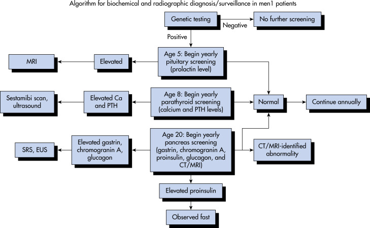
  • MEN I (PPP [pituitary, pancreas, parathyroid])3,4
    1. Diagnostic criteria generally include two MEN-associated tumors, one tumor in a patient with a family history, or positive genetic testing
    2. Primary hyperparathyroidism (parathyroid adenoma or hyperplasia) is the most common manifestation and can cause hypercalcemia (urolithiasis, GI disturbance, bone pain, neuropsychiatric disturbances) and also affect bone density
    3. Pancreatic neuroendocrine tumors cause symptoms related to their secretory properties or metastases
      1. Gastrinoma: Peptic ulcers, diarrhea, esophageal symptoms (see “Gastrinoma”)
      2. Insulinoma: Hypoglycemic symptoms upon fasting and after exercise (see “Insulinoma”)
      3. Glucagonoma: Necrolytic migratory erythema (a blistering skin lesion), diabetes/glucose intolerance, weight loss
      4. VIPoma: Diarrhea, hypokalemia, decreased gastric acid
      5. Somatostatinoma: Hyperglycemia, cholelithiasis, diarrhea, abdominal pain, weight loss
      6. Nonfunctioning tumors can metastasize (frequently to the liver), a frequent cause of death in MEN I
    4. Pituitary tumors may cause compressive symptoms, such as visual field defects or hypopituitarism (see “Pituitary Adenoma”). Hormonal secretion may also cause symptoms.
      1. Prolactinoma (most common pituitary tumor in MEN I): Menstrual irregularities, hypogonadism, gynecomastia, and/or galactorrhea (see “Prolactinoma”)
      2. Somatotroph adenomas (producing growth hormone): Gigantism or acromegaly (frontal bossing, increased shoe/hat size, hyperglycemia, hyperhidrosis) depending on patient age (see “Acromegaly”)
      3. Corticotroph adenomas (producing adrenocorticotropic hormone): Cushingoid features such as weight gain, moon facies, hyperglycemia, bone loss, proximal muscle weakness, hypertension, hypokalemia (see “Cushing Disease and Syndrome”)
      4. Nonfunctioning (nonsecretory) tumors
    5. Other manifestations: Carcinoid tumors, collagenomas, angiofibromas, meningiomas, lipomas
  • MEN IIA:5,6
    1. Medullary thyroid carcinoma (MTC) is a tumor of the thyroid’s calcitonin-secreting C cells. It can present with a neck mass, as well as flushing and diarrhea due to elevated calcitonin levels.
    2. Primary hyperparathyroidism: See “MEN I.” Less aggressive in “MEN IIA.”
    3. Pheochromocytoma is an adrenal tumor producing catecholamines, which can lead to life-threatening hypertensive crises.
  • MEN IIB:5,6
    1. MTC
    2. Pheochromocytoma
    3. Oral mucosal neuromas
Etiology

Tumor development facilitated by the previously described genetic mutations (Table E1).

TABLE E1 Tumors in Multiple Endocrine Neoplasia Syndromes

Type (Chromosomal Location)Tumors (Estimated Penetrance)
MEN 1 (11q13)Parathyroid adenoma (90%)
Enteropancreatic tumor (30%-70%)
Gastrinoma (40%)
Insulinoma (10%)
Nonfunctioning and PPoma (20%-55%)
Glucagonoma (<1%)
VIPoma (<1%)
Pituitary adenoma (30%-40%)
Prolactinoma (20%)
Somatotropinoma (10%)
Corticotropinoma (<5%)
Nonfunctioning (<5%)
Associated Tumors
Adrenal cortical tumor (40%)
Pheochromocytoma (<1%)
Bronchopulmonary NET (2%)
Thymic NET (2%)
Gastric NET (10%)
Lipomas (30%)
Angiofibromas (85%)
Collagenomas (70%)
Meningiomas (8%)
MEN 2 (10 cen- 10q11.2)
MEN 2AMTC (90%)
Pheochromocytoma (50%)
Parathyroid adenoma (20%-30%)
MTC onlyMTC (100%)
MEN 2B (also known as MEN 3)MTC (>90%)
Pheochromocytoma
Associated abnormalities (40%-50%)
Mucosal neuromas
Marfanoid habitus
Medullated cornel nerve fibers
Megacolon

MEN, Multiple endocrine neoplasia; MTC, medullary thyroid carcinoma; NET, neuroendocrine tumor; PPoma, pancreatic polypeptidoma; VIPoma, vasoactive intestinal peptidoma.

Modified from Thakker RV: Multiple endocrine neoplasia type 1. In Jameson JL et al (eds): Endocrinology: adult and pediatric, Philadelphia, 2016, Saunders.

Diagnosis

Differential Diagnosis

Tumors associated with MEN may occur sporadically.

Workup

  • MEN I: Note that opinions vary regarding the aggressiveness and frequency of screening for MEN-associated tumors. Fig. E1 illustrates an algorithm for screening and management of MEN1 syndromes.
    1. Genetic testing: Offered to patients meeting MEN I clinical criteria or to patients in whom there is high suspicion, as well as to first-degree relatives of MEN I patients.
    2. Hyperparathyroidism screening: Parathyroid hormone (PTH) and calcium annually.
    3. Pancreatic tumor screening: Annual pancreas imaging (endoscopic ultrasonography, computed tomography [CT], or MRI) and annual biochemical testing (glucose, gastrin, vasoactive intestinal peptide [VIP], glucagon, insulin, pancreatic polypeptide, chromogranin A). Notably, Gallium-68 DOTATATE PET-CT is an exciting new tool for neuroendocrine tumor localization, though it is not typically utilized in screening at this time.
    4. Pituitary tumor screening: Insulin-like growth factor 1 (IGF-1) and prolactin annually and pituitary MRI every 3 to 5 yr.
    5. Carcinoid tumor screening: Chest CT or MRI every 2 yr.
  • MEN IIA:
    1. Genetic testing: At-risk members of families with known RET mutations should undergo screening for the specific mutation. Patients with MTC should have genetic testing of the tumor and genetic testing based on results. Cutaneous lichen amyloidosis (a skin finding) should prompt testing; Hirschsprung disease may also prompt testing.2
    2. Hyperparathyroidism screening: PTH and calcium annually
    3. Pheochromocytoma: Plasma catecholamines and metanephrines annually for screening, age to begin depending on specific mutation; CT and MRI may localize; I-123 and PET (possibly including gallium-68 DOTATATE PET-CT) also helpful for localization.7
    4. MTC screening: Depends on risk category; physical exam, neck ultrasound, and calcitonin level yearly; carcinoembryonic antigen may also be indicated.5,6
  • MEN IIB:
    1. Genetic testing: See “MEN IIA”
    2. Pheochromocytoma: See “MEN IIA”
    3. MTC screening: Although there may be a role for monitoring, prophylactic thyroidectomy is typically performed early

Figure E1 Algorithm for Screening and Management in Multiple Endocrine Neoplasia Type 1 (Men1) Syndromes

From Townsend CM et al: Sabiston textbook of surgery, ed 21, St Louis 2022, Elsevier.

Laboratory Tests

See “Workup.”

Imaging Studies

See “Workup.”

Treatment

No therapy available at this time to reverse the underlying genetic cause. Treatment (both medical and surgical) focuses on tumor prevention and management.

Nonpharmacologic Therapy

  • MEN I:3,4
    1. For hyperparathyroidism, surgical parathyroidectomy is indicated in patients with significant hypercalcemia, osteoporosis, renal disease, or nephrolithiasis or found to be at high risk for nephrolithiasis. Hyperparathyroidism may be treated surgically with parathyroidectomy.
    2. Pituitary tumors may be surgically excised via transsphenoidal approach; notably, medical therapy is first line for prolactinoma.
    3. Pancreatic tumor treatment is extremely variable. Gastrinoma is frequently complicated by duodenal metastases, which are difficult to treat surgically, so treatment choice varies between centers. Ulcers caused by gastrinoma may require endoscopic or surgical management. Surgery is preferred for insulinoma, VIPoma, and glucagonoma. Nonfunctioning tumors may be treated surgically depending on tumor size and location. Conservative management for insulinoma may involve frequent carbohydrate intake. Cytotoxic chemotherapy and tyrosine kinase inhibitors are options for patients with metastatic disease. Peptide receptor radionucleotide therapy (PRRT) is an emerging and exciting therapeutic modality for metastatic disease.
  • MEN IIA:
    1. MTC is generally treated surgically. Prophylactic thyroidectomy is considered based on genetic mutation. Almost all children with MEN IIA will require thyroidectomy (screening as previously described). Patients should undergo ultrasound and total thyroidectomy with cervical lymph node dissection. Extent of further neck dissection depends on metastases and may be guided by calcitonin levels. A more palliative surgical approach is considered in the setting of advanced disease.5,6
    2. Hyperparathyroidism: Resection of only enlarged glands with intraoperative PTH monitoring is preferred.
    3. Pheochromocytoma is treated surgically. Unilateral adrenalectomy is preferred, although many patients will develop a contralateral pheochromocytoma. Preoperative blood pressure control with alpha blockade is key. If present, pheochromocytoma must be removed before thyroidectomy.7
  • MEN IIB:
    1. MTC: Prophylactic thyroidectomy offered in childhood.5,6
    2. Pheochromocytoma: See “MEN IIA.”
Acute General Rx

  • MEN I:3,4
    1. Hyperparathyroidism causes hypercalcemia that may be treated with intravenous (IV) hydration, diuretics, and bisphosphonates. Early vitamin D repletion prevents bone destruction postoperatively due to the “hungry bone syndrome.”
    2. Gastrinoma may lead to peptic ulcers requiring IV proton-pump inhibitor (PPI).
    3. Supportive care (glucose for hypoglycemia caused by insulinoma; fluids and electrolytes for hypovolemia caused by diarrhea from VIPoma or gastrinoma) is required.
  • MEN IIA:
    1. Hyperparathyroidism: See “MEN I.”
    2. Pheochromocytoma: Preoperative blood pressure control is key; alpha blockers are first line. Phenoxybenzamine irreversibly blocks alpha adrenergic receptors; doxazosin may also be helpful. Calcium channel blockers can be utilized. There is concern that unopposed beta blockade may allow for alpha-medicated vasoconstriction.7
  • MEN IIB:
    1. Pheochromocytoma: See “MEN IIA.”
Chronic Rx

  • MEN I:3,4
    1. Hyperparathyroidism may be amenable to agonists of the calcium-sensing receptor (cinacalcet), although surgery is preferred in patients who meet surgical criteria and are operative candidates.
    2. Pancreatic tumors may benefit from medical therapy.
      1. Gastrinoma: PPI or H2 antagonists, somatostatin agonists (such as octreotide)
      2. Insulinoma: Diazoxide or somatostatin agonists
      3. Glucagonoma and VIPoma: Somatostatin agonists
    3. Adjunctive medical therapy can be considered for pituitary tumors, although surgical treatment is first-line with the exception of prolactinoma (for which dopamine agonists are first line). Radiation therapy and/or medical therapy are considered in cases of incomplete resection or regrowth.
      1. Prolactinoma: Dopamine agonists (bromocriptine or cabergoline). Temozolomide is a chemotherapeutic agent for refractory cases.
      2. Somatotroph adenoma: Somatostatin or dopamine agonists, growth hormone receptor antagonist
      3. Corticotroph adenoma: Antiadrenal agents, adrenal enzyme blocker (such as ketoconazole), somatostatin or dopamine agonists, glucocorticoid receptor blockers
  • MEN II A:
    1. Hyperparathyroidism: See “MEN I.”
    2. MTC: Tyrosine kinase inhibitors and chemotherapy may help treat metastatic disease. After thyroidectomy, levothyroxine is indicated (thyroid-stimulating hormone suppression not required).5,6
  • MEN IIB

MTC: See “MEN II A.”

Disposition

Most workup can be done as an outpatient (multidisciplinary team); inpatient stays may be required due to acute complications or for surgical procedures.

Complementary & Alternative Medicine

MEN is unlikely to be amenable to this.

Referral

Patients with MEN should be followed by a multispecialty team, including endocrinologists, endocrine surgeons, and genetic counselors.

Pearls & Considerations

Comments

MEN is a group of genetic syndromes that requires close monitoring and intervention to prevent and treat tumor development, as well as genetic counseling of patients and family members.

Prevention

Prevention focuses on screening for MEN in persons at risk and early identification of MEN-associated tumors.

Patient & Family Education

Related Content

Acromegaly (Related Key Topic)

Cushing Disease and Syndrome (Related Key Topic)

Gastrinoma (Related Key Topic)

Hyperparathyroidism (Related Key Topic)

Insulinoma (Related Key Topic)

Pheochromocytoma (Related Key Topic)

Pituitary Adenoma (Related Key Topic)

Prolactinoma (Related Key Topic)

Thyroid Carcinoma (Related Key Topic)

Related Content

    1. Giusti F. : Multiple endocrine neoplasia type 1 Pagon R.A., editors : GeneReviews. University of Washington-Seattle, 2015.
    2. Marquard J., Eng C. : Multiple endocrine neoplasia type 2 Pagon R.A., editors : GeneReviews. University of Washington-Seattle, 2015.
    3. Thakker R.V. : Multiple endocrine neoplasia type 1 Jameson J.L., editors : Endocrinology: adult and pediatric. Saunders-Philadelphia:2566-2593, 2016.
    4. Thakker R.V. : Clinical practice guidelines for multiple endocrine neoplasia type 1 (MEN1)J Clin Endocrinol Metab. ;97(9):2990-3011, 2012.
    5. Tacon L.J. : Multiple endocrine neoplasia type 2 and medullary thyroid carcinoma Jameson J.L., editors : Endocrinology: adult and pediatric. Saunders-Philadelphia:2594-2605, 2016.
    6. Wells S.A. : Revised American Thyroid Association guidelines for the management of medullary thyroid carcinomaThyroid. ;25(6):567-610, 2015.
    7. Lenders J.W. : Pheochromocytoma and paraganglioma: an endocrine society clinical practice guidelineJ Clin Endocrinol Metab. ;99(6):1915-1942, 2014.