section name header

Basic Information

AUTHORS: Brett Patrick, MD and Mark F. Brady, MD, MPH, MMSc and Fred F. Ferri, MD

Definition

Pheochromocytomas are catecholamine-producing tumors that originate from the chromaffin cells of the adrenergic system. While they generally secrete both norepinephrine and epinephrine, norepinephrine is usually the predominant amine.

Synonym

Paraganglioma

ICD-10CM CODES
C74.9Malignant neoplasm of adrenal gland, unspecified
C75.9Malignant neoplasm of endocrine gland, unspecified
E27.5Adrenomedullary hyperfunction
Epidemiology & Demographics

  • Incidence: 0.05% of population; peak incidence in 30s and 40s.
  • Approximately 25% of patients with apparently sporadic pheochromocytoma may be carriers of mutations.
  • Approximately 25% of pheochromocytomas are familial and associated with genetic disorders (Table 1). Pheochromocytoma is a feature of two disorders with an autosomal dominant pattern of inheritance:
    1. Multiple endocrine neoplasia (MEN) type 2
    2. von Hippel-Lindau disease: Angioma of the retina, hemangioblastoma of the central nervous system, renal cell carcinoma, pancreatic cysts, and epididymal cystoadenoma
  • Pheochromocytomas occur in 5% of patients with neurofibromatosis type 1.

TABLE 1 Autosomal Dominant Syndromes Associated With Pheochromocytoma and Paraganglioma

SyndromeGeneGene LocusProtein ProductProtein FunctionGene MechanismTypical Tumor Location
SDHD (familial paraganglioma type 1)SDHD11q23SDH D subunitATP productionTumor suppressorSkull base and neck; occasionally adrenal medulla, mediastinum, abdomen, pelvis
Familial paraganglioma type 2SDHAF211q13.1Flavination cofactorATP productionTumor suppressorSkull base and neck; occasionally abdomen and pelvis
SDHC (familial paraganglioma type 3)SDHC1q21SDH C subunitATP productionTumor suppressorSkull base and neck
SDHB (familial paraganglioma type 4)SDHB1p36.1-35SDH B subunitATP productionTumor suppressorAbdomen, pelvis and mediastinum; rarely adrenal medulla, skull base, and neck
MEN-1MEN-111q13MeninTranscription regulationTumor suppressorAdrenal medulla
MEN-2A and MEN-2BRET10q11.2RETTyrosine kinase receptorProtooncogeneAdrenal medulla, bilaterally
Neurofibromatosis type 1NF117q11.2NeurofibrominGTP hydrolysisTumor suppressorAdrenal-periadrenal
von Hippel-Lindau diseaseVHL3p25-26VHLTranscription elongation suppressionTumor suppressorAdrenal medulla, bilaterally; occasionally paraganglioma
Familial pheochromocytomaFP/TMEM1272q11Transmembrane proteinRegulation of the mTORC1 signaling complexTumor suppressorAdrenal medulla

ATP,Adenosine triphosphate; GTP, guanosine triphosphate; MEN, multiple endocrine neoplasia; mTORC1, mammalian target of rapamycin complex 1; RET, “rearranged during transfection” proto-oncogene; SDH, succinate dehydrogenase; VHL, von Hippel-Lindau disease.

Associated with maternal imprinting.

From Melmed S: Williams textbook of endocrinology, ed 12, Philadelphia, 2011, Saunders.

Physical Findings & Clinical Presentation

  • Hypertension: Can be sustained (55%) or paroxysmal (45%).
  • Headache (80%): Usually paroxysmal in nature and described as “pounding” and severe.
  • Palpitations (70%): Can be present with or without tachycardia.
  • Hyperhidrosis (60%): Most evident during paroxysmal attacks of hypertension.
  • Physical examination may be entirely normal if done in a symptom-free interval; during a paroxysm the patient may demonstrate marked increase in both systolic and diastolic pressure, profuse sweating, visual disturbances (caused by hypertensive retinopathy), dilated pupils (from catecholamine excess), paresthesias in the lower extremities (caused by severe vasoconstriction), tremor, and tachycardia.
  • Orthostatic hypotension is common among patients with pheochromocytoma due to reduction of blood volume and desensitization of adrenergic receptors by the chronic excess of catecholamines.
  • Box E1 summarizes features suggestive of pheochromocytoma.

BOX E1 Features Suggestive of Pheochromocytoma

Hypertension, Persistent or Paroxysmal

  • Markedly variable blood pressures (± orthostatic hypotension)
  • Sudden paroxysms (± subsequent hypertension) in relation to:
    • Stress: Anesthesia, angiography, parturition
    • Pharmacologic provocation: Histamine, nicotine, caffeine, β-blockers, glucocorticoids, tricyclic antidepressants
    • Manipulation of tumors: Abdominal palpation, urination
  • Rare patients persistently normotensive
  • Unusual settings
  • Childhood, pregnancy, familial
  • Multiple endocrine adenomas: Medullary carcinoma of the thyroid (MEN-2), mucosal neuromas (MEN-2B) von Hippel-Lindau disease
  • Neurocutaneous lesions: Neurofibromatosis
Associated Symptoms

  • Sudden spells with headache, sweating, palpitations, nervousness, nausea, vomiting
  • Pain in chest or abdomen
Associated Signs

  • Sweating, tachycardia, arrhythmia, pallor, weight loss

From Zipes DP: Braunwald’s heart disease: a textbook of cardiovascular medicine, ed 11, Philadelphia, 2019, Elsevier.

Etiology

  • Catecholamine-producing tumors that are usually located in the adrenal medulla.
  • Specific mutations of the RET protooncogene cause familial predisposition to pheochromocytoma in MEN-2.
  • Mutations in the von Hippel-Lindau tumor suppressor gene (VHL gene) cause familial disposition to pheochromocytoma in von Hippel-Lindau disease.
  • Recently identified genes for succinate dehydrogenase subunit D (SDHD) and succinate dehydrogenase subunit B (SDHB) predispose carriers to pheochromocytoma and globus tumors.

Diagnosis

Differential Diagnosis

  • Anxiety disorder
  • Thyrotoxicosis
  • Amphetamine or cocaine abuse
  • Carcinoid
  • Essential hypertension
  • The differential diagnosis of pheochromocytoma-type spells is summarized in Box 2

BOX 2 Differential Diagnosis of Pheochromocytoma-Type Spells

Endocrine Causes

  • Carbohydrate intolerance
  • Hyperadrenergic spells
  • Hypoglycemia
  • Pancreatic tumors (e.g., insulinoma)
  • Pheochromocytoma
  • Primary hypogonadism (menopausal syndrome)
  • Thyrotoxicosis
Cardiovascular Causes

  • Angina
  • Cardiovascular deconditioning
  • Labile essential hypertension
  • Orthostatic hypotension
  • Paroxysmal cardiac arrhythmia
  • Pulmonary edema
  • Renovascular disease
  • Syncope (e.g., vasovagal reaction)
Psychologic Causes

  • Factitious (e.g., drugs, Valsalva maneuver)
  • Hyperventilation
  • Severe anxiety and panic disorders
  • Somatization disorder
Pharmacologic Causes

  • Chlorpropamide-alcohol flush
  • Combination of a monoamine oxidase inhibitor and a decongestant
  • Illegal drug ingestion (cocaine, phencyclidine, lysergic acid diethylamide)
  • Sympathomimetic drug ingestion
  • Vancomycin (red man syndrome)
  • Withdrawal of adrenergic-inhibitor
Neurologic Causes

  • Autonomic neuropathy
  • Cerebrovascular insufficiency
  • Diencephalic epilepsy (autonomic seizures)
  • Migraine headache
  • Postural orthostatic tachycardia syndrome
  • Stroke
Other Causes

  • Carcinoid syndrome
  • Mast cell disease
  • Recurrent idiopathic anaphylaxis
  • Unexplained flushing spells

From Melmed S et al: Williams textbook of endocrinology, ed 14, Philadelphia, 2020, Elsevier.

Workup

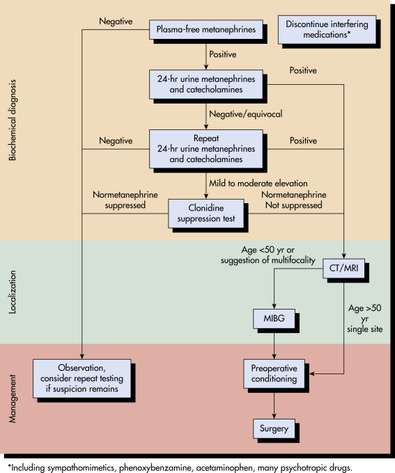
Laboratory evaluation and imaging studies to locate the neoplasm (Fig. 1). Anatomic and functional imaging studies that can be used to localize pheochromocytomas are summarized in Table E2. Misdiagnosis of pheochromocytoma is common. Correct interpretation of biochemical tests and imaging is crucial to a correct diagnosis.

Figure 1 Algorithm for the diagnosis, localization, and management of pheochromocytoma.

Initial plasma-free metanephrine testing can effectively exclude the diagnosis if the result is negative. A 24-h urine collection for catecholamines and their metabolites is generally performed twice, with cutoffs approximately twice the upper limit of normal being criteria for positivity (see Table 3). Clonidine suppression testing can be used for the small fraction of patients in whom the diagnosis remains uncertain after urine testing. Localization with computed tomography (CT) or magnetic resonance imaging (MRI) follows biochemical confirmation of the diagnosis, with metaiodobenzylguanidine (MIBG) scanning performed for younger patients and those otherwise at risk for multifocal disease. Phenoxybenzamine is given in escalating doses for at least 2 wk before surgery.

From Townsend CM et al: Sabiston textbook of surgery, ed 21, St Louis, 2022, Elsevier.

TABLE E2 Anatomic and Functional Imaging Studies That Can Be Used to Localize Pheochromocytomas and Paragangliomas

Imaging ModalityMRICT18F-FDG68Ga-DOTA Peptide18F-FDA18F-DOPAMIBG
AdvantagesAnatomic detailAnatomic detailWidely available, preferred functional imaging testMost sensitive functional imaging study for all sites of disease, potential for therapy with avid diseaseSensitive functional imaging for primary tumor (except skull base and neck)Most sensitive functional imaging study for skull base and neck paragangliomasPotential for therapy if disease avid tumor is not resectable
DisadvantagesLower specificityLower specificityLower sensitivity and higher false-positive rate as compared with other functional imaging studiesNot widely availableNot widely availableNot widely availableLower sensitivity than other functioning imaging studies
Best clinical setting to use imaging studiesInitial localization of tumorInitial localization of tumorRule out metastatic disease or multiple primaryRule out metastatic disease or multiple primary; SDHx mutation carriers; when planning treatment with peptide receptor radionuclide therapyRule out metastatic disease or multiple primaryPatients with primary skull base and neck paragangliomas; multiple and metastatic diseaseWhen planning treatment with 131I-MIBG

CT, Computed tomography; DOPA, dihydroxyphenylalanine; DOTA, 1,4,7,10-tetraazacyclododecane-1,4,7,10-tetraacetic acid; FDA, fluorodopamine; FDG, fluorodeoxyglucose; MIBG, metaiodobenzylguanidine; MRI, magnetic resonance imaging; SDH, succinate dehydrogenase.

From Cameron JL, Cameron AM: Current surgical therapy, ed 12, Philadelphia, 2017, Elsevier.

Laboratory Tests

  • Although there is no consensus on the best test, plasma-free metanephrines have been suggested as the test of first choice for excluding the tumor. Elevated plasma concentrations of normetanephrine or metanephrine have a sensitivity of up to 100%, but the specificity is markedly lower (85%).
  • 24-h urine collection will also show increased metanephrines (90% sensitivity, 95% specificity); the accuracy of the 24-h urinary levels for metanephrines can be improved by indexing urinary metanephrine levels by urine creatinine levels.
  • Cutoff values for biochemical diagnosis of pheochromocytoma are summarized in Table 3.
  • Medications that may increase measured levels of fractionated catecholamines and metanephrines are summarized in Box 3.

BOX 3 Medications That May Increase Measured Levels of Fractionated Catecholamines and Metanephrines

  • Tricyclic antidepressants (including cyclobenzaprine)
  • Levodopa
  • Drugs containing adrenergic receptor agonists (e.g., decongestants)
  • Amphetamines
  • Buspirone and antipsychotic agents
  • Prochlorperazine
  • Reserpine
  • Withdrawal from clonidine and other drugs (e.g., illicit drugs)
  • Illicit drugs (e.g., cocaine, heroin)
  • Ethanol

From Melmed S et al: Williams textbook of endocrinology, ed 14, Philadelphia, 2020, Elsevier.

TABLE 3 Cutoff Values for Biochemical Diagnosis of Pheochromocytoma

Cutoff Value
TestmolgDefinitionsSensitivity (%)Specificity (%)
Plasma-free metanephrine03 nmol/L59 μg/LPaired test, positive result if either or both values are elevated9985-89
Plasma-free normetanephrine06 nmol/L110 μg/L
Urinary total metanephrines66 μmol/day13 mg/day71996
Urinary epinephrine191 nmol/day35 μg/day29996
Urinary norepinephrine1005 nmol/day170 μg/day50996
Urinary dopamine4571 nmol/day700 μg/day8100
Urinary total metanephrines and catecholamines-Grouped test, positive result if any one of the following three urinary values is elevated: Total metanephrines, epinephrine, norepinephrine, dopamine8899
Urinary vanillylmandelic acid40 μmol/day79 mg/day6495
Clonidine suppression testPositive result = elevated level after clonidine and fall of <4096100
Plasma-free normetanephrine061 nmol/L112 μg/L

When it is performed twice, 24-h urine testing of urinary total metanephrines and catecholamines (grouped test) is highly sensitive and highly specific.

From Townsend CM et al: Sabiston textbook of surgery, ed 21, St Louis, 2022, Elsevier.

Imaging Studies

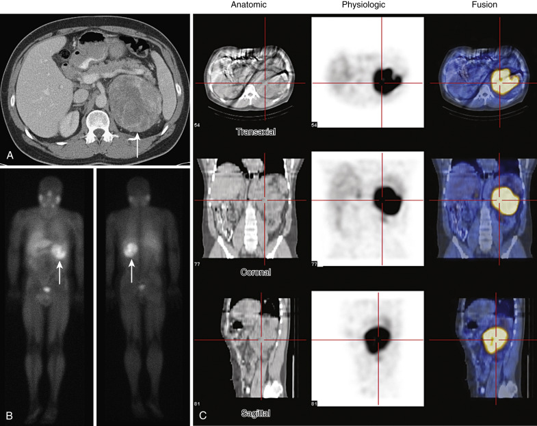
  • Abdominal computed tomography (CT) scan (Fig. E2) with and without contrast (88% sensitivity) is useful in locating pheochromocytomas >0.5 inch in diameter (90% to 95% accurate).
  • MRI with contrast: Pheochromocytomas demonstrate a distinctive MRI appearance (up to 100% sensitivity); MRI may become the diagnostic imaging modality of choice.
  • Scintigraphy with 131 or 1-123 I-MIBG (up to 100% sensitivity) (see Fig. E2): This norepinephrine analog localizes in adrenergic tissue; it is particularly useful in locating extraadrenal pheochromocytomas.
  • 6-[18F]Fluorodopamine positron emission tomography is reserved for cases in which clinical symptoms and signs suggest pheochromocytoma, and results of biochemical tests are positive but conventional imaging studies cannot locate the tumor. It is also used for identification of metastatic disease.

Figure E2 Computed Tomography (CT) and 123I-Metaiodobenzylguanidine (123I-MIBG) Imaging of a 44-Yr-Old Man

He presented with a 9-yr history of hypertension and recent onset of head throbbing, chest pressure, and abdominal pain. The 24-h urine studies were abnormal: Norepinephrine, 900 μg (normal, <170); epinephrine, 28 μg (normal, <35); dopamine, 468 μg (normal, <700); and total metanephrines, 17,958 μg (normal, <1000). A, Axial CT image with contrast shows a large, partially vascular, and partially necrotic left adrenal tumor (arrow). B, 123I-MIBG whole-body scan shows a large focus of increased radiotracer uptake in the left upper abdomen (arrow) that corresponds to the mass seen on the CT image; no other abnormal uptake is seen. C, 123I-MIBG and single-photon emission computed tomography (SPECT) fusion images correlate the images seen on CT (anatomic) with those seen on 123I-MIBG (physiologic) in the axial, coronal, and sagittal planes. After α- and β-adrenergic blockade, a 13.5-cm × 12-cm × 9-cm, 680-g pheochromocytoma was removed.

From Melmed S: Williams textbook of endocrinology, ed 12, Philadelphia, 2011, Saunders.

Treatment

General Rx

Laparoscopic adrenalectomy (surgical resection for both benign and malignant disease):

  • Preoperative stabilization with combination of alpha-adrenergic blocking agents (phenoxybenzamine, prazosin, doxazosin, or terazosin), β-blocker, and liberal fluid and salt intake starting 10 to 14 days before surgery. β-Blockers should be avoided until patients receive adequate alpha-adrenergic blockade for several days to avoid hypertensive crisis due to unopposed alpha stimulation. Amlodipine or verapamil can be added to β-blockers if blood pressure control is still inadequate. Table 4 describes orally administered drugs to treat pheochromocytoma.
  • Hypertensive crisis preoperatively and intraoperatively can be controlled with nitroprusside. Table 5 summarizes intravenously administered drugs used to treat pheochromocytoma.

TABLE 5 Intravenously Administered Drugs Used to Treat Pheochromocytoma

AgentDosage Range
For Hypertension
PhentolamineAdminister a 1-mg IV test dose, then 2- to 5-mg IV boluses as needed or continuous infusion.
NitroprussideIV infusion rates of 2 μg/kg of body weight per min are suggested as safe. Rates >4 μg/kg/min may lead to cyanide toxicity within 3 h. Doses >10 μg/kg/min are rarely required, and the maximal dose should not exceed 800 μg/min.
NicardipineInitiate therapy at 5 mg/h; the IV infusion rate may be increased by 2.5 mg/h q15min up to a maximum of 15 mg/h.
For Cardiac Arrhythmia
LidocaineInitiate therapy with an IV bolus of 1-1.5 mg/kg (75-100 mg); additional boluses of 0.5-0.75 mg/kg (25-50 mg) can be given q5-10min if needed up to a maximum of 3 mg/kg. Loading is followed by maintenance IV infusion of 2-4 mg/min (30-50 μg/kg/min) adjusted for effect and settings of altered metabolism (e.g., heart failure, liver congestion) and as guided by blood level monitoring.
EsmololAn initial IV loading dose of 0.5 mg/kg is infused over 1 min, followed by a maintenance infusion of 0.05 mg/kg/min for the next 4 min. Depending on the desired ventricular response, the maintenance infusion may then be continued at 0.05 mg/kg/min or increased stepwise (e.g., by 0.1 mg/kg/min increments to a maximum of 0.2 mg/kg/min), with each step being maintained for 4 min.

IV, Intravenous.From Melmed S: Williams textbook of endocrinology, ed 12, Philadelphia, 2011, Saunders.

TABLE 4 Orally Administered Drugs Used to Treat Pheochromocytoma

DrugInitial Dosage, mg/day (Maximum)Side Effects
α-Adrenergic Blocking Agents
Phenoxybenzamine10 (100)Postural hypotension, tachycardia, meiosis, nasal congestion, diarrhea, inhibition of ejaculation, fatigue
Prazosin1 (20)First-dose effect, dizziness, drowsiness, headache, fatigue, palpitations, nausea
Terazosin1 (20)First-dose effect, asthenia, blurred vision, dizziness, nasal congestion, nausea, peripheral edema, palpitations, somnolence
Doxazosin1 (20)First-dose effect, orthostasis, peripheral edema, fatigue, somnolence
Combined α- and β-Adrenergic Blocking Agent
Labetalol200 (1200)Dizziness, fatigue, nausea, nasal congestion, impotence
Calcium Channel Blocker
Nicardipine sustained-release30 (120)Edema, dizziness, headache, flushing, nausea, dyspepsia
Catecholamine Synthesis Inhibitor
α-Methyl-ρ-l tyrosine (metyrosine)1000 (4000)Sedation, diarrhea, anxiety, nightmares, crystalluria, galactorrhea, extrapyramidal symptoms

Given once daily unless otherwise indicated.

Given in two doses daily.

Given in three or four doses daily.

From Melmed S: Williams textbook of endocrinology, ed 12, Philadelphia, 2011, Saunders.

Pearls & Considerations

Comments

  • Obtaining a detailed family history is important because 25% of pheochromocytomas are familial.
  • Screening for pheochromocytoma should be considered in patients with any of the following:
    1. Malignant hypertension
    2. Poor response to antihypertensive therapy
    3. Paradoxic hypertensive response
    4. Hypertension during induction of anesthesia, parturition, surgery, or thyrotropin-releasing hormone testing
    5. Hypertension associated with imipramine or desipramine
    6. Neurofibromatosis (increased incidence of pheochromocytoma)
  • All patients with pheochromocytoma should be screened for MEN-2 and von Hippel-Lindau disease with the pentagastrin test, serum parathyroid hormone, ophthalmoscopy, MRI of the brain, CT scan of the kidneys and pancreas, and ultrasonography of the testes.
  • In patients with pheochromocytoma, routine analysis for mutations of RET, VHL, SDHD, and SDHB is indicated to identify pheochromocytoma-associated syndromes.
Related Content

Pheochromocytoma (Patient Information)

Hypertension (Related Key Topic)

Suggested Readings

    1. Himes C.P. : Perioperative evaluation and management of endocrine disordersMayo Clin Proc. ;95(12):2760-2774, 2020.
    2. Lenders J.W. : Pheochromocytoma and paraganglioma: an endocrine society clinical practice guidelineJ Clin Endocrinol Metab. ;99:1915-1942, 2014.
    3. Neumann H.P.H. : Pheochromocytoma and paragangliomaN Engl J Med. ;381(6):552-565, 2019.
    4. PDQ® Adult Treatment Editorial Board: PDQ pheochromocytoma and paraganglioma treatment, Bethesda, MD, 2020, National Cancer Institute. Available at https://www.cancer.gov/types/pheochromocytoma/hp/pheochromocytoma-treatment-pdq. Accessed January 9, 2021.