section name header

Basic Information

AUTHORS: Amanda Jones, MD and Anthony Sciscione, DO

Definition

A pituitary adenoma is a benign neoplasm of the anterior lobe of the pituitary. Pituitary adenomas may cause symptoms, either by excess secretion of hormones or by a local mass effect as the tumor impinges on other nearby structures (e.g., the optic chiasm, hypothalamus, or pituitary stalk). Pituitary adenomas are classified by their size, function, and features that characterize their appearance. Microadenomas are <10 mm in size, macroadenomas are 10 mm in size, and giant adenomas are 40 mm in size. Depending on the anterior pituitary cell type of origin of the tumor, a pituitary adenoma may be secretory or nonsecretory.

  • Nonsecretory pituitary adenomas are those in which the neoplasm is a space-occupying lesion whose secretory products do not cause a specific disease state.
  • Secretory pituitary adenomas (Table 1) can cause endocrine manifestations related to the hormone they are secreting.

TABLE 1 Types of Pituitary Adenomas Based on Hormones Secreted

Adenoma TypeHormonesPossible Clinical Manifestations
Somatotroph adenomaGrowth hormoneGigantism, acromegaly
MammosomatotrophGrowth hormone + prolactinGigantism or acromegaly along with galactorrhea or amenorrhea
Corticotroph adenomaAdrenocorticotropic hormoneCushing disease
Lactotroph adenomaProlactinWomen: Reproductive or sexual dysfunction, galactorrhea, amenorrhea, ovulatory disorders
Men: Erectile dysfunction, decreased libido
Gonadotroph adenomaFollicle-stimulating hormone or luteinizing hormoneMost are clinically silent and usually present due to mass effect
Thyrotroph adenomaThyroid-stimulating hormoneHyperthyroidism
PlurihormonalGrowth hormone, prolactin, thyroid-stimulating hormone, various hormonal combinationsMost are clinically silent
Null cell adenomaNo hormonesNo symptoms of hormonal excess, usually present due to mass effect

Most common type of adenoma (40% of cases).

From Jankovic J et al: Bradley and Daroff’s neurology in clinical practice, ed 8, Philadelphia, 2022, Elsevier.

Endocrine manifestations of secretory adenomas include:

  • Acromegaly, a disease state characterized by a pituitary adenoma with somatotroph cell origin that secretes growth hormone (GH); notably, a significant majority of somatotroph adenomas are clinically silent
  • Galactorrhea, which is the result of a prolactinoma with lactotroph cell origin that secretes prolactin (PRL)
  • Cushing disease, a disease state of hypersecretion of adrenocorticotropic hormone (ACTH) due to corticotroph cell origin of the tumor
  • Hyperthyroidism due to a thyrotropin-secreting pituitary adenoma, due to a thyrotroph cell origin which secretes primarily thyroid-stimulating hormone (TSH); notably, thyrotroph adenomas may also present as nonsecretory sellar masses
ICD-10CM CODE
D35.2Benign neoplasm of pituitary gland
Epidemiology & Demographics
Classification (by Hormone Secreted)

  • No hormone: 30%
  • PRL only: 35%
  • GH only: 20%
  • PRL and GH: 7%
  • ACTH: 7%
  • Luteinizing hormone (LH), follicle-stimulating hormone (FSH), TSH: 1%
Incidence & Prevalence

  • Pituitary adenomas: Up to 15% of all intracranial neoplasms; 3% to 27% at autopsy series. The prevalence of pituitary adenomas has increased to 100 cases/100,000 over the past decades, likely as a result of enhanced awareness and improved diagnostic imaging and hormone assays. True prevalence is likely underestimated due to nonfunctioning pituitary adenomas going undiagnosed until they are large. There is a higher prevalence in females.
  • Prolactinomas: Up to 20% in women with unexplained primary or secondary amenorrhea.
  • GH-secreting pituitary adenoma: Prevalence of 3.3 to 13.7 cases/100,000. They account for 8% to 16% of pituitary tumors and are most common in men over age 50.
  • Thyrotropin-secreting pituitary adenoma: 1% of pituitary adenomas with a slight female:male predominance of 1.7:1.
  • Corticotropin-secreting pituitary adenomas: Female:male predominance of 8:1 but overall uncommon diagnosis, accounting for 2% to 6% of adenomas.
Physical Findings & Clinical Presentation

Most individuals remain asymptomatic.

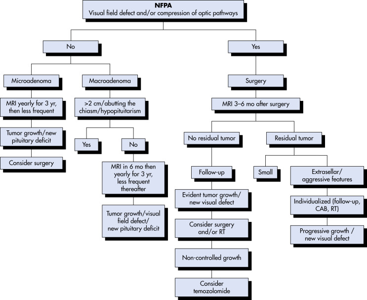
All sellar masses can cause visual defects by compression of the optic chiasm (bitemporal hemianopsia (Fig. E1) or headaches.

Figure E1 The Visual Pathway, Which Extends from the Eyes to the Occipital Cortex, is Vulnerable Throughout its Entire Course

Lesions at Specific Sites Produce Characteristic Visual Field Defects. Conversely, the Defects Indicate the Lesion's Location and Give Hints to its Etiology and Expected Associated Findings. (A) Optic Nerve Lesions Typically Produce an Ipsilateral Scotoma. (B) Optic Chiasm Lesions from below, When Small (Smaller Ring), Cause a Superior Bitemporal Quadrantanopsia. (C) When Large (Larger Ring), Optic Chiasm Lesions Result in Bitemporal Hemianopia. (D) Cerebral Lesions that Disrupt the Optic Tract Produce Contralateral Homonymous Hemianopia. (E) Lesions that Interfere with the Occipital Cortex Also Yield Contralateral Homonymous Hemianopia, Sometimes with Macular Sparing. Although the Determination of Visual Fields Serves as a Highly Reliable Sign of Localized Neurologic Disease, Their Graphic Representation is One of Clinical Neurology’s Most Confusing Exercises. In the Standard Manner, These Sketches Portray Visual Field Defects as Solid or Crosshatched Areas from the Patient’s Perspective. for Example, the Sketch of the Left Homonymous Hemianopia (D) Portrays the Abnormal Areas on the Left Side of Each Circle-as When the Patient Looks at the Paper. The Sketch of Cerebral Optic Tract Pathways Portrays the Tracts as Though a Picture Had Been Taken of the Patient’s Brain from above. In Contrast, Cts and Mris Traditionally Show the Left Side of the Brain on the Right Side of the Image. Medical Illustrations Should Include a Notation to Orient the Reader.

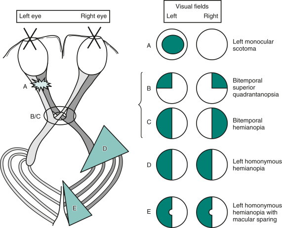
From Kaufman DM et al: Kaufman’s clinical neurology for psychiatrists, ed 9, Philadelphia, 2023, Elsevier.

Prolactinomas

  • Females:
    1. Galactorrhea
    2. Amenorrhea
    3. Oligomenorrhea with anovulation
    4. Infertility
    5. Estrogen deficiency and associated osteopenia
    6. Decreased vaginal lubrication
  • Males:
    1. Large tumors more common as a result of delayed diagnosis
    2. Possible impotence, decreased libido, or hypogonadism
    3. Galactorrhea rare because males lack the estrogen-dependent breast growth and differentiation
GH-Secreting Pituitary Adenoma-Acromegaly

  • Coarse facial features
  • Oily skin
  • Prognathism
  • Carpal tunnel syndrome
  • Osteoarthritis
  • History of increased hat, glove, or shoe size
  • Decreased exercise capacity
  • Visual field deficits
  • Diabetes mellitus
Corticotropin-Secreting Pituitary Adenoma-Cushing Disease

  • Usually present when the tumor is small (1 to 2 mm)
  • 50% of the tumors are <5 mm
  • Other symptoms:
    1. Truncal obesity
    2. Round facies (moon face)
    3. Dorsocervical fat accumulation (buffalo hump)
    4. Hirsutism
    5. Acne
    6. Menstrual disorders
    7. Hypertension
    8. Striae
    9. Bruising
    10. Thin skin
    11. Hyperglycemia
Thyrotropin-Secreting Pituitary Adenoma

  • In males, larger, more invasive, and more rapidly growing tumors that present later in life
  • Other symptoms: Thyrotoxicosis, goiter, visual impairment
Nonsecretory Pituitary Adenomas (Endocrine Inactive Pituitary Adenoma)

  • Usually large at the time of diagnosis
  • Symptoms:
    1. Bitemporal hemianopsia as a result of compression of the optic chiasm
    2. Hypopituitarism from compression of the pituitary gland
    3. Hypogonadism in men and in premenopausal women
    4. Cranial nerve deficits caused by extension into the cavernous sinus
    5. Hydrocephalus from extension into the third ventricle, compressing the foramen of Monro
    6. Diabetes insipidus resulting from compression of the hypothalamus or pituitary stalk (a rare complication)
Etiology

Benign neoplasms of epithelial origin. Genetic mutations of MEN1, Gs-Alpha, and AIP are related to the development of pituitary adenomas.

Diagnosis

Differential Diagnosis
Prolactinoma

  • Pregnancy
  • Postpartum puerperium
  • Primary hypothyroidism
  • Breast disease
  • Breast stimulation
  • Drug ingestion (especially phenothiazines, antidepressants, haloperidol, methyldopa, reserpine, opiates, amphetamines, and cimetidine)
  • Chronic renal failure
  • Liver disease
  • Polycystic ovarian disease
  • Chest wall disorders
  • Spinal cord lesions
  • Previous cranial irradiation
Acromegaly

  • Ectopic production of GH-releasing hormone from a carcinoid or other neuroendocrine tumor
Cushing Disease

  • Diseases that cause ectopic sources of ACTH overproduction (including small cell carcinoma of the lung, bronchial carcinoid, intestinal carcinoid, pancreatic islet cell tumor, medullary thyroid carcinoma, or pheochromocytoma)
  • Adrenal adenomas, adrenal carcinoma
  • Nelson syndrome
Thyrotropin-Secreting Pituitary Adenomas

  • Primary hypothyroidism
Nonsecretory Pituitary Adenoma

  • Nonneoplastic mass lesions of various etiologies (e.g., infectious, granulomatous, cystic, pituitary hyperplasia)
  • Other sellar tumors (e.g., craniopharyngioma, meningioma, pituicytoma)
  • Metastases to the hypothalamus or pituitary gland (e.g., breast cancer in females, lung cancer in males)
Workup

  • Pituitary adenomas should be identified at an early stage so that effective treatment can be implemented.
  • Screening tests for functional pituitary adenomas are described in Table 2.

TABLE 2 Screening Tests for Functional Pituitary Adenomas

DisorderTestComments
AcromegalyIGF-1 OGTT with GH obtained at 0, 30, and 60 minInterpret IGF-1 relative to age- and gender-matched controls. Normal subjects should suppress GH to <1 μg/L.
ProlactinomaSerum PRL levelA level >500 μg/L is pathognomonic for macroprolactinoma. If >200 μg/L, prolactinoma is likely.
Cushing disease24-h UFC nighttime salivary cortisol dexamethasone (1 mg) at 11 P.M. and fasting plasma cortisol measured at 8 A.M. ACTH assayEnsure that urine collection is total and accurate by measuring urinary creatinine. Free salivary cortisol reflects circadian rhythm, and elevated levels may indicate Cushing disease. Normal subjects suppress to <1.8 μg/dl. Distinguishes adrenal adenoma from ectopic ACTH or Cushing disease.
TSH-secreting tumorTSH measurement free T4 by dialysis total T3If T4 or T3 is elevated and TSH is measurable or elevated, a TSH-secreting tumor may be present.

ACTH, Adrenocorticotropic hormone; GH, growth hormone; IGF-1, insulin-like growth factor type 1; OGTT, oral glucose tolerance test; PRL, prolactin; T3, triiodothyronine; T4, thyroxine; TSH, thyroid-stimulating hormone; UFC, urinary free cortisol.

Risperidone may result in prolactin levels >200 μg/L.

From Melmed S et al: Williams textbook of endocrinology, ed 12, Philadelphia, 2011, Saunders.

Prolactinoma

First step: Measurement of basal PRL levels (practitioners should be aware of discriminatory values in their own institutions).

  • Elevated PRL levels are correlated with tumor size.
  • Level >200 ng/ml indicates likely prolactinoma, with levels of 100 to 200 ng/ml being equivocal and possibly associated with medications or other sources.
  • Basal PRL levels between 20 and 100 suggest a microadenoma but can be due to common medications (estrogen, antidepressants, metoclopramide, Aldomet, and others) or recent breast stimulation.
  • Basal level <20 ng/ml is usually considered normal. Each laboratory should develop its own normative values, however, and practitioners should refer to these values.
  • Threshold level for obtaining imaging such as MRI should be developed by individual providers depending on the level of specificity and sensitivity desired.
Acromegaly

  • First screening tests are the measurement of the serum insulin-like growth factor I level, postprandial serum GH, and thyrotropin-releasing hormone (TRH) stimulation test.
  • Follow with an oral glucose tolerance test.
  • Failure to suppress serum GH to <2 ng/ml with an oral load of 100 g glucose is considered conclusive.
  • A GH-releasing hormone level >300 ng/ml is indicative of an ectopic source of GH.
Cushing Disease

  • Measurement of late-night salivary cortisol level is the best screening test.
  • Normal or slightly elevated corticotropin levels ranging from 20 to 200 pg/ml; normal is 10 to 50 pg/ml (normative data should be developed by each institution for its population).
  • Level <10 pg/ml usually indicates an autonomously secreting adrenal tumor.
  • Level >200 pg/ml suggests an ectopic corticotropin-secreting neoplasm.
  • Cushing disease can be assessed by absence of cortisol suppression with the low-dose dexamethasone test but with the presence of cortisol suppression after the high-dose test. As a method to distinguish Cushing disease from an ectopic source of ACTH, this test is robust.
  • 24-h urine collection should demonstrate an increased level of cortisol excretion.
Thyrotropin-Secreting Pituitary Adenoma

  • Highly sensitive thyrotropin assays, which evaluate the presence of thyrotoxicosis, are one way to detect a thyrotropin-secreting tumor.
  • Free alpha subunit is secreted by >80% of tumors, with the ratio of the alpha subunit to thyrotropin <1.
  • With central resistance to thyroid hormone, ratio is <1, and the sella is normal.
  • Laboratory tests show elevated serum levels of both T3 and T4.
Nonsecretory Pituitary Adenoma

  • Visual field testing
  • Assessment of the pituitary and organ function to determine if there is hypopituitarism or hypersecretion of hormones (even if the effects of hypersecretion are subclinical)
  • TRH to provoke secretion of FSH, LH, and LH-beta-subunit; will not elicit response in normal persons
  • Exclusion of Klinefelter syndrome in patient with long-standing primary hypogonadism, elevated gonadotropin levels, and enlargement of the sella
Imaging Studies

Study of choice: MRI of the pituitary and hypothalamus. If an MRI shows the tumor impinging on the optic chiasm, then formal visual field testing is indicated.

  • When evaluating Cushing disease, small size at the onset of symptoms noted.
  • MRI, in this case, only 60% sensitive at best and may yield false-positive results.
  • CT scan only when MRI is unavailable or is otherwise contraindicated.

Treatment

Nonpharmacologic Therapy
Surgery

  • Selective transsphenoidal resection of the adenoma (Table 3) is the treatment of choice for acromegaly, Cushing disease, and thyrotropin-secreting pituitary adenomas, all of which tend to be microadenomas at the time of onset of symptoms.
  • Macroadenomas, such as the nonsecretory pituitary adenoma, may also be surgically removed, but risk of recurrence is greater with these tumors and adjunctive therapy such as irradiation may also be necessary.
  • Bilateral adrenalectomy has been performed in patients with Cushing disease after failure of other therapies; complications that may occur include requiring lifelong hormone replacement or Nelson syndrome (rapid enlargement of pituitary tumor due to adrenal resection).

TABLE 3 Transsphenoidal Pituitary Surgery

Primary Indications
General
Visual tract or central nervous system compression arising from within sella
Relief of compressive hypopituitarism by presenting, residual, or recurrent tumor tissue
Tumor recurrence after surgery or irradiation
Pituitary hemorrhage
Cerebrospinal fluid leak
Resistance to medical therapy
Intolerance of medical therapy
Personal choice
Desire for immediate pregnancy with macroadenoma
Requirement for diagnostic tissue histology
Specific
Acromegaly
Cushing disease
Clinically nonfunctioning macroadenoma
Prolactinoma
Nelson syndrome
TSH-secreting adenoma
Side Effects
Transient
Diabetes insipidus
Cerebrospinal fluid leak and rhinorrhea
Inappropriate ADH secretion
Arachnoiditis
Meningitis
Postoperative psychosis
Local hematoma
Arterial wall damage
Epistaxis
Local abscess
Pulmonary embolism
Narcolepsy
Permanent (up to 10%)
Diabetes insipidus
Total or partial hypopituitarism
Visual loss
Inappropriate ADH secretion
Vascular occlusion
CNS damage: Oculomotor palsy, hemiparesis, encephalopathy
Nasal septum perforation
Surgery-Related Mortality (up to 1%)
Brain, hypothalamic
Vascular damage
Postoperative meningitis
Cerebrospinal fluid leak
Pneumocephalus
Acute cardiopulmonary disease
Anesthesia-related
Seizure

ADH, Antidiuretic hormone; CNS, central nervous system; TSH, thyroid-stimulating hormone.

From Melmed S et al: Williams textbook of endocrinology, ed 12, Philadelphia, 2011, Saunders.

Radiotherapy

  • Radiotherapy is used primarily as adjuvant treatment. It is reserved for patients who have not responded to surgical treatment and who still have symptoms of the adenoma.
  • Used with varying degrees of success in all types of pituitary adenomas.
  • Radiotherapy complications include long-term hypopituitarism (40% of patients) and secondary neoplasms (1.5% of patients).
Acute General Rx
Prolactinoma

  • For prolactinomas, initial therapy is generally dopamine agonists. Bromocriptine, a dopamine analogue, is generally given orally in divided doses of 1.5 to 10 mg. Cabergoline is given once or twice weekly. It is better tolerated and more effective than bromocriptine for tumor shrinkage but more expensive.
  • Side effects of these treatments include orthostatic hypotension, nausea, and dizziness and may be avoided by beginning with low-dose therapy.
  • Other compounds include pergolide mesylate, a long-acting ergot derivative with dopaminergic properties, as well as other nonergot derivatives.
Acromegaly

  • Somatostatin analogues: Octreotide, lanreotide administered as monthly injections.
  • Cabergoline or bromocriptine may also be used. They have modest activity but can be administered orally and are less expensive than somatostatin analogues.
  • Pegvisomant may also be used to normalize IGF-1 levels.
Cushing Disease

  • Ketoconazole, which inhibits the cytochrome P-450 enzymes involved in steroid biosynthesis, is effective in managing mild to moderate disease in daily oral doses of 600 to 1200 mg.
  • Metyrapone and aminoglutethimide may be used to control hypersecretion of cortisol but are generally used when preparing a patient for surgery or while waiting for a response to radiotherapy.
Thyrotropin-Secreting Pituitary Adenoma

  • Ablative therapy with either radioactive iodide or surgery is indicated.
  • Treatment directed to the thyroid alone may accelerate growth of the pituitary adenoma.
  • Octreotide has been shown to be effective in doses similar to those used for acromegaly.
Nonsecretory Pituitary Adenoma

  • There is no role for medical therapy at this time.
  • Surgery and radiotherapy may be indicated. An algorithm for the management of nonfunctioning pituitary adenomas is described in Fig. E2.

Figure E2 Suggested algorithm for the management of patients with NFPAs.

CBA, Cabergoline; NFPA, nonfunctioning pituitary adenoma; MRI, magnetic resonance imaging; RT, radiotherapy.

From Drummond JB et al: Non-functioning pituitary adenomas. [Updated 2018 Nov 28.] In Feingold KR et al (eds): Endotext [Internet], South Dartmouth, 2000, MDText.

Chronic Rx

For all pituitary adenomas:

  • Careful follow-up is necessary. Patients undergoing transsphenoidal microsurgical resection should be seen in 4 to 6 wk to ensure that the adenoma has been completely removed and that any endocrine hypersecretion is resolved.
  • If there is good clinical response, patient should be monitored yearly for recurrence and to follow the level of the hypersecreted hormone.
  • Patients who have undergone irradiation should experience close follow-up with backup medical therapy, because response to radiotherapy may be delayed; incidence of hypopituitarism also increases with time.
  • Surgical resection is not indicated in pituitary incidentalomas that are microadenomas, as only 10% will experience tumor growth.
  • Approximately 30% of surgically resected adenomas have persistent or progressive postoperative growth for several decades. Tumors particularly prone to invasive growth and recurrence include those arising from sparsely granulated somatotrophs, silent corticotrophs, corticotroph Crooke cells, and lactotrophs in middle-aged or older men.
Related Content

Pituitary Adenoma (Patient Information)

Acromegaly (Related Key Topic)

Amenorrhea (Related Key Topic)

Cushing Disease and Syndrome (Related Key Topic)

Galactorrhea (Related Key Topic)

Prolactinoma (Related Key Topic)

Suggested Readings

    1. Melmed S. : Pituitary-tumor endocrinopathiesN Engl J Med. ;382:937-950, 2020.
    2. Molitch M.E. : Diagnosis and treatment of pituitary adenomas: a reviewJAMA. ;317(5):516-524, 2016.
    3. Trouillas J. : In search of a prognostic classification of endocrine pituitary tumorsEndocr Pathol. ;25(2):124-132, 2014.