section name header

Basic Information

AUTHOR: Fred F. Ferri, MD

Definition

Insulinoma is a pancreatic insulin-secreting tumor that leads to inappropriately elevated plasma insulin or proinsulin levels with suppression of hepatic glucose output and subsequent hypoglycemia, especially during periods of fasting.

ICD-10CM CODES
C25.4Malignant neoplasm of endocrine pancreas
D13.7Benign neoplasm of endocrine pancreas
D37.7Neoplasm of uncertain or unknown behavior of other digestive organs (pancreas)
Epidemiology & Demographics
Incidence

One case per 250,000 persons annually. 90% of insulinomas are benign.

Predominant Sex & Age

Insulinomas occur in both sexes (approximately 60% in women) and at all ages. In a Mayo Clinic series, the median age at diagnosis was 50 yr in sporadic cases, but 23 yr in patients with multiple endocrine neoplasia, type 1 (MEN-1).

Physical Findings & Clinical Presentation

Symptoms typically occur in the morning before breakfast (i.e., fasting hypoglycemia as opposed to reactive hypoglycemia, which is not commonly associated with insulinoma).

Neuroglycopenic Symptoms%
Various combinations of diplopia, blurred vision, sweating, palpitations, or weakness85
Confusion or abnormal behavior80
Unconsciousness or amnesia53
Grand mal seizures12
Adrenergic Symptoms%
Sweating43
Tremulousness23
Hunger, nausea12
Palpitations10
Etiology, Pathology, & Pathophysiology

  • Insulinomas are almost always solitary. Malignant insulinomas account for 5% of the total; they tend to be larger (6 cm). Metastases are usually to the liver (47%), regional lymph nodes (30%), or both.
  • Insulinomas are evenly distributed in the head, body, and tail of the pancreas; ectopic insulinomas are rare (1% to 3%). Tumor size: 5% are 0.5 cm, 34% are 0.5 to 1 cm, 53% are 1 to 5 cm, and 8% are >5 cm.
  • Histologic classification includes insulinoma in 86% of patients, adenomatosis in 5% to 15%, nesidioblastosis in 4%, and hyperplasia in 1%. Adenomatosis consists of multiple macroadenomas or microadenomas and occurs especially in patients with MEN-1. Nesidioblastosis is also a diffuse lesion in which islet cells form as buds on ductular structures.
Diagnosis

Differential Diagnosis (Of Fasting Hypoglycemia)

Hyperinsulinism:

  • Insulinoma
  • Nonpancreatic tumors
  • Severe congestive heart failure
  • Severe renal insufficiency in non-insulin-dependent diabetes

Hepatic enzyme deficiencies or decreased hepatic glucose output (primarily in infants and children):

  • Glycogen storage diseases
  • Endocrine hypofunction
  • Hypopituitarism
  • Addison disease
  • Liver failure
  • Alcohol abuse
  • Malnutrition

Exogenous agents:

  • Sulfonylureas, biguanides
  • Insulin
  • Other drugs (aspirin, pentamidine)

Functional fasting hypoglycemia:

  • Autoantibodies to insulin receptor or insulin
Laboratory Tests

  • An overnight fasting blood sugar level combined with a simultaneous plasma insulin, proinsulin, and/or C peptide level will establish the existence of fasting organic hypoglycemia in 60% of patients. Table E1 describes biochemical patterns in patients with various causes of hyperinsulinemic hypoglycemia.
  • If single overnight fasting glucose and insulin levels are nondiagnostic, a 72-h fast is usually done with blood glucose and insulin levels determined at 2- to 4-h intervals. A total of 75% of patients with insulinoma develop symptoms and a blood sugar level of <40 mg/dl by 24 h, 92% to 98% develop these by 48 h, and virtually all patients develop them by 72 h. The test is considered positive for insulinoma if the plasma insulin/glucose ratio is more than 0.3. If at any point the patient becomes symptomatic, plasma insulin and glucose values should be obtained and IV glucose should be administered.
  • The Endocrine Society guidelines for diagnosis of hypoglycemic disorders is based on glucose level <55 mg/dl (<3.1 mmol/L), elevated C-peptide level 0.61 ng/ml (0.2 nmol/L). Elevated insulin level 18 pmol/L, proinsulin level >5 pmol/L, and suppressed β-hydroxy-butyric acid level.
  • An “amended” insulin-glucose ratio that accounts for the normal variation in insulin secretion according to prevailing glycemia has been shown to improve diagnostic accuracy of insulinomas. The “amended” insulin-glucose ratio is derived from the simple insulin-glucose ratio by subtracting 30 mg/dl (1.7 mmol/L) from the measured glucose concentrations.

Table E1 Interpretation of Laboratory Results and Differential Diagnosis in Patients With Whipple Triad

DiagnosisGlucose (mg/dl)Insulin (mIu/mL)C-Peptide (nmol/L)Proinsulin (pmol/L)Anti-insulin or Anti-insulin Receptor Antibody (+/-)Circulating Oral Hypoglycemic Agents (Sulfonylureas, Meglitinides)Beta Hydroxy-ButyratePancreatic Mass (Islet Cell Tumor)Timing of Hypoglycemia
Insulinoma<5530.25-No2.7YesFasting
NIPHS, postgastric bypass hypoglycemia<5530.25-No2.7NoPostprandial
Surreptitious insulin administration<55>>>3<0.2<5-No2.7NoWith administration of inappropriate insulin
Oral hypoglycemic administration<5530.25-Yes2.7NoWith administration of oral agents
Insulin autoimmune hypoglycemia<55>>>3>>>0.2>>>5+No2.7NoFasting
IGF mediated<55<3<0.2<5-No2.7NoFasting
IGF mediated<55<3<0.2<5-No>2.7NoFasting

IGF, Insulin-like growth factor; NIPHS, noninsulinoma pancreatogenous hypoglycemia syndrome.

Laboratory findings consistent with insulinoma should prompt evaluation for islet cell tumor. In the minority of cases, the pancreatic islet cell tumor may be difficult to localize preoperatively.

From Townsend CM et al: Sabiston textbook of surgery, ed 21, St Louis, 2022, Elsevier.

Imaging Studies

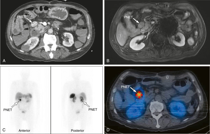
  • Abdominal CT scan (Fig. E1) or MRI detects half to two thirds of insulinomas (abdominal ultrasound is not effective); should be done only after laboratory tests for insulinoma have confirmed the diagnosis
  • Intraoperative ultrasound
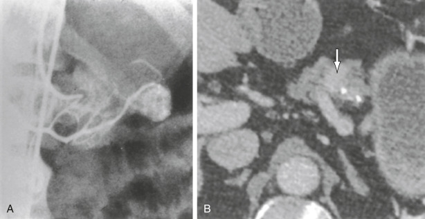
  • Arteriography (Fig. E2)
  • Octreotide scan (see Fig. E1)

Fig E1 Patient with a Pancreatic Neuroendocrine Tumor (Pnet)

(A) Computed Tomography (CT) Scan Demonstrating a Hyperenhancing Lesion Adjacent to the Head of the Pancreas (Arrow). (B) Same Lesion Shown on Magnetic Resonance Imaging. (C) Same Lesion Shown on Somatostatin Receptor Scintigraphy, Anterior and Posterior View; Note the Nonprecise Anatomic Localization and the Physiologic Uptake of Tracer in the Kidneys, Liver, and Spleen. (D) Same Lesion on Single-Photon Emission CT/CT; Note the Better Anatomic Localization and Clear Identification of Nonphysiologic Tracer Uptake.

From Townsend CM et al: Sabiston textbook of surgery, ed 21, St Louis, 2022, Elsevier.

Fig E2 Arteriographic Demonstration of an Insulinoma

(A) Selective Injection into the Specific Dorsal Pancreatic Artery Demonstrates the Tumor Precisely. (B) Insulinoma with Triphasic Enhancement on Computed Tomography. The Mass in the Pancreatic Body (Arrow) Demonstrates Early and Prolonged Enhancement with Washout During the Portal Venous Phase; Note that the Maximal Difference in Enhancement Between the Tumor and Normal Pancreas Occurs During the Pancreatic Phase (Shown).

From Townsend CM et al: Sabiston textbook of surgery, ed 21, St Louis, 2022, Elsevier.

Treatment

Nonpharmacologic Therapy

  • Enucleation of single insulinoma
  • Partial pancreatectomy for multiple adenomas
Acute General Rx

  • Carbohydrate administration
  • Diazoxide directly inhibits insulin release and has an extrapancreatic, hyperglycemic effect that enhances glycogenolysis
  • Lanreotide and octreotide (somatostatin analogues)
  • Streptozotocin
Referral

To an endocrinologist and then to an endocrine surgeon

Related Content

Hypoglycemia (Related Key Topic)