section name header

Basic Information ⬇

AUTHOR: Muhammad Ubaid Hafeez, MD

Definition

Unruptured intracranial aneurysms (UIAs) are acquired aneurysms found at bifurcations of major arteries. Most are found in the circle of Willis, with 85% being in the anterior circulation. Rupture of these aneurysms results in more than 80% to 85% of nontraumatic subarachnoid hemorrhages (SAH).

Synonyms

Berry aneurysms

Saccular aneurysms

UIAs

ICD-10CM CODE
I67.1Cerebral aneurysm, nonruptured
Classification

A classification of intracranial aneurysms is summarized in Box E1.

BOX E1 Classification of Intracranial Aneurysms

By Morphology

Saccular

Fusiform

Dissecting

By Pathogenesis

Genetic

Hemodynamic (arteriovenous malformation, contralateral carotid ligation, etc.)

Infection (bacterial, fungal)

Trauma

Atherosclerosis

Neoplasm (primary or metastatic)

Radiation

Drug abuse

Connective tissue disorders

Vasculopathy

Miscellaneous conditions (moyamoya disease, etc.)

By Size

<3 mm (small)

3-6 mm (small)

7-12 mm (small-medium)

13-25 mm (large)

>25 mm (giant)

By Location
Internal Carotid Artery
  1. Cavernous
  2. Carotid cave
  3. Paraclinoid
  4. Ophthalmic
  5. Superior hypophyseal
  6. Posterior communicating
  7. Anterior choroidal
  8. Carotid bifurcation
Anterior Cerebral Artery
  1. A1
  2. Anterior communicating
  3. A1-2
  4. A2 and distal
Middle Cerebral Artery
  1. M1
  2. Bifurcation
  3. M2 and distal
Vertebral Artery
  1. Posterior inferior cerebellar artery
Basilar Artery
  1. Trunk
  2. Fenestration
  3. Anterior inferior cerebellar artery
  4. Superior cerebellar artery
  5. Basilar apex
Posterior Cerebral Artery
  1. P1
  2. P2 and distal

From Jankovic J et al: Bradley and Daroff’s neurology in clinical practice, ed 8, Philadelphia, 2022, Elsevier.

Epidemiology & Demographics
Prevalence

The prevalence of UIAs is approximately 3% in a population without comorbidity and with a mean age of 50 yr. Most UIAs are likely never discovered, and the incidence of rupture is very low at about 0.25%. However, about 50% of aneurysms that are discovered are found after they have ruptured and resulted in SAH.1

Predominant Sex & Age

UIAs are three times more common in women and are more likely to occur in those aged 60 and older. In the pediatric population, UIAs are twice as common in males.1,2

Peak Incidence

The incidence of UIAs is currently unknown.

Risk Factors (Box E2

Modifiable risk factors include cigarette smoking and hypertension. Nonmodifiable risk factors include female sex, older age, family history, and genetic factors.

  • Risk factors that increase the risk of SAH are also relevant to consider, as aneurysm rupture results in SAH. These factors include smoking, heavy alcohol use, high-dose estrogen, cocaine use, and low body mass index.1,2

BOX E2 Risk Factors and Associated Conditions for Intracranial Aneurysms

  • Hypertension
  • Tobacco smoking
  • Autosomal dominant inherited polycystic kidney disease
  • Ehlers-Danlos syndrome, vascular type (formally called type IV)
  • Fibromuscular dysplasia
  • Coarctation of the aorta
  • Moyamoya syndrome
  • Pseudoxanthoma elasticum
  • α1-Antitrypsin deficiency
  • Systemic lupus erythematosus
  • Sickle cell anemia
  • Bacterial endocarditis
  • Fungal infection
  • Neurofibromatosis type 1
  • Tuberous sclerosis
  • Pheochromocytoma
  • Arteriovenous malformations
  • Anomalous carotid-vertebrobasilar anastomoses
  • Cocaine use

From Jankovic J et al: Bradley and Daroff’s neurology in clinical practice, ed 8, Philadelphia, 2022, Elsevier.

Genetics

No specific genetic mutations have been shown to cause UIAs. However, more than 19 single nucleotide polymorphisms (SNPs) have been associated with sporadic aneurysms. SNPs on the EDNRA, CDKN2B, and SOX17 genes located on chromosomes 4, 8, and 9 have been found to have the strongest association with developing UIAs.1

  • Genetic disorders that increase the risk of UIAs include autosomal dominant polycystic kidney disease, Marfan syndrome, Ehlers-Danlos syndrome type IV, multiple endocrine neoplasia type I, neurofibromatosis type 1, and hereditary hemorrhagic telangiectasia. 1 Less commonly, aneurysms can occur with systemic lupus erythematosus, fibromuscular dysplasia, sickle-cell disease, and coarctation of the aorta.1,2
Physical Findings & Clinical Presentation

UIAs often remain asymptomatic and undetected for long periods of time but also may present with nonspecific symptoms such as headaches or vertigo. A phase of aneurysm growth usually precedes any cases of rupture. Small aneurysms (<5 mm) are likely to remain asymptomatic, and those that grow to a large size may compress surrounding structures and present with specific neurologic deficits. Examples include third cranial nerve palsy due to an aneurysm of the posterior communicating or basilar arteries or cavernous sinus syndrome due to aneurysms in the cavernous sinus. In rare cases, emboli may originate from aneurysms and cause transient ischemic attacks or infarctions in other regions.

Etiology

The development of aneurysms is a multifactorial process that involves hemodynamic stress, inflammation, and molecular changes that result in a weakened arterial wall. Saccular aneurysms form as protrusions that are often thin-walled and lack tunica media. Fusiform aneurysms are less common and form due to dilatation of the artery itself. It is currently thought that hemodynamic stress causes endothelial dysfunction and starts the process of arterial wall inflammation and remodeling, while other factors such as smoking and connective tissue disorders are contributory.3

Diagnosis ⬆ ⬇

Workup

  • The presence of UIAs should be investigated in those with a history of SAH.
  • Patients with significant family history (>2 first-degree relatives with UIA or SAH), autosomal dominant polycystic kidney disease, coarctation of the aorta, or microcephalic osteodysplastic primordial dwarfism should be offered screening with magnetic resonance angiography (MRA) or computed tomographic angiography (CTA).1,2
Laboratory Tests

Laboratory tests are unrevealing when investigating UIAs.

Imaging Studies

  • Most UIAs are discovered incidentally on brain MRI or CT. However, MRA and CTA have been shown to detect smaller aneurysms and are the tests of choice in screening for UIAs. Patients with contraindications to contrast administration can be screened with MRA.
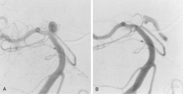
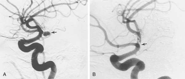
  • Digital subtraction angiography (Figs. E1 and E2) is used when surgical treatment of UIA is considered.

Figure E1 (A) Subtraction Carotid Angiogram Shows a 4- × 6-mm Berry Aneurysm (Arrow) Originating from the Distal Internal Carotid Artery

(B) Postoperative Carotid Angiogram Shows Clip Placement (Arrow) with Total Obliteration of the Aneurysm.

From Townsend CM et al: Sabiston textbook of surgery, ed 21, St Louis, 2022, Elsevier.

Figure E2 (A) Subtraction Vertebral Angiogram Shows a Basilar Tip Aneurysm

(B) Subtracted Vertebral Angiogram after the Placement of Coils Demonstrates Excellent Obliteration of the Aneurysm and Preservation of Adjacent Vessels.

From Townsend CM et al: Sabiston textbook of surgery, ed 21, St Louis, 2022, Elsevier.

Treatment ⬆ ⬇

Nonpharmacologic Therapy

  • Reduction of risk factors such as smoking.4
  • Regular physical exercise is associated with a reduced risk of aneurysm formation.5 However, intense exercise has been reported as a potential trigger for rupture.5,6 Patients, in general, are advised to stay physically active due to small population attributable risk and perceived cardiovascular benefits.5,6
Acute General Rx

  • The main surgical treatments for UIAs are endovascular coiling and surgical clipping. Endovascular flow-diverting stents and Woven Endo Bridge (WEB) devices have recently emerged as treatment options in select cases. The decision for surgical or endovascular treatment is based on the location, size, morphology, growth rate, prior history of ruptured aneurysms, associated neurologic deficits, family history, and comorbid conditions.2
  • In the landmark study International Study of Unruptured Intracranial Aneurysms, the cumulative 5-yr risk of rupture for aneurysms of <7 mm was 0% in anterior circulation and 2.5% in posterior circulation. In real world experience and as seen in other studies, the risk of rupture is not completely absent for aneurysms <7 mm.8,9 Currently, it is thought that stable, small UIAs of <3 mm can be monitored conservatively by serial imaging, whereas patients who present with headaches or nerve palsies should be treated surgically. Beyond this, a detailed discussion of the risks and benefits and an individualized treatment plan is necessary.2,3,7-9
  • Patients with known UIAs presenting with severe headaches or symptoms of nerve compression should raise high suspicion for development of SAH.
Chronic Rx

  • Blood pressure management with a goal of normal blood pressure.2
  • Aspirin use has been associated with lower risk of aneurysm rupture based on nonrandomized observational studies.2
Disposition

The risk of rupture for patients managed conservatively is related to age, smoking status, blood pressure, size, growth rate, location of the aneurysm, and history of previous SAH. The risk for rupture according to size and location as is summarized in Table E1.7 Currently, it is recommended that those with family history, multiple UIAs, or a high number of risk factors should be monitored for aneurysm growth.

TABLE E1 Risk of Rupture Over 5 Year (%) According to International Study of Unruptured Intracranial Aneurysms

Type Of Aneurysm<7 mm and No Prior Sah<7 mm and Prior Sah7-12 mm13-24 mm>24 mm
Carotid-cavernous0003.06.4
Anterior circulation01.52.614.540.0
Posterior circulation2.53.414.518.450.0

SAH, Subarachnoid hemorrhage.

From Townsend CM et al: Sabiston textbook of surgery, ed 21, St Louis, 2022, Elsevier.

Referral

Vascular neurology or neurosurgery

Pearls & Considerations ⬆ ⬇

Prevention

Decreasing modifiable risk factors such as smoking

Patient & Family Education

American Association of Neurological Surgeons https://www.aans.org/Patients/Neurosurgical-Conditions-and-Treatments/Cerebral-Aneurysm

Related Content

Subarachnoid Hemorrhage (Related Key Topic)

Related Content ⬆

  1. Brown R.D., Broderick J.P. : Unruptured intracranial aneurysms: epidemiology, natural history, management options, and familial screeningLancet Neurol. ;13(4):393-404, 2014.doi:10.1016/s1474-4422(14)70015-8
  2. Thompson B.G. : Guidelines for the management of patients with unruptured intracranial aneurysms: a guideline for healthcare professionals from the American Heart Association/American Stroke AssociationStroke. ;46(8):2368-2400, 2015.
  3. Signorelli F. : Hemodynamic stress, inflammation, and intracranial aneurysm development and rupture: a systematic reviewWorld Neurosurg. ;115:234-244, 2018.
  4. Can A. : Association of intracranial aneurysm rupture with smoking duration, intensity, and cessationNeurology. ;89(13):1408-1415, 2017.
  5. Vlak M.H. : Trigger factors and their attributable risk for rupture of intracranial aneurysms: a case-crossover studyStroke. ;42(7):1878-1882, 2011.doi:10.1161/strokeaha.110.606558
  6. Vlak M.H.M. : Independent risk factors for intracranial aneurysms and their joint effectStroke. ;44(4):984-987, 2013.doi:10.1161/STROKEAHA.111.000329
  7. Wiebers DO: Unruptured intracranial aneurysms: natural history and clinical management. Update on the international study of unruptured intracranial aneurysms, Neuroimaging Clin North Am 16(3):383-390, vii, 2006.
  8. Burkhardt J.K. : Management of small incidental intracranial aneurysmsNeurosurg Clin North Am. ;28(3):389-396, 2017.
  9. Malhotra A. : Growth and rupture risk of small unruptured intracranial aneurysms: a systematic reviewAnn Internal Med. ;167(1):26-33, 2017.