section name header

Basic Information

AUTHOR: Anthony G. Thomas, DO, FACP

Definition

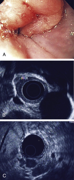
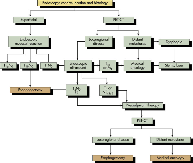
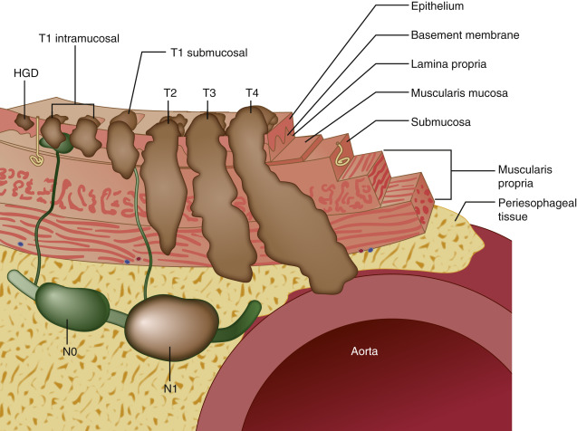
Esophageal tumors include benign and malignant neoplasms of the esophageal mucosa and wall. Carcinomas of the esophageal epithelium, both squamous cell carcinoma and adenocarcinoma (including adenoacanthoma, mucoepidermoid, and adenoid cystic), are the most common tumors of the esophagus. Rare esophageal tumors include both malignant (spindle cell, small cell, sarcoma, lymphoma, melanoma, and choriocarcinoma) and benign neoplasms (leiomyoma, papilloma, and fibrovascular polyps). One can also develop metastatic disease from a cancer that originated in another organ, but this is very rare. Breast cancer, lung cancer, and melanoma would be the most likely culprits. Other cancers can directly extend to the esophagus from the larynx, pharynx, lung, thyroid, or stomach. Approximately 15% of esophageal tumors arise in the proximal esophagus, 50% in the middle third of the esophagus, and 35% in the lower third. Tumors involving the esophageal-gastric junction are usually staged and treated as esophageal cancers if the tumor epicenter is no more than 2 cm into the proximal stomach. Tumors in the upper two thirds are usually squamous cell cancers, and tumors in the lower third are usually adenocarcinomas. The incidence of superficial esophageal cancers is increasing. These invade no deeper than the submucosa.

ICD-10CM CODES
C15.XMalignant neoplasm of the esophagus (X defines location)
C15.3Malignant neoplasm of upper third of esophagus
C15.4Malignant neoplasm of middle third of esophagus
C15.5Malignant neoplasm of lower third of esophagus
D00.2Carcinoma of esophagus, in situ
Epidemiology & Demographics
Incidence

It is the eighth most common cancer worldwide and the seventh leading cause of cancer deaths. Rates are increasing every decade and are highest in the Asian esophageal cancer belt, extending from the Caspian Sea to northern China, with certain high-incidence pockets in Finland, Ireland, southeast Africa, and northwest France. Incidence has increased six-fold since 1975. Foods including cured meats and spicy regional fare likely play a role in certain areas. Increasing substance abuse with tobacco and alcohol also coincides with a rise in esophageal cancers. Rates of squamous cell carcinoma are decreasing while those of adenocarcinomas are dramatically increasing. The direct causes of squamous cell carcinoma most commonly include tobacco and alcohol abuse. Epithelial dysplasia usually occurs, which progresses to carcinoma in situ. Adenocarcinomas are usually the result of gastroesophageal reflux disease (GERD) and obesity. The mucosa of the esophagus undergoes intestinal metaplasia. Genetic alterations occur and perpetuate during proliferation.

Prevalence

In the U.S., there were an estimated 16,000 new cases and 15,000 deaths are reported yearly, making it the seventh leading cause of death by cancer among men. The majority of cases are diagnosed at an advanced stage (unresectable or metastatic disease).

Race, Sex, & Age Predominance

In the U.S., squamous cell esophageal cancer is more common among African Americans compared to whites, whereas adenocarcinoma more common in whites. The overall male:female ratio is 3 to 4:1; the highest male:female ratio is in the Hispanic population. Usually develops in fifth to seventh decades and associated with lower socioeconomic status.

Genetics

Increasing evidence shows that genetics may play a role by increasing susceptibility to esophageal cancer. One well-identified disease associated with esophageal cancer is tylosis (focal nonepidermolytic palmoplantar keratoderma), linked to loss of heterozygosity on chromosome 17q. Familiar clustering of Barrett esophagus and the recent identification of germline mutations in affected sibling pairs support a genetic link to esophageal adenocarcinoma. Also, up to 35% of esophageal adenocarcinomas can have overexpression of HER2, which can lead to the use of targeted therapy (trastuzumab) in the treatment plan.

Clinical Presentation

Symptoms and signs:

  • Dysphagia (74%): Initially with solid foods, gradually progresses to semisolids and liquids; latter signs usually indicate incurable disease with tumor involving more than 60% of the esophageal circumference. It may be felt as chest pain
  • Unintentional weight loss of short duration. Loss >10% body mass predicts poor outcome
  • Hoarseness: Suggests recurrent laryngeal nerve involvement
  • Odynophagia and halitosis: Unusual symptoms
  • Cervical adenopathy: Usually involving supraclavicular lymph nodes
  • Dry cough: Suggests tracheal involvement
  • Aspiration pneumonia: Caused by fistula between the esophagus and trachea
  • Iron deficiency anemia: Related to chronic GI blood loss
  • Massive hemoptysis or hematemesis from the invasion of vascular structures
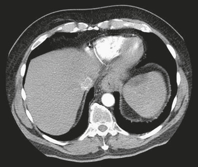
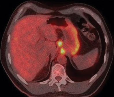
  • Advanced disease spreads to lymph nodes, liver, lungs, peritoneum, and pleura
  • Hypercalcemia: Associated with squamous cell carcinoma from secretion of a parathyroid-like tumor peptide

Clinical findings:

  • 50% to 60% of patients present with inoperative disease (locally advanced/metastatic).
Etiology

The pathogenesis of esophageal cancers is attributable to chronic recurrent oxidative damage from any of the following etiologic agents, which cause inflammation, and esophagitis, increased cell turnover, and, ultimately, initiation of the carcinogenic process.

Etiologic Agents

Squamous cell carcinoma

  • Excess alcohol consumption is strongly associated with squamous cell esophageal cancer in the U.S.; hard liquor is associated with a higher incidence than wine or beer
  • Tobacco and alcohol synergistically increase risk for squamous cell cancer
  • Other ingested carcinogens:
    1. Nitrates (converted to nitrites): South Asia, China
    2. Smoked opiates: Northern Iran
    3. Fungal toxins in pickled vegetables
    4. Betel nut chewing
  • Mucosal damage
    1. Long-term exposure to extremely hot tea (>70° C [158° F])
    2. Ingestion of lye or extremely acidic solutions, including reflux of stomach acid
  • Radiation-induced strictures
  • Achalasia: Incidence of esophageal cancer is seven times greater in this population
  • Host susceptibility as a result of precancerous lesions:
    1. Plummer-Vinson syndrome (Paterson-Kelly): Glossitis with iron deficiency
    2. Congenital hyperkeratosis and pitting of palms and soles (tylosis)
  • Human papillomavirus infection (types 16 and 18) has been detected in squamous cell carcinoma of the esophagus, sometimes associated with p53 tumor suppressor gene mutations
  • Possible relationship with prolonged bisphosphonates (10 prescriptions, or >3 yr use)
  • Possible association with celiac sprue or dietary deficiencies molybdenum, selenium, zinc, vitamin A
Adenocarcinoma

  • The incidence of adenocarcinoma is continually rising.
  • Smoking increases risk of adenocarcinoma, particularly in patients with Barrett.
  • Obesity, hiatal hernia, and diets lacking in fresh fruit and vegetables and high in fat (particularly from red meat and processed foods) increase risk.
  • Chronic GERD leading to Barrett metaplasia and adenocarcinoma via immune cell infiltration and production of inflammatory mediators and reactive oxygen species. The annual rate of transformation from Barrett to adenocarcinoma is <0.5%.
  • Helicobacter pylori infection may reduce risk of adenocarcinoma but can increase risk of lymphoma.

Diagnosis

Differential Diagnosis

  • Achalasia
  • Scleroderma of the esophagus
  • Diffuse esophageal spasm
  • Esophageal rings and webs
Laboratory Tests

CBC, blood chemistry, and liver enzymes should be obtained at diagnosis. No biomarkers are currently recommended to diagnose, monitor, or predict outcomes. While both the CEA and CA-19-9 can be elevated in patients with esophageal cancer (up to 70%), the sensitivity is low (18% to 35%) and there is no proven predictive value.

Imaging Studies

Imaging studies are important not only for diagnosis but for accurate staging:

  • Esophagogastroduodenoscopy (EGD) (Fig. E1) should be performed initially to visualize all tumors and allow histopathologic confirmation.
  • Endoscopic inspection of the larynx, trachea, and bronchi may identify concomitant cancers of head, neck, and lung (“triple endoscopy”).
  • Endoscopic ultrasound (EUS) (Fig. E1) appears to be the most accurate method for locoregional staging: To determine the depth of tumor invasion and to assess for and possibly obtain fine needle aspiration biopsies of suspicious lymph nodes. EUS is superior to computed tomography (CT) (Fig. 3) or PET for assessment of both T and N status. It is highly accurate for celiac nodal status, though slightly lower for other regional lymph nodes due to difficulty accessing the node without traversing the tumor. Obstructing lesions may preclude EUS assessment.
  • PET CT has become the standard of care along with EUS, for the most accurate staging. These modalities can determine tumor spread for preoperative staging. Obtaining the PET/CT scan before EUS has several advantages. The PET/CT scan may demonstrate distant metastatic disease, eliminating the need for the patient to undergo EUS. The PET/CT scan may also identify a suspicious lymph node that can be specifically examined and sampled during the EUS procedure (Fig. E2).
  • Bronchoscopy should be considered for proximal and middle third esophageal tumors to assess for direct tracheal invasion.
  • CT scans of the chest and abdomen are useful for restaging patients after initial therapy.
Figure 3 Computed Tomography of the Chest Showing Abnormal Thickening of the Esophagus Suggestive of Malignancy

From Cameron JL, Cameron AM: Current surgical therapy, ed 12, Philadelphia, 2017, Elsevier.

Figure E1 Esophageal cancer.

A, Endoscopic picture of malignant esophageal stricture. B, EUS image showing T3 lesion. C, Malignant celiac lymphadenopathy. EUS, Endoscopic ultrasound.

From Cameron JL, Cameron AM: Current surgical therapy, ed 10, Philadelphia, 2011, Saunders.

Figure E2 Fused Transaxial Positron Emission Tomography/Computed Tomography Image Demonstrating Increased Fluorodeoxyglucose Activity in a Gastroesophageal Junction Tumor and Celiac Lymphadenopathy

From Townsend CM et al: Sabiston textbook of surgery, ed 21, St Louis, 2022, Elsevier.

Staging

Table 1 describes the TNM staging system for cancer of the esophagus from the American Joint Committee on Cancer Criteria. The depth of invasion of the tumor defines the T status (Fig. 4). High-grade dysplasia includes malignant cells confined to the epithelium by the basement membrane and is by definition noninvasive (Tis). T1a tumors invade the lamina propria or muscularis mucosa, whereas T1b tumors invade into the submucosa. T2 tumors invade the muscularis propria, and T3 tumors invade the adventitia but not surrounding structures. T4a tumors invade adjacent structures that are usually resectable (diaphragm, pleura, and pericardium). T4b tumors invade adjacent structures that are typically unresectable (trachea and aorta). Fig. 5 illustrates an algorithm for staging thoracic esophageal cancer.

TABLE 1 TNM Staging System for Cancer of the Esophagus (American Joint Committee on Cancer Criteria)

Primary Tumor (T)
TXPrimary tumor cannot be assessed
T0No evidence of primary tumor
TisHigh-grade dysplasia
T1Tumor invades lamina propria, muscularis mucosae, or submucosa
T1aTumor invades lamina propria or muscularis mucosae
T1bTumor invades submucosa
T2Tumor invades muscularis propria
T3Tumor invades adventitia
T4Tumor invades adjacent structures
T4aResectable tumor invading pleura, pericardium, or diaphragm
T4bUnresectable tumor invading other adjacent structures, such as aorta, vertebral body, trachea, etc.
Lymph Node (N)
NXRegional lymph nodes cannot be assessed
N0No regional lymph node metastasis
N1Metastasis in 1-2 regional lymph nodes
N2Metastasis in 3-6 regional lymph nodes
N3Metastasis in 7 or more regional lymph nodes
Distant Metastasis (M)
MXMetastasis cannot be assessed
M0No distant metastasis
M1Distant metastasis

TNM, tumor, node, metastases.

(1) At least maximal dimension of the tumor must be recorded and (2) multiple tumors require the T(m) suffix.

High-grade dysplasia includes all noninvasive neoplastic epithelia that was formerly called carcinoma in situ.

Number must be recorded for total number of regional nodes sampled and total number of reported nodes with metastasis.

From Edge S et al (eds): AJCC cancer staging manual, ed 7, New York, 2010, Springer.

Figure 5 An algorithm for staging a thoracic esophageal cancer patient.

!!flowchart!!

Metastatic disease must always be confirmed by pathologic evaluation of the tissue in question. PET-CT, Positron emission tomography-computed tomography.

From Sellke FW et al: Sabiston & Spencer surgery of the chest, ed 9, Philadelphia, 2016, Elsevier.

Figure 4 Tumor classification for esophageal carcinoma as defined by depth of invasion.

HGD, High-grade dysplasia.

From Townsend CM et al: Sabiston textbook of surgery, ed 21, St Louis, 2022, Elsevier.

Treatment

All Stages of Esophageal Cancer

Although the histology of esophageal cancer can differ, most studies have combined tissue types in exploring treatment options. The histology develops secondary to differing pathogenesis and causative agents with tumor biology likely playing a role through varying mutations. The likelihood of response and prognosis can differ as well. However, as there is a lack of data on how histology should dictate the treatment approach, we generally attack this cancer in a uniform manner as most studies have suggested the optimum benefit be obtained through a neoadjuvant chemoradiation approach followed by surgery when applicable in up to stage III disease.

Combination Therapy: Chemoradiotherapy Followed by Surgical Resection

Induction chemotherapy, given before chemoradiation for patients with locally advanced but still possibly resectable disease, has been administered with good results. However, no studies have shown this approach superior to chemoradiation alone. Standard fractionation 3D-RT is utilized in chemoradiotherapy.

  • Chemotherapy is most often given with concurrent radiotherapy. Chemotherapy acts as a radiosensitizer and makes tumor cells more vulnerable to the effects of ionizing radiation, thus improving tumoricidal effects on cancer. Neoadjuvant chemoradiotherapy followed by surgery is the most common approach for patients with resectable disease but is employed primarily for patients with stage IIA or higher disease. Five-yr survival is improved with a neoadjuvant approach (39%) versus surgery alone (16%). Several trials have now shown that preoperative therapy improves survival among patients with potentially curable esophageal or esophagogastric junction cancer. Neoadjuvant chemotherapy alone is another option for locally advanced disease, but results are not as good as with neoadjuvant chemoradiotherapy.
  • Chemoradiotherapy followed by surgery should be offered to late stage I (T1bN0 or higher), stage II, and stage III esophageal cancer patients as the current standard of care. In several studies, this approach significantly improved local control, reduced recurrence, and reduced mortality compared with surgery alone in patients with resectable esophageal cancer. This also increases the chance of an R0 resection. Trimodality therapy is the preferred treatment for most esophageal cancers.
  • Chemoradiotherapy alone may be offered as definitive treatment for patients who are not surgical candidates, and some of these patients may be cured.
  • Combination chemotherapy using a platinum doublet (platinum agent plus second agent) can achieve significant tumor reduction in 30% to 60% of patients. Cisplatin, oxaliplatin, or carboplatin is usually given with 5-FU (5-fluorouracil) or paclitaxel to obtain the desired tumoricidal effects. Other chemotherapeutic agents with activity in esophageal cancer include docetaxel, irinotecan, and epirubicin. The latter two are not used with concurrent radiotherapy secondary to increased toxicities.
  • Capecitabine in combination with either cisplatin or oxaliplatin is as effective as 5-FU in the neoadjuvant or definitive treatment setting.
  • Neoadjuvant chemoradiotherapy can result in a 50% pCR for patients with squamous pathology and 25% pCR for those with adenocarcinoma. Consequently, the need for resection in these patients is unclear. To establish a CR short of resection, the patient would need EGD with EU and biopsies. In patients whom surgery is a high risk secondary to comorbidities, those with squamous pathology may opt to defer on surgery if all biopsies are negative.
  • Complications of chemoradiotherapy primarily include mucositis, nausea, vomiting, diarrhea, myelosuppression, nephrotoxicity, ototoxicity, neurotoxicity with peripheral neuropathy, esophageal stricture, esophageal rupture, trachea-esophageal fistula (6%), and radiation pneumonitis. These can occur to varying degrees and are certainly more significant in the elderly and those with significant comorbidities.
  • Postoperative adjuvant chemoradiotherapy should be offered to node-positive patients who underwent surgical resection without neoadjuvant chemoradiotherapy.
Surgical Resection

  • Surgical resection of both squamous cell and adenocarcinoma of the middle esophagus and lower third of the esophagus is an acceptable initial modality for local and resectable disease (early stage I and stage II) in the absence of widespread metastases detected by CT-PET and transesophageal ultrasound (T1 and T2 tumors). Gastric pull-through or colonic interposition typically is used to provide luminal continuity.
  • The optimal timing of surgery following neoadjuvant chemoradiotherapy is approximately 5 to 7 wk, though some patients may require more time to improve nutrition.
  • Endoscopic mucosal resection may replace radical surgical resection in patients with dysplasia and some small early tumors with no lymph node involvement (Tis or T1a), but a recent Cochrane review found no studies comparing endoscopic resection vs. surgery. Endoscopic mucosal resection may be beneficial for patients who are poor surgical candidates. It may be performed in conjunction with ablative therapies, including radiofrequency ablation, thermal ablation techniques, laser ablation, argon plasma coagulation, or photodynamic therapy. Electrocoagulation (electrofulguration) is also being used and may aid in the relief of esophageal blockage.
  • Complications of surgery, including endoscopic resection and local therapy:
    1. Esophageal rupture.
    2. Anatomic fistula (usually with colon interposition, subphrenic abscesses).
    3. Respiratory complications.
    4. Cardiovascular complications are most common, including myocardial infarction, cerebrovascular accident, and pulmonary embolism.
    5. Mortality is lower and clinical outcomes are better at high-volume hospitals and with minimally invasive surgery.
    6. Hybrid minimally invasive esophagectomy has been reported to have a lower incidence of intraoperative and postoperative major complications, specifically pulmonary complications, than open esophagectomy, without compromising overall and disease-free survival over a 3-yr period.1

Despite adequate preoperative staging, 25% of patients initially treated with surgical resection will have microscopically positive resection margins and are upstaged at the time of surgery. This has led to the majority of patients receiving neoadjuvant chemoradiotherapy as demonstrated in the CROSS Trial. The median disease-free survival for this group of patients was significantly prolonged as compared with the surgery-alone group. Death from recurrent cancer was decreased by 9% in the neoadjuvant group as well. The benefit of neoadjuvant therapy on survival was consistent regardless of histologic subtype.

For patients who receive no neoadjuvant therapy and undergo resection and are found to have positive margins or node-positive disease, adjuvant therapy is strongly recommended in an attempt to prevent progression. There is likely a benefit for chemoradiotherapy with positive margins, but the role of chemotherapy alone vs. chemoradiotherapy in node-positive patients is unclear. If combination therapy is likely to be tolerated, it should be considered.

Pretreatment Patient Preparation

The patient needs to stop smoking and drinking alcohol if at all possible. Before neoadjuvant or definitive chemoradiotherapy, the patient should have placement of an intravenous access device and a feeding tube (J-tube is preferable before surgical resection).

Radiation Therapy

  • Squamous cell carcinoma is more radiosensitive than adenocarcinoma. Radiotherapy achieves good local control but is generally only used as monotherapy in a palliative mode for obstructive symptoms in patients with unresectable or advanced cancer or those with multiple comorbidities that limit treatment. It is best used for cervical esophageal tumors, but response rates are best when combined with chemotherapy.
  • Radiotherapy in the preoperative/neoadjuvant setting is taken to a total dose of 40 to 50 Gy. For definitive therapy, the dose range is 60 to 66.6 Gy.
  • Palliative radiotherapy for bone metastasis is also effective.
  • Complications of radiotherapy: Can best be avoided by 3D conformal therapy.
    1. Esophageal stricture, fistula formation, radiation-induced pulmonary fibrosis, and transverse myelitis are the most common adverse events.
    2. Radiotherapy-induced cardiomyopathy and skin changes are rare but can occur. It is difficult to shield the heart from radiation as these organs are positioned so close.
  • Intensity modulated radiation therapy (IMRT) can also be used for the treatment of esophageal cancers. It is associated with a more favorable toxicity profile. Very few studies have been completed to date using chemotherapy with IMRT and is therefore not considered a standard approach at this time for neoadjuvant therapy.
  • Brachytherapy: This is also an option for patients in a palliative mode. It provides high-dose radiation to a localized area and may prevent the need for a stent in patients with dysphagia. Its use is very limited in prior irradiated tissue for fear of fistula and perforation of the esophagus.
  • Laser therapy and stents can also be considered for palliative care.
Treatment of Unresectable, Locally Advanced, or Metastatic Disease

  • Combination chemotherapy regimens as a rule have a higher response rate than single-agent therapy. Response rates can be as high as 50% but that does not always translate into prolonged survival.
  • Neoadjuvant chemotherapy regimens can also be utilized in locally advanced or metastatic settings. Cisplatin is probably the most active agent, and this combined with 5-FU can yield response rates shown in several studies of up to 50%. The most widely used regimen in both squamous cell and adenocarcinoma is FOLFOX. If a taxane is added to this regimen in the metastatic setting, the triple drug regimen can lead to a prolongation in disease progression by about 2 mo, which may translate into prolonged survival. However, patients have to be carefully chosen for a triple drug regimen because of increased toxicity. Other active doublet regimens combine cisplatin with irinotecan, etoposide, or gemcitabine. Capecitabine can be substituted for 5-FU in these regimens.
  • Trifluridine/tipiracil is another option for metastatic disease in the third-line setting. Fixed-dose combination tablets comprising trifluridine, an antineoplastic nucleoside analog, and tipiracil, a thymidine phosphorylase inhibitor, are available. In the TAGS trial, this combination improved both PFS and OS compared to best supportive care. However, OS was improved by only 2 mo, not a major breakthrough.
Targeted Molecular Therapy

Two drugs have been approved for adenocarcinoma of the esophagus. Ramucirumab is a VEGF inhibitor, and trastuzumab is effective in cancers with overexpression of HER2. There is no apparent role for either drug in squamous cell carcinoma.

  • Ramucirumab is a recombinant monoclonal IgG1 antibody that is a vascular endothelial growth factor receptor 2 (VEGFR-2) antagonist approved in 2014. It inhibits ligand proliferation and migration of endothelial cells and ultimately inhibits angiogenesis. Indicated for second-line therapy in with paclitaxel or as third line monotherapy.
  • Trastuzumab, in combination with oxaliplatin and 5-FU (FOLFOX), is considered the standard of care as first-line therapy for metastatic esophageal cancer in patients with HER2 overexpressing adenocarcinoma. This was approved based on the results of the ToGA trial (2010). Approximately 22% of adenocarcinomas will overexpress the type II epidermal growth factor receptor HER2. The overall response rate is 47%.
  • Fam-trastuzumab deruxtecan-nxki (Enhertu) was approved in 2021 for the treatment of metastatic gastric and gastroesophageal adenocarcinoma as monotherapy for patients who have received a prior trastuzumab-based regimen. This is an HER2-directed antibody and topoisomerase inhibitor conjugate drug that was approved based on the result of the Phase II DESTINY-Gastric 01 trial, which demonstrated statistically significant ORR and OS in patient who progressed on at least two prior chemo regimens, including trastuzumab, 5-FU, and a platinum-containing regimen.
  • Entrectinib and larotrectinib are also approved for neurotrophic-tropomyosin receptor kinase (NTRK) gene fusion-positive tumors. These are selective tyrosine kinase inhibitors. The NTRK gene fusion and amplification is much more common in adenocarcinoma, but in both types of histology, less than 1% of tumors have this molecular marker.
Immunotherapy in Esophageal Cancer

  • Drugs constitute a class that act as an immune checkpoint inhibitor that targets programmed death ligand 1 (PD-1).
  • Patients at high risk of recurrent disease following neoadjuvant chemoradiation and surgery who undergo an R0 resection have been found to benefit from 1 yr of adjuvant immunotherapy with nivolumab by the CheckMate 577 Trial reported in the New England Journal of Medicine in April 2021. Nivolumab significantly improved disease-free survival versus placebo.
  • Studies have been done using nivolumab and pembrolizumab with varying response rates of 10% to 25% in patients who have progressive/metastatic disease and failed traditional therapy with at least one prior chemotherapy regimen. Both pembrolizumab and nivolumab are now approved in the U.S. for treatment of esophageal cancer. These appear to be most effective in patients with squamous cell histology, but adenocarcinomas also respond regardless of PD-L1 status, as noted in the KEYNOTE-181 study. A recent open-label phase 3 trial of adults with previously untreated, unresectable, recurrent, or metastatic esophageal squamous cell carcinoma revealed that both first-line treatment with nivolumab plus chemotherapy and first-line treatment with nivolumab plus ipilimumab resulted in significantly longer overall survival than chemotherapy alone in patients with advanced esophageal squamous-cell carcinoma with no new safety signals identified.2
  • Patients with MSI-H- and dMMR-positive tumors are more likely to respond to immunotherapy.
Follow-Up Care

The majority of recurrences develop within 12 mo of diagnosis. Clinical monitoring, lab tests, imaging, and endoscopic evaluations where appropriate (especially in Barrett esophagus), are performed for postoperative surveillance, without clear benefit of earlier detection or decreased mortality. For patients who have undergone definitive therapy, endoscopic surveillance should be performed every 3 mo for the first yr, and then at least annually. Palliative procedures such as endoscopic dilation, endoscopic ablation, endoscopic mucosal resection, photodynamic therapy, brachytherapy, feeding tube insertion, or placement of expandable metal stents or polyvinyl prostheses to bypass tumors have all been used for unresectable patients. The morbidity and mortality associated with resection in patients with advanced disease and/or for palliation argues against offering surgery to most of these patients.

Survivorship

  • Overall 5-yr survival for all stages at presentation is 15% (39% for localized disease, 21% for regional disease, and 4% for distant disease).
  • Endoscopic therapy for highly selected stage 0 or stage I patients with disease limited to the submucosa may have 5-yr survival rates of 70% to 90%.
  • Surgical resection without neoadjuvant treatment: 5-yr survival rate is 5% to 30%, with higher survival (45%-50%) in early-stage cancers.
  • Radiation therapy without chemotherapy or surgery: 5-yr survival rate of 6% to 20%.
  • Chemoradiotherapy without surgery: 5-yr survival up to 30%.
  • Combined trimodality treatment: 45% to 50% 5-yr survival rates (all stages of disease).
  • Patients with metastatic disease have median survivals of less than 1 yr with palliative chemotherapy.
Referral

  • To gastroenterologist for endoscopy for patients with dysphagia, odynophagia, unexplained weight loss, or for palliative care
  • To medical oncologist for evaluation of preoperative chemotherapy and care of the metastatic patient
  • To radiation oncologist for palliative therapy if tumor is actively bleeding, unresectable, or if obstruction is present
  • To hospice if appropriate

Pearls & Considerations

Comments

More than 50% of patients with esophageal cancer are diagnosed when the disease is metastatic or unresectable.

Prevention

  • A diet high in fruits, vegetables, and antioxidants may be associated with lower risk of esophageal cancer.
  • Avoid tobacco and excessive alcohol use.
  • Avoid ingested toxins known to cause esophageal cancers.
  • Aspirin may have a chemopreventive role in Barrett esophagus, but is only currently recommended for patients with other (e.g., cardiac) indications.
  • No evidence that vitamins, Chinese herbs, or green tea prevents esophageal cancer.
  • Screening the general population is not recommended. If Barrett esophagus is detected, regularly scheduled surveillance endoscopies are necessary, with consideration for radiofrequency or other ablation therapy if dysplasia is detected.
Patient & Family Education

Provide education and support about the likely prognosis because most esophageal cancers are diagnosed at an advanced stage.

Related Content

Esophageal Cancer (Patient Information)

Barrett Esophagus (Related Key Topic)

Related Content

    1. Mariette C. : Hybrid minimally invasive esophagectomy for esophageal cancerN Engl J Med. ;380(2):152-162, 2019.
    2. Doki Y. : Nivolumab combination therapy in advance esophageal squamous-cell carcinomaN Engl J Med. ;386(5):449-462, 2022.
    3. Rustgi A.K., El-Serag H.B. : Esophageal carcinomaN Engl J Med. ;371:2499-2509, 2014.