section name header

Basic Information

AUTHOR: Kausik Umanath, MD, MS, FACP FASN

Definition

Renal artery stenosis (RAS) is progressive narrowing of the renal artery, which is most commonly due to atherosclerosis or fibromuscular dysplasia (FMD). RAS is an important, potentially reversible cause of hypertension, ischemic nephropathy, and destabilizing cardiac syndromes. RAS increases the risk of renal artery occlusion via progressive stenosis.

Synonyms

RAS

Fibromuscular disease

FMD

Renovascular disease

ICD-10CM CODES
Q27.1Congenital renal artery stenosis
I70.1Atherosclerosis of renal artery
I15.0Renovascular hypertension
I77.9Disorder of arteries and arterioles, unspecified
Epidemiology & Demographics

  • Atherosclerotic renal artery stenosis:
    1. Atherosclerotic renal artery stenosis (ARAS) accounts for 90% of cases. The true prevalence of ARAS is unknown.1
      1. General population autopsy studies: 10% to 27% over 50 yr; hypertensive patients, 0.2% to 5%.
      2. In a general population older than 65 yr, the prevalence is 6.8% by Doppler ultrasound (5.5% of women, 9.1% of men; 6.7% African Americans, and 6.9% White). Bilateral disease was present in 12% of affected individuals.
      3. In patients with malignant hypertension, the prevalence is 43% in White patients and 7% in African American patients. In patients with mild hypertension, the prevalence is <1%.
      4. In patients with peripheral artery disease, the prevalence is 22% to 59%.
    2. Fibromuscular dysplasia:
      1. FMD accounts for 10% of chronic RAS and is typically encountered in women (90%). FMD was previously thought to be a disease of the young and healthy, with few risk factors. Recent data suggest that the average age of onset of hypertension is 43 yr. Bilateral renal artery involvement is seen in 25% to 35% of cases.2
Physical Findings & Clinical Presentation

Progressive RAS:

  • RAS should be considered in any White female patients <30 yr with hypertension not attributed to any other cause, any patients >50 yr with new-onset refractory hypertension, or patients with stable hypertension that has abruptly and/or significantly worsened.
  • RAS most often presents as a clinically asymptomatic finding. Manifestations can include renovascular hypertension, ischemic nephropathy, or flash pulmonary edema.
  • Flash pulmonary edema in the absence of cardiac disease most often indicates severe bilateral RAS.
  • Renovascular hypertension should be considered in individuals with resistant hypertension (lack of blood pressure control on a medication regimen that includes 3 maximally dosed medications, one of which is a diuretic).
  • Ischemic nephropathy should be considered if kidney function is rapidly deteriorating or kidney sizes are decreasing on serial imaging, particularly with bilateral disease.
  • Acute elevations of serum creatinine (>30%) after starting ACE inhibitors or angiotensin receptor blockers (ARBs) may be seen in bilateral disease.
  • A bruit heard in either upper quadrant on abdominal auscultation may suggest RAS. Patients with ARAS will often have bruits heard in other vascular beds.
Etiology

  • Atherosclerosis: ARAS has similar risk factors to atherosclerosis in other vascular beds, including family history, smoking, diabetes, hypertension, and hyperlipidemia. ARAS most often involves the ostium and proximal third of the main renal artery.
  • FMD (Figs. 1 and 2): Etiology is unknown. Classified into three categories based on the affected layer of arterial wall: Medial (>90%), intimal (<10%), and adventitial (<1%). FMD typically involves the distal main renal artery and intrarenal branches.

Figure 1 Magnetic resonance angiogram.

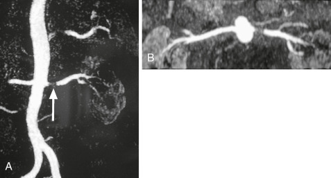
A, Coronal maximum intensity projection (MIP) of a patient with significant left renal artery stenosis (white arrow). B, Axial MIP of the same patient.

From Adam A et al: Grainger & Allison’s diagnostic radiology, ed 5, 2007, Churchill Livingstone; and Grant LA: Grainger & Allison’s diagnostic radiology essentials, ed 2, 2019, Philadelphia, Elsevier.

Figure 2 Fibromuscular dysplasia.

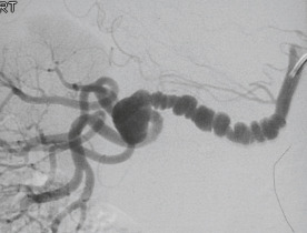
On a selective right anterior oblique (RAO) digital subtraction angiogram, the characteristic saccular dilations and weblike stenoses are clearly evident.

From Adam A et al: Grainger & Allison’s diagnostic radiology, ed 5, 2007, Churchill Livingstone; and Grant LA: Grainger & Allison’s diagnostic radiology essentials, ed 2, 2019, Philadelphia, Elsevier.

Pathogenesis

  • Pathogenesis of FMD is unknown. FMD involves abnormal constrictions and dilations of the renal artery leading to a typical “string of beads” appearance on angiography.
  • The pathogenesis of atherosclerotic RAS is similar to that of atherosclerosis in other vascular beds.
  • The pathogenesis of renovascular hypertension is related to the neurohormonal cascade resulting from renal ischemia. Renal hypoperfusion or ischemia secondary to arterial stenosis produces an increase in plasma renin and secondary elevations of angiotensin II, with consequent vasoconstriction, aldosterone elevation, sodium retention, and renal potassium wasting. Hypertension results and can be self-sustaining, even in the case of unilateral RAS due to hypertensive damage of the contralateral kidney3.
  • The pathogenesis of ischemic nephropathy is due to the activation of numerous mechanisms of tissue injury resulting in progressive renal parenchymal loss and fibrosis.
  • “Flash” or sudden-onset pulmonary edema, a manifestation usually seen in bilateral ARAS, results from sodium and water retention and upregulation of the sympathetic nervous system2.
Natural History

  • RAS caused by FMD rarely causes renal artery occlusion or ischemic nephropathy.
  • ARAS rarely progresses to total occlusion. One study of serial ultrasounds of patients with RAS demonstrated that only 5% of those with >60% stenosis progressed to total occlusion in 1 yr, and an additional 11% progressed to total occlusion by 2 yr.

Diagnosis

Screening

American College of Cardiology and American Heart Association (ACC/AHA) guidelines for identification of patients who should be screened for RAS:

  • Onset of hypertension at age <30 yr or severe hypertension at age >55 yr
  • Clinical findings that suggest secondary hypertension as opposed to essential hypertension in the absence of a more likely cause of secondary hypertension, such as pheochromocytoma
  • Malignant hypertension: Hypertension with coexistent evidence of acute end-organ damage (acute renal failure, acute decompensated heart failure, new visual or neurologic disturbance, and/or advanced retinopathy)
  • Accelerated hypertension: Sudden and persistent worsening of previously controlled hypertension
  • Resistant hypertension: Full doses of a 3-drug regimen that includes a diuretic
  • Sudden unexplained pulmonary edema
  • New acute kidney injury following initiation of an ACE inhibitor or ARB
  • Unexplained atrophic kidney or variation in size of kidneys by >1.5 cm, with smaller kidney affected by RAS
Laboratory Tests

  • Urinalysis.
  • Basic chemistry panel including sodium, potassium, blood urea nitrogen, and serum creatinine.
  • Renal vein renin sampling determines whether one or both kidneys are overproducing renin in cases of suspected renovascular hypertension (RVH). A positive result has strong predictive value. However, 50% of patients with no evidence for lateralization can still respond to unilateral intervention.
  • Renal vein sampling is typically performed at specialized hypertension centers.
Imaging Studies

Renal duplex Doppler ultrasonography, CT angiography (CTA), and magnetic resonance angiography (MRA) are effective diagnostic screening methods.1 The choice of imaging modality will depend on the availability of the diagnostic tool, experience and local accuracy of each modality, patient characteristics including body size, renal function, history of contrast media allergy, and presence of prior vascular stents.

Renal Doppler Ultrasound

  • Renal Doppler ultrasound is a noninvasive, inexpensive screening study and can determine whether a stenosis of >60% is present.
  • Abnormal results in RAS:
    1. Peak systolic velocity (PSV) >250 cm/sec
    2. Peak diastolic velocity >150 cm/sec
    3. Renal-to-aortic ratio (RAR) >3.5
    4. Acceleration time >100 msec
    5. Exact definitions of a positive renal Doppler ultrasound for RAS varies by institution
  • Compared to renal angiography, Doppler studies have shown varied performance, with a sensitivity of 84% to 98% and a specificity of 62% to 99%.
  • Limitations include high user variability. Valid test performance requires technicians who perform a high volume of these tests to reduce user error. If significant user variability exists at certain institutions, renal Doppler ultrasound may be excluded from the diagnostic algorithm. Patients with a large body habitus may have limited imaging results. It is not as sensitive or specific as MR or CT methods. Detection of RAS is limited to identifying whether stenosis of 60% to 99% is present.
  • Duplex Doppler ultrasound is a good modality for monitoring patency following therapeutic, vascular stent insertion.
Magnetic Resonance Angiography (MRA)

  • MRA provides good visualization of main and accessory renal arteries. The test has high sensitivity (90%-100%) and specificity (76%-94%). MRA is superior to renal Doppler ultrasonography and equivalent to CTA.3
  • Benefits include the lack of exposure to radiocontrast media or radiation.
  • Limitations include claustrophobia, high cost, and inability to image within a previously placed metallic stent.
Computed Tomography Angiography (CTA)

  • CTA is rapid and effective. When compared with angiography, this modality has a sensitivity of 59% to 96% and a specificity of 82% to 99%. It is superior to renal Doppler ultrasonography and equivalent to MRA. Radiocontrast media uptake in kidneys can also be used to estimate the viability of affected kidneys.3
  • CTA has good spatial resolution and can detect restenosis through metal stents.
  • CTA can be used more easily than MRA in obese or claustrophobic patients.
  • Limitations include radiation exposure and potentially nephrotoxic, iodinated radiocontrast exposure to patients with chronic kidney disease. Heavily calcified arteries may appear narrower than in actuality.
Direct Angiography

  • IV digital subtraction angiography (DSA) has an 88% sensitivity and 90% specificity. It is the gold standard for anatomic diagnosis of RAS. It is not a first-line screening tool and is recommended after a positive noninvasive test. DSA is used when there is a high clinical suspicion for RAS with inconclusive noninvasive tests and when the decision has already been made that correction of stenosis will produce clinical benefit. DSA allows for angioplasty with stenting during the same procedure. This modality is reserved for patients with a high likelihood of intervention.
  • Limitations include its invasive nature, that is, requirement for intraaortic catheterization, which may lead to aortic and/or renal artery trauma and/or aortic dissection, rupture, thrombosis, or embolization.
  • Procedure requires a certified interventionalist (radiologist, cardiologist, nephrologist, or vascular surgeon).
  • Procedure requires iodinated contrast that must be used cautiously with patients who have chronic kidney disease. Carbon dioxide contrast imaging and limiting image acquisition number reduce kidney injury risk.
Nuclear Renography (Without captopril)

A noninvasive test conducted when renal atrophy is present to document differential kidney function. This test is useful when determining whether to revascularize an atrophic kidney or to remove it. Atrophic kidneys with <20% differential renal function are unlikely contributing to overall renal functional decline but may still contribute to renovascular hypertension.

Treatment

Pharmacologic Therapy

  • Because of RAAS activation, ACE inhibitors or ARB are recommended and well-tolerated (92%) as treatment of RVH. Kidney function should be monitored carefully when initiating or titrating these medications, particularly when bilateral RAS (78% tolerability) or unilateral stenosis with a solitary kidney is present to avoid precipitating acute kidney injury.
  • Diuretics should be considered in patients with congestive heart failure or flash pulmonary edema.
  • Antiplatelet therapy and statin therapy.
Nonpharmacologic Therapy

  • If blood pressure is not controlled by medications alone, flash pulmonary edema occurs, or kidney function rapidly declines, referral for renal angiography, angioplasty, or stenting may be indicated.
  • Angioplasty without stenting is insufficient for atherosclerotic lesions due to a high failure rate or a high rate of restenosis.
  • Occasionally, a kidney that has lost function from RAS may cause refractory hypertension. This situation would be discovered only by renal vein renin sampling in a patient with unilateral renal atrophy. In such cases, nephrectomy of the atrophied kidney may be the best method to control blood pressure.
  • Renal artery bypass is rarely required.
  • If RAS is suspected as the cause of flash pulmonary edema or rapidly declining kidney function, bilateral disease is likely. If confirmed by angiography, both renal arteries should be stented.
  • Hypertension is rarely cured with revascularization of RAS due to longstanding hypertension. The goal of intervention is blood pressure control.
  • The ACC/AHA guidelines for clinical indications of renal artery revascularization in the presence of significant stenosis include the following:
    1. Accelerated, resistant, or malignant hypertension (class IIa)
    2. Hypertension with unilateral small kidney (class IIa)
    3. Hypertension with intolerance to medication (class IIa)
    4. Treatment of cardiac destabilization syndromes such as unexplained heart failure exacerbations or episodes of flash pulmonary edema (class I) and refractory or unstable angina (class IIa)
    5. Progressive chronic kidney disease with bilateral RAS or RAS associated with a solitary functioning kidney (class IIa)
  • Many patients who meet these criteria will not have a beneficial response to renal revascularization. Careful patient selection for angiography and intervention is recommended.
  • Multiple randomized controlled trials (e.g., ASTRAL, CORAL, DRASTIC, and STAR) have shown no benefit of revascularization versus medical therapy, with end points of blood pressure control, renal function, and cardiovascular events.2,4,5 Therefore the decision to refer a patient for revascularization should be made by specialists skilled and practiced in RAS.
Fibromuscular Dysplasia

  • Medical therapy should include an ACE inhibitors or ARB to control blood pressure unless severe bilateral disease is present (rare).
  • RVH from FMD is often cured by renal artery revascularization because many younger patients do not have background essential hypertension.
  • In most cases, patients should be referred for renal artery angioplasty regardless of whether blood pressure can be controlled medically.
  • Stenting is not appropriate in patients with FMD because angioplasty alone usually yields a durable result. In addition, recurrence of FMD is common, and the presence of stents may hinder additional interventions.6
  • Renal artery bypass may be necessary in patients in whom FMD recurs multiple times or in whom angioplasty failed to yield an improvement in blood pressure.
Acute General Rx

The treatment of RAS is targeted to the clinical presentation:

  • Asymptomatic disease requires no treatment.
  • Patients with chronic hypertension, in the setting of incidentally discovered RAS, require only antihypertensive medical therapy.
  • When RVH is suspected, initial therapy is antihypertensive therapy, specifically medications that block the renin-angiotensin-aldosterone system (RAAS). As discussed later, failure to control blood pressure despite an adequate antihypertensive regimen is an indication to consider renal revascularization.
  • Patients with ischemic nephropathy should be considered for intervention only if the renal function is declining rapidly or flash pulmonary edema occurs in the setting of bilateral RAS.
Disposition & Referral

  • Patients with uncontrolled hypertension on multiple agents should be referred for management by a hypertension specialist.
  • Percutaneous intervention for ARAS must be reserved for selected patients until further data are available.

Pearls & Considerations

Related Content

Renal Artery Stenosis (Patient Information)

Hypertension (Related Key Topic)

Related Content

    1. Seddon M., Saw J. : Atherosclerotic renal artery stenosis: review of pathophysiology, clinical trial evidence, and management strategiesCan J Cardiol. ;27:468-480, 2011.
    2. Weber B.R. : Renal artery stenosis: epidemiology and treatmentInt J Nephrol Renovasc Dis. ;7:169-181, 2014.
    3. Lao D. : Atherosclerotic renal artery stenosis, diagnosis and treatmentMayo Clin Proc. ;86(7):649-657, 2011.
    4. Tuttle K.R. : Effects of stenting for atherosclerotic renal artery stenosis on eGFR and predictors of clinical events in the CORAL trialClin J Am Soc Nephrol. ;11(7):1180-1188, 2016.
    5. Cooper C.J. : Stenting and medical therapy for atherosclerotic renal-artery stenosisN Engl J Med. ;370:13-22, 2014.
    6. Trinquart L. : Efficacy of revascularization for renal artery stenosis caused by fibromuscular dysplasia: a systematic review and meta-analysisHypertension. ;56(3):525-532, 2010.