section name header

Basic Information ⬇

AUTHOR: Daniel K. Asiedu, MD, PhD, FACP

Definition

Severe and invasive diseases are caused by Candida infection. More than 15 different Candida spp. cause disease in humans, but at least 95% of invasive disease is caused by C. albicans, C. glabrata, C. tropicalis, C. parapsilosis, and C. krusei. These organisms cause serious disease referred to as invasive candidiasis. Invasive candidiasis embodies a variety of diseases caused by hematogenous spread of Candida to multiple viscera (e.g., kidney, brain, heart). These diseases include candidemia, disseminated candidiasis, meningitis, and endophthalmitis. Invasive candidiasis is a significant cause of morbidity and mortality for certain groups of patients. Further, invasive candidiasis is a very common fungus in patients with COVID-19. This coinfection can be associated with severe illness and death.

Synonym

Systemic candidiasis

ICD-10CM CODES
B37.89Other sites of candidiasis
B37.1Pulmonary candidiasis
B37.2Candidiasis of skin and nail
B37.5Candidal meningitis
B37.6Candidal endocarditis
B37.7Candidal sepsis
B37.9Candidiasis unspecified
Epidemiology & Demographics
Incidence

  • Invasive candidiasis is the most common fungal disease among hospitalized patients in the developed world. It is an important nosocomial infection. It affects over 250,000 people worldwide each year and causes more than 50,000 deaths.
  • In the U.S., Candida spp. cause 8% to 10% of nosocomial bloodstream infections (fourth most common bloodstream infection). C. albicans is the most common cause of candidemia, but other non-albicans spp. have been implicated in recent yrs. These include C. glabrata, C. parapsilosis, C. tropicalis, and C. krusei. Incidence rates of candidemia are between 2 and 14 cases per 100,000 persons.
Prevalence

No data available.

Predominant Sex & Age

Equal between males and females; all ages are susceptible.

Risk Factors

Prolonged hospitalization and intensive care unit (ICU) stay, use of broad-spectrum antibiotics, prolonged indwelling of catheters (especially central venous catheters), acute and chronic renal failure, surgery requiring general anesthesia, cancer (e.g., solid neoplasms), transplantation (bone marrow or solid organ), recent chemotherapy/radiation therapy, use of immunosuppressive drugs, parenteral alimentation, use of internal prosthetic devices, organ transplant, hemodialysis, mechanical, surgical procedures

Physical Findings & Clinical Presentation

  • History
    1. Fever unresponsive to broad-spectrum antibiotics
    2. History of prolonged indwelling intravenous (IV) catheter
    3. A personal history of any of the risk factors listed earlier
  • Physical findings (general)
    1. Fever
    2. Hypotension
    3. Generalized malaise
    4. Tachycardia
    5. Change in mental status
    6. Signs of multiorgan system failure
  • Specific diseases
    1. Candidemia
      1. A positive blood culture is the gold standard for the diagnosis of candidemia. Obtain blood cultures in patients suspected to have candidemia. Candida spp. must be isolated from at least one blood culture. A positive culture for Candida should be investigated thoroughly because of the increased risk of morbidity and mortality. Attributable mortality to candidemia in adults is 15% to 20%.
      2. Most common manifestation of invasive candidiasis.
      3. Physical examination may include fever, macronodular skin lesions, septic shock, Candida endophthalmitis.
    2. Disseminated candidiasis
      1. Seen in patients with neutropenia who have undergone cytotoxic chemotherapy for a hematologic malignancy
      2. Associated with multiple deep-organ infections or failure
      3. Blood culture positive
      4. Fever not responding to broad-spectrum antibiotics
      5. Physical examination: Discrete erythematous or palpable rash, sepsis/septic shock
    3. Endophthalmitis
      1. Iatrogenic/accidental or traumatic fungal infection of the eye (exogenous) or hematogenous seeding of the eye (endogenous); C. albicans accounts for about 90% of cases of endogenous endophthalmitis.
      2. Starts as choroidal lesion, progresses to vitreitis and endophthalmitis and eventually blindness.
      3. Physical examination shows fever. An early funduscopic examination by an ophthalmologist should be performed in all patients with candidemia. Funduscopic examination may show large and off-white cotton ball-like lesions with indistinct borders. Patients usually present with decreased visual acuity and occasional pain.
    4. Candida infection of the central nervous system
      1. Meningitis: Candida can spread hematogenously to the meninges during craniotomy or through ventriculoatrial/peritoneal shunts. Culture cerebrovascular fluid to establish diagnosis.
      2. Commonly found in long-term ICU patients.
      3. May manifest as meningitis, mycotic aneurysms, change in mental status.
      4. Physical examination reveals fever, neck rigidity, confusion, headache, and coma.
    5. Candidal musculoskeletal infections
      1. Candida infects the skeletal system, especially the joints as a result of trauma, joint injections, and other surgical interventions, such as IV drug use (hematogenous seeding).
      2. Previously uncommon; now relatively common probably because of increased frequency of candidemia and disseminated candidiasis.
      3. Knee and vertebral column (especially lumbosacral vertebral disks and vertebral bodies, which can lead to vertebral osteomyelitis, with or without diskitis) are involved.
      4. Physical examination is usually unremarkable but may show tenderness over involved area, fever, erythema, bone deformity, weight loss, and sometimes a draining fistulous tract.
    6. Candidal infections of the heart
      1. Usually found in patients with artificial heart valves, IV drug users, and patients with an indwelling central venous catheter.
      2. May manifest as infective endocarditis, myocarditis, or pericarditis.
      3. Physical examination reveals fever, hypotension, tachycardia, new or changing murmur, and signs and symptoms of heart failure.
    7. Hepatosplenic candidiasis (chronic systemic candidiasis)
      1. Seen in patients with hematologic malignancy and neutropenia; usually develops during recovery from a neutropenic state (normally after undergoing myeloablative chemotherapy).
      2. On examination, patients have low-grade fever, right upper quadrant pain, palpable/tender liver, splenomegaly, and rarely jaundice.
      3. MRI/US/Computed tomography (CT) may reveal multiple focal abnormalities in the liver, spleen, and kidneys.
    8. Candida peritonitis
      1. Associated with GI surgery: Perforations, acute necrotizing pancreatitis, peritoneal dialysis. Pancreatic abscess, gangrenous cholecystitis, and common bile duct obstruction are other GI manifestations of Candida infection. C. albicans is the commonly isolated species in intraabdominal Candida infection.
      2. Clinical manifestations include fever, chills, abdominal pain; nausea, vomiting, constipation.
      3. Physical examination reveals abdominal distention, abdominal pain, absent bowel sounds.
    9. Other forms of invasive candidiasis
      1. Candida splenic abscess.
      2. Candida cholecystitis.
      3. Renal candidiasis.
      4. Mediastinitis: Usually occurs after thoracic surgery. Clinical manifestations include chest wall erythema, sternal instability, and fever.
      5. Empyema: Common in patients with malignancies.
      6. Pneumonia (rare).
      7. Septic arthritis.
Etiology

  • Fig. 1 illustrates the pathogenesis of invasive candidiasis.
  • Several species of Candida exist in nature.
  • Medically significant include:
    1. C. albicans: Together with C. glabrata, they account for 70% to 80% of Candida in invasive candidiasis.
    2. C. glabrata: Together with C. albicans, they account for 70% to 80% of Candida in invasive candidiasis.
    3. C. parapsilosis: Associated with indwelling vascular catheters and prosthetic devices.
    4. C. tropicalis: Especially in leukemic patients.
    5. C. krusei: Resistant to fluconazole and ketoconazole.
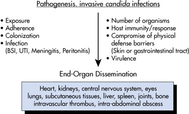
Figure 1 Pathogenesis of invasive candidiasis.

BSI, Bloodstream infection; UTI, urinary tract infection.

From Cherry JD: Feigin and Cherry’s pediatric infectious diseases, ed 8, Philadelphia, 2019, Elsevier.

Diagnosis ⬆ ⬇

Differential Diagnosis

  • Sepsis (bacterial)
  • Septic shock
  • Cryptococcosis
  • Aspergillosis
Laboratory Tests

  • Laboratory studies are nonspecific. It is often necessary to perform several diagnostic tests to achieve maximum accuracy.
  • High index of suspicion is needed.
  • Candidemia/disseminated candidiasis: Candidemia represents the tip of the iceberg with respect to the more invasive forms of candidiasis. Central lines often contribute to the propagation of candidemia. From the blood, infection can spread to almost any organ.
    1. Blood culture is the gold standard of diagnosis. They are helpful but have low positive yield. Only 40% to 60% of patients with infection have positive culture. 95% of blood cultures that are positive for Candida spp. become positive within 96 hours. Candida in a blood culture is not a contamination, and the source of the infection should be sought.
    2. Diagnosis also can be made from normally sterile sites. Specific species identification is necessary because only 10% of known Candida spp. produce disease in humans.
    3. The T2 magnetic resonance assay of whole blood can be performed on blood samples even after initiation of antifungal therapy.
    4. Serum (1,3) beta-D-glucan detection assay: High specificity and high positive predictive value. It can also be used for diagnosing invasive candidiasis when blood cultures are negative.
    5. New techniques allow for quick identification of Candida spp. in blood. These include:
      1. Peptide nucleic acid fluorescence in-situ hybridization (PAN-FISH).
      2. Matrix-assisted laser desorption ionization time-of-flight mass spectrometry (MALDI TOF MS).
  • Hepatosplenic candidiasis (focal):
    1. Elevated serum alkaline phosphatase.
Imaging Studies

  • Imaging studies are generally not required or useful.
  • Ultrasound is useful for diagnosing hepatosplenic abscess. “Bull’s eye or target lesions” are observed in the liver and spleen.
  • CT scanning may be used to diagnose hepatosplenic candidiasis, as well as intraabdominal/renal abscesses.
  • Transesophageal echocardiogram is useful to rule in or rule out Candida endocarditis.

Treatment ⬆ ⬇

Treatment Plans
Candidemia

General principles:

  • For documented candidemia, 2 wk of antifungal treatment after the first negative blood culture is recommended.
  • In nonneutropenic patients, a dilated funduscopic examination in the first week of treatment is recommended.
  • In neutropenic patients, delay above until neutrophil recovery because characteristic ocular findings may be delayed.
  • Treatment depends on whether the patient is neutropenic or not.
    1. Nonneutropenic adult patients: The initial treatment is with an echinocandin (e.g., caspofungin 70 mg loading dose, then 50 mg/day IV or micafungin 100 mg/day IV). Alternatives include fluconazole 800 mg as loading dose, then 400 mg/day for at least 2 wk after clinical improvement or negative blood culture. Amphotericin B is equally efficacious.
    2. Neutropenic adult patients: An echinocandin is the drug of choice (e.g., caspofungin) 70 mg IV loading dose, then 50 mg/day IV or micafungin 100 mg/day IV or anidulafungin 200 mg IV loading dose, then 100 mg IV all for at least 2 wk after clear blood culture and after clinical improvement. An alternative is fluconazole.
    3. Preferred oral step-down therapy: Neutropenic and nonneutropenic clinically stable patients can be transitioned to oral fluconazole (400 mg [6 mg/kg] daily) after about 5 to 7 days if they have:
      1. Candida-susceptible fluconazole and
      2. Negative blood cultures
Disseminated Candidiasis

Fluconazole is the drug of choice.

Disseminated Candidiasis with End-Organ Infection

  • Treatment is the same as for candidemia of nonneutropenic patients. In most cases, therapy is prolonged for at least 4 to 6 wk.
  • The echinocandins are the first-line therapy.
Osteomyelitis or Septic Arthritis

  • Fluconazole 400 mg IV or PO or
  • Caspofungin 50 mg/day IV or
  • Micafungin 100 mg/day IV
Endocarditis

  • Caspofungin 50 to 150 mg/day or
  • Micafungin 100 to 150 mg/day or
  • Anidulafungin 100 to 200 mg/day
Myocarditis

  • Lipid-based amphotericin B 3 to 5 mg/kg/day or
  • Caspofungin 150 mg/day or micafungin 100 mg/day
Esophagitis

  • Caspofungin 50 mg/day IV
  • Fluconazole 200 to 400 mg/day
Pericarditis

Lipid-based amphotericin B 3 to 5 mg/kg/day

Surgical Care

Includes:

  • Drainage
  • Removal of any foreign bodies
  • Surgical debridement
  • Organ-specific care (e.g., valve replacement for endocarditis, splenectomy for splenic abscess, or vitrectomy for fungal endophthalmitis)
Disposition

  • Several factors affect prognosis: Infection site, degree of immune suppression, and how quickly diagnosis and therapy are initiated
  • Overall mortality rate: 30% to 40%
Referral

  • Always involve an infectious disease specialist.
  • Referral to specialist will depend on the organ involved. For example:
    1. Endocarditis will require a cardiothoracic surgeon.
    2. Endophthalmitis will require an ophthalmologist.
Follow-Up Care

  • Prolonged periods, mainly in the hospital, of antifungal treatment may be necessary.
  • Closely monitor patients on amphotericin B because of the high incidence of side effects. Check basic metabolic panel, magnesium, and CBC at least twice a week.

Pearls & Considerations ⬆ ⬇

Prevention

Basic preventive measures are similar to those used for nosocomial infections. This includes:

  • Maximizing hand hygiene recommendations:
    1. Handwashing
    2. Using alcohol/chlorhexidine solution
  • Adhering strictly to recommendations for placement and care of central lines and catheters
  • Judicious use of antimicrobials
    1. Notes on Candida auris:
      1. The Centers for Disease Control and Prevention issued warnings in 2016 about the emergence of C. auris, a multidrug-resistant Candida sp. It is resistant to fluconazole and amphotericin B in 93% and 35% of patients, respectively.
      2. It has caused invasive health care-associated infections in many countries and has high mortality rates.
      3. Initial treatment is with echinocandin. Patient should be closely followed with cultures.
      4. Special infection control precautions should be followed for patients infected with or colonized by C. auris.
Prophylaxis

Antifungal prophylaxis should be limited to patients in whom it has proved beneficial: Patients with GI anastomotic leakage, patients undergoing transplantation of the pancreas or small bowel, selected patients undergoing liver transplantation who are at high risk for candidiasis, and extremely low-birth-weight neonates in settings with a high incidence of neonatal candidiasis.

Patient & Family Education

  • Inform them about the risk factors for invasive candidiasis.
  • Inform them of the seriousness of the disease and the associated high morbidity/mortality rates, thus requiring aggressive treatment.
  • Side effects and toxicities associated with treatment.
Related Content

Candidiasis (Patient Information)

Candidiasis, Cutaneous (Related Key Topic)

Suggested Readings ⬆

    1. Kullberg B.J. : Invasive candidiasisN Engl J Med. ;373, 2015.
    2. Pappas P.G. : Invasive candidiasisNat Rev Dis Primers. ;4, 2018.