section name header

Basic Information

AUTHORS: Lisa Bird, MD and Nima R. Patel, MD, MS

Definition

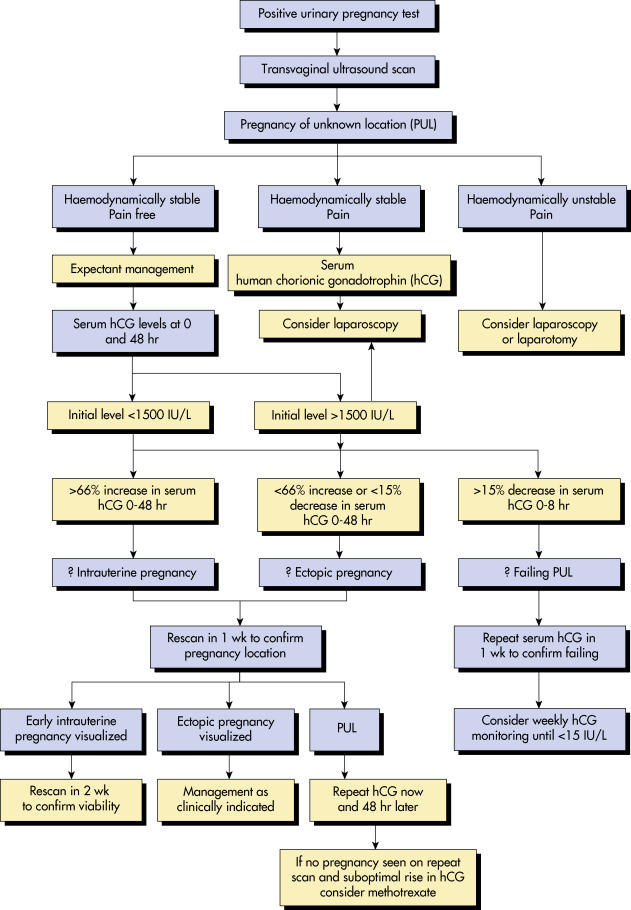
An ectopic pregnancy (EP) occurs when a fertilized ovum implants outside the endometrial cavity.1

Synonyms

Tubal pregnancy (97%)

Interstitial (cornual) pregnancy (1% to 2%)

Ovarian pregnancy (1% to 3%)

Abdominal pregnancy (0.03% to 1%)

Cervical pregnancy (0.5%)

Cesarean scar pregnancy (1% to 3%, 6% of all EP in women with prior cesarean)

Heterotopic pregnancy (1/4000 to 1/30,000, but 1/100 after in vitro fertilization)1

ICD-10CM CODES
O00.9Ectopic pregnancy, unspecified
O00.0Abdominal pregnancy
O00.1Tubal ectopic pregnancy
O00.2Ovarian ectopic pregnancy
O00.8Other ectopic pregnancy
Epidemiology & Demographics

  • 1% to 2% of pregnancies; represents 3% of pregnancy-related deaths.2
  • Most common cause of death in first trimester.
  • African American patients have a 1.5-fold higher risk.
Prevalence (In U.S.)

Accounts for up to 18% of women presenting to the emergency room with vaginal bleeding or abdominal pain.1 Currently over 100,000 reported cases/yr.

Risk Factors

Prior ectopic, altered tubal anatomy, prior pelvic infection (pelvic inflammatory disease, tuboovarian abscess, salpingitis), prior sterilization procedure, prior tuboplasty or tubal surgery, prior cesarean delivery, current intrauterine device use, assisted reproductive techniques, infertility, cigarette use, age >35, multiple lifetime sexual partners, diethylstilbestrol (DES) exposure in utero. Half of patients with EP have no risk factors.1

Physical Findings & Clinical Presentation

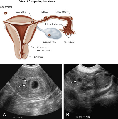
  • The classic presentation of EP includes the triad of abnormal vaginal bleeding, pelvic pain, and an adnexal mass
  • Abdominal tenderness: 95%
  • Adnexal tenderness: 87% to 99%
  • Amenorrhea or abnormal vaginal bleeding: 75%
  • Peritoneal signs: 71% to 76%
  • Adnexal mass: 33% to 53%
  • Enlarged uterus: 6% to 30%
  • Shoulder pain: 10%
  • Tissue passage: 6% to 7%
  • Shock: 2% to 17%
Etiology

  • Anatomic obstruction to zygote passage
  • Abnormalities in tubal motility
  • Transperitoneal migration of the zygote

Diagnosis

Differential Diagnosis

  • Corpus luteum cyst
  • Rupture or torsion of ovarian cyst, especially hemorrhagic
  • Threatened or incomplete abortion
  • Pelvic inflammatory disease/tuboovarian abscess
  • Appendicitis
  • Degenerating uterine fibroids
  • Endometrioma or dermoid cyst
  • Hydrosalpinx
  • Heterophile antibodies1
Workup

  • Consider in all patients with a positive pregnancy test and abdominopelvic pain
    1. Fig. 1 describes a diagnostic approach to suspected EP.
  • Transvaginal ultrasound (Fig. E2 [bottom]).
    1. Intrauterine gestational sac with a yolk sac should be visible between 5 and 6 wk of gestation regardless of whether there are one or multiple gestations. If uncertain dating, use quantitative human chorionic gonadotropin (qhCG) levels to guide expected findings.
  • 70% EP are tubal. Fig. E2 (top) describes potential sites of ectopic implantations.
  • Obtain qhCG level. Consider progesterone level if ultrasound inconclusive. Type and screen if presents with vaginal bleeding. Give Rhogam if Rh-negative status on initial presentation of vaginal bleeding with positive pregnancy test even if diagnosis inconclusive.
  • Laparoscopy (Fig. E3) in equivocal situations and possibly for treatment.
  • If pregnancy is not desired or determined to be nonviable, uterine aspiration can identify intrauterine location of chorionic villi.

Figure 1 Algorithm for managing suspected ectopic pregnancy.

!!flowchart!!

From Magowan BA: Clinical obstetrics & gynecology, ed 4, Philadelphia, 2019, Elsevier.

Figure E2 Top, Schematic Drawing Depicting Implantation Sites of Ectopic Pregnancies

A and B, Heterotopic Pregnancy. This Pregnant Patient Presented with Vaginal Bleeding at 5 to 6 wk of Gestational Age. Bottom, A, Transverse Transvaginal Ultrasound (TVUS) Image of the Uterus Reveals an Intrauterine Gestational Sac Containing a Yolk Sac. Note Small Subchorionic Hemorrhage (Arrows), Most Likely Accounting for the Vaginal Bleeding. B, Sagittal TVUS Image of the Right Adnexa Reveals an Echogenic Tubal Ring (Arrow) Clearly Separate from the Right Ovary (OV), Which was Surgically Confirmed to Be an Ectopic Pregnancy.

From Fielding JR et al: Gynecologic imaging, Philadelphia, 2011, Saunders.

Figure E3 An ectopic pregnancy in the ampulla of the fallopian tube.

From Magowan BA et al: Clinical obstetrics & gynaecology, ed 4, Philadelphia, 2019, Elsevier.

Laboratory Tests

  • qhCG: Check on initial presentation to enable interpretation of initial ultrasound. If qhCG >6000 mIU/ml, should see intrauterine pregnancy (IUP) on abdominal scan; qhCG >3500 mIU/ml for transvaginal ultrasound. Inability to see IUP at or above these levels raises concern for ectopic. Multiple gestation may take longer to visualize, however, due to increased qhCG levels.1
  • 25% to 50% of EP presents as a pregnancy of unknown location (PUL), in which initial ultrasound does not show a pregnancy in the uterus or the fallopian tube. Serial measurement of qhCG, obtained every 48 h, can help distinguish between an IUP, resolving miscarriage, or EP. The expected rate of qhCG rise varies by the initial starting qhCG. For an initial qhCG level <1500 mIU/ml, the expected rate of increase is 49%, compared to 40% for an initial qhCG level between 1500 and 3000 mIU/ml, and a 33% increase for an initial qhCG >3000 mIU/ml. However, some EPs will have a normal-appearing qhCG rise, and some will still rupture despite decreasing qhCG.1
  • A single progesterone level <5 ng/ml has 99% specificity to predict nonviable pregnancy with inconclusive sonographic findings, but this cannot distinguish EP from failed IUP.
  • Dropping hemoglobin/hematocrit may be associated with tubal rupture with internal hemorrhage or possible miscarriage.
Imaging Studies

  • Ultrasound: Presence of an intrauterine yolk sac makes EP extremely unlikely. However, if the patient used assisted reproductive technologies, a heterotopic pregnancy (EP with concurrent IUP) is possible. Repeat ultrasound within 10 to 14 days after presentation may identify the location of a pregnancy that was not identified initially.
  • Findings on ultrasound in EP include:
    1. Empty uterus (i.e., no yolk sac or fetal pole; a pseudosac in uterus may appear similar to a gestational sac)
    2. Adnexal mass ( “ring of fire” appearance, can typically distinguish EP appearance from corpus luteum by tubal vs ovarian location)
    3. Enlarged hysterotomy scar with thin anterior uterine wall, may bulge beyond uterine contour
    4. Free fluid in cul-de-sac or Morison’s pouch
    5. Yolk sac and/or fetal pole in tube or extrauterine cardiac activity in adnexa

Treatment

Nonpharmacologic Therapy

Surgery performed via laparoscopy is preferred; however, laparotomy is appropriate if patient is very unstable or if poor visualization during laparoscopy. Ruptured EP is managed surgically, as life-threatening intraabdominal hemorrhage may ensue. Transfuse blood as indicated.

  • Salpingostomy, removing EP with tubal conservation, has benefit if patient only has one remaining tube preoperatively and desires future fertility, but will require postoperative serial monitoring of qhCG. Otherwise, randomized controlled trials suggest there is no difference in fecundity rate between salpingostomy and salpingectomy.
  • Salpingectomy, removal of affected fallopian tube, is the standard surgical procedure and preferred in the following circumstances:
    1. Ruptured tube
    2. Recurrent EP in the same tube
    3. Uncontrolled hemorrhage
    4. Future fertility not desired
  • Direct injection of chemotherapy into a nontubal EP by laparoscopy, ultrasound guidance, or hysteroscopy. Direct injection of methotrexate (MTX), and possibly KCl if cardiac activity, may be performed when the pregnancy is in a high-morbidity location, such as the cervix, cesarean delivery scar, or cornua.2,3 A possible algorithm for use of MTX vs. surgery for ectopic pregnancy is illustrated in Fig. 4.
  • Cervical and cesarean scar pregnancies may be managed with a combination of MTX administration, laparoscopic and/or hysteroscopic techniques, ultrasound-guided suction curettage, uterine artery embolization (UAE), laparotomy, and possible hysterectomy. Vasopressin and UAE reduce bleeding. Systemic MTX as monotherapy is not recommended for cesarean scar EP.3
  • Ovarian EP is managed surgically. Interstitial EP is also more safely managed surgically due to proximity to uterine vessels in case of rupture.2
  • Expectant management is only appropriate in asymptomatic cases with significant spontaneous qhGC decrease from an initial low level.
Figure 4 Possible Algorithm for Use of Methotrexate Vs

Surgery for Ectopic Pregnancy. HCG, Human Chorionic Gonadotropin; Quant, Quantitative; TVUS, Transvaginal Ultrasound. (From Gershenson DM Et Al: Comprehensive Gynecology, Ed 8, Philadelphia, 2022, Elsevier.)

Acute General Rx

  • Medical management with MTX, a folic acid antagonist, is a safe option for tubal EP if the patient is stable. Check labs prior to administration (CBC, comprehensive metabolic panel). Future fertility is not affected.
  • Absolute contraindications:
    1. Hemodynamically unstable; ruptured EP
    2. Patient unable to comply with follow-up
    3. Immunodeficiency
    4. Intrauterine pregnancy
    5. Medical contraindication to MTX includes clinically relevant hepatic or renal disease, as well as preexisting blood dyscrasias (thrombocytopenia, leukopenia, or significant anemia), as MTX is directly toxic to hepatocytes and renally excreted
    6. Breastfeeding, active pulmonary disease, or active peptic ulcer disease
  • Relative contraindications:
    1. EP >4 cm mass as imaged by transvaginal ultrasound
    2. qhCG >5000 mIU/ml (up to 14% failure rate)
    3. Presence of embryonic cardiac activity (high failure rate)
    4. Refusal to accept blood products in case of failure and subsequent hemorrhage from rupture
  • Most common regimen is single-dose MTX at a dose of 50 mg/m2 per body surface area and is administered intramuscularly on day 1. May require second dose or surgical intervention if qhCG increases or plateaus (<15% drop) when comparing values from day 4 and 7. If qhCG does not decrease after two doses, consider surgical management.
  • Success rate of single-dose protocol approximately 88% vs. multidose protocol, which is 92%. Single dose is associated with less adverse effects. Initial serum qhCG level is the best prognostic indicator of treatment success of single-dose protocol.
  • Rupture during MTX treatment ranges from 7% to 14%.
  • Other MTX regimens include multidose protocol ± leucovorin rescue.
  • Avoid intercourse and heavy exercise to avoid trigger of rupture, alcohol due to symptom masking, and folic acid (prenatal vitamins) due to decreased MTX effectiveness, as well as sun exposure and NSAIDs due to increased toxicity until MTX treatment is complete.2
  • Methotrexate adverse effects include nausea, vomiting, stomatitis, diarrhea, elevated LFTs, abdominal pain, and nephrotoxicity. Rare adverse effects of pneumonitis or alopecia can occur.
  • Ultrasound surveillance of resolution is not indicated because findings do not predict rupture or time to resolution.1
  • The FDA recommends avoiding pregnancy until at least 1 ovulatory cycle after MTX.1 Some experts recommend delaying pregnancy for at least 3 mo after MTX due to prolonged persistence in human tissue.1
Chronic Rx

Persistent EP results from residual trophoblastic tissue or secondary implantation after salpingostomy. There is a 5% incidence of persistent EP with conservative treatment.

Disposition

If diagnosed and treated early (before rupture), prognosis is excellent for good recovery. Monitor qhCG weekly and use reliable contraception until qhCG is negative. With subsequent pregnancies, perform early ultrasound to confirm IUP and follow qhCG as indicated. There is a 10% recurrence rate for EP; however, this rate increases to 25% after two prior EPs.1

Referral

Should obtain gynecologic consultation if EP is suspected.

Pearls & Considerations

Any patient who presents with vaginal bleeding, abdominal pain, or both with a positive pregnancy test and no prior documented IUP needs to be assessed for ectopic pregnancy. Do not assume that contraceptive use means pregnancy test will be negative.

Prevention

Absolute risk of EP is reduced by use of contraception, so although likelihood of EP is increased with IUD failure, the risk of EP is still lower overall due to contraceptive effectiveness.2 Use of condoms reduces tubal exposure to infection, which also lowers future risk.

Related Content

Ectopic Pregnancy (Patient Information)

Spontaneous Abortion (Related Key Topic)

Vaginal Bleeding During Pregnancy (Related Key Topic)

Related Content

    1. Barnhart K.T., Franasiak J.M. : ACOG Practice Bulletin Number 193: tubal ectopic pregnancyObstet Gynecol. ;131(3):e91-e103, 2018.
    2. Ectopic pregnancy Hoffman B.L., editors : Williams gynecology. ed 4McGraw Hill, New York, 2020.https://accessmedicine.mhmedical.com/content.aspx?bookid=2658&sectionid=220754945
    3. Society for Maternal-Fetal Medicine (SMFM), Miller R., Gyamfi-Bannerman C., Publications Committee : Electronic address: pubs@smfm.org. Society for Maternal-Fetal Medicine Consult Series #63: Cesarean scar ectopic pregnancyAm J Obstet Gynecol. ;227(3):B9-B20, 2022.doi:10.1016/j.ajog.2022.06.024