section name header

Basic Information

AUTHORS: Anthony Sciscione, DO and Kathryn G. Vollum, MD

Definition

Fibrocystic breast disease (FBD), or fibrocystic breast changes (FBC), is a category of benign breast lesions that includes:

  • Microcystic and macrocystic changes
  • Fibrosis
  • Mild and moderate hyperplasia
  • Sclerosing adenosis
  • Apocrine metaplasia
  • Fibroadenoma
  • Papilloma
  • Papillomatosis
  • Radial scar

Nonmalignant breast lesions can be divided into nonproliferative, proliferative without atypia, and atypical hyperplasia. Nonproliferative lesions such as simple breast cysts and galactoceles, are considered not to be at increased risk for breast cancer (RR = 1.17). Proliferative lesions, including ductal hyperplasia, intraductal papillomas, sclerosing adenosis, radial scars, and fibroadenomas, are considered markers for a small increase in risk of progressing to breast cancer (proliferation without atypia RR = 1.76, with hyperplastic atypia RR = 3.93), and patients should be counseled on risk reduction.

Synonyms

Cystic changes

Chronic cystic mastitis

Fibrocystic breast changes

Mammary dysplasia

Fibrocystic mastopathy

Diffuse cystic mastopathy

ICD-10CM CODES
N60.01Solitary cyst of right breast
N60.02Solitary cyst of left breast
N60.09Solitary cyst of unspecified breast
N60.11Diffuse cystic mastopathy of right breast
N60.12Diffuse cystic mastopathy of left breast
N60.19Diffuse cystic mastopathy of unspecified breast
N60.3Fibrosclerosis
Epidemiology & Demographics

  • FBC affects 30% to 60% of reproductive-age women.
  • Most commonly seen in women ages 30 to 50 yr.
  • Prevalence of various benign breast changes in the postmenopausal cohort is increased by a factor of 1.7 in those receiving exogenous estrogen and progestins.
Etiology

The cause of FBC is unknown but is thought to be influenced by reproductive hormones, estrogen in particular. Because the majority of FBC is found in healthy breasts, it is regarded as a nonpathologic process.

Diagnosis

Differential Diagnosis

See Table E1.

TABLE E1 Typical U.S. Characteristics of Solid Breast Lesions

BenignMalignant
ShapeOval/ellipsoidVariable
AlignmentWider than deep aligned parallel to tissue planesDeeper than wide
MarginsSmooth/thin echogenic pseudocapsule with 2-3 gentle lobulationsIrregular or spiculated echogenic “halo”
EchotextureVariable-to-intense hyperechogenicityLow-level
Marked hypoechogenicity
Homogeneity of internal echoesUniformNonuniform
Lateral shadowingPresentAbsent
Posterior effectMinimum attenuation/posterior enhancementAttenuation with obscured posterior margins
Other signsCalcification
Microlobulation
Intraductal extension
Infiltration across tissue planes and increased echogenicity of surrounding fat

From Sutton D: Textbook of radiology and imaging, ed 7, 1998, Churchill Livingstone; and Grant LA: Grainger & Allison’s diagnostic radiology essentials, ed 2, 2019, Elsevier.

From Sutton D: Textbook of radiology and imaging, ed 7, 1998, Churchill Livingstone; and Grant LA: Grainger & Allison’s diagnostic radiology essentials, ed 2, 2019, Elsevier.

Physical Findings & Clinical Presentation

  • Lumpy or ropy breast texture
  • Nodular areas
  • Tenderness of breasts and/or nipples
  • Dominant mass more often in the upper outer quadrants
  • Symptoms tend to be affected by changes in the menstrual cycle
  • Symptoms can be aggravated in postmenopausal women who initiate hormone therapy
  • Nipple discharge

For differentiation of breast masses, see Table E2. Characteristics of breast masses suspect for cancer (90% sensitivity, 40% to 60% specificity) are:

  • Fixed mass
  • Poorly defined mass
  • Hard (scirrhous) mass

TABLE E2 Differentiation of Breast Masses

CharacteristicCystic DiseaseBenign AdenomaMalignant Tumor
Patient age25-60 yr10-55 yr25-85 yr
NumberOne or moreOneOne
ShapeRoundRoundIrregular
ConsistencyElastic, soft to hardFirmStony hard
DelimitationWell delimitedWell delimitedPoorly delimited
MobilityMobileMobileFixed
TendernessPresentAbsentAbsent
Skin retractionAbsentAbsentPresent

From Swartz MH et al: Textbook of physical diagnosis, ed 7, Philadelphia, 2014, Saunders.

Workup

  • Rule out breast carcinoma if breast mass, thickening, discharge, and/or pain are present.
  • Perform biopsy of suspected area for histologic confirmation.
Imaging Studies

Mammography and ultrasound studies required:

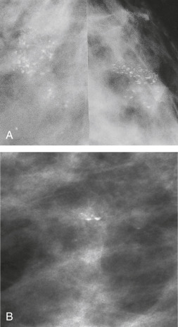
  • For mammographic changes (suspicious densities, microcalcifications [Fig. E1], architectural distortion): Careful evaluation, including possibly biopsy to exclude breast cancer
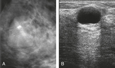
  • Ultrasound study (Fig. E2): To evaluate the presence of a solid vs. cystic mass
  • MRI is not recommended for routine evaluation of breast pain or fibrocystic changes

Figure E1 Fibrocystic Change

A, On the Cc View, the Calcifications Appear as Round “smudge” Shadows. B, On the Lateral View, “teacups” Representing the Layering of Calcific Material in the Dependent Portion of Microcysts are Seen.

From Adam A et al: Grainger & Allison’s diagnostic radiology, ed 5, 2007, Churchill Livingstone; Sutton D: Textbook of radiology and imaging, ed 7, 1998, Churchill Livingstone; and Grant LA: Grainger & Allison’s diagnostic radiology essentials, ed 2, 2019, Elsevier.

Figure E2 Cyst

A, A Well-Defined, Rounded Mass with an Associated Lucent Halo. B, Ultrasound. The Absence of Internal Echoes and Posterior Enhancement is Diagnostic of a Cyst.

From Adam A et al: Grainger & Allison’s diagnostic radiology, ed 5, 2007, Churchill Livingstone; and Grant LA: Grainger & Allison’s diagnostic radiology essentials, ed 2, 2019, Elsevier.

Treatment

Nonpharmacologic Therapy

Treatment is symptomatic:

  • Reassurance
  • Supportive bra
  • OTC pain medications including NSAIDs and Tylenol
  • Reduced intake of methylxanthines (coffee, chocolate), although this is not as effective as previously thought
  • Decreasing vitamin E or salt has not proved to be effective
  • Periodic physician examination to monitor patients with FBC who have pronounced nodular features
  • Aspiration for palpable or symptomatic cysts (NOTE: Cysts often recur; repeat aspiration is not always required unless pain is a problem)
Pharmacologic Treatment

For breast pain:

  • Danocrine (Danazol): This is the only FDA-approved drug for breast pain. Moderate success has been reported, but the side effect profile is significant due to androgenic effects.
  • Bromocriptine: This inhibits prolactin, and its effects are better than a placebo, but side effects such as light-headedness and gastrointestinal symptoms limit its use.
  • Tamoxifen: This has been found to reduce breast pain in 70% of affected women with cyclic breast pain, but the side effects of hot flashes and vaginal dryness interfere with compliance.
  • Pharmacologic therapy is usually tapered down after 3 to 6 mo of treatment.
Follow-Up

  • Evaluate carefully to exclude breast cancer; then offer reassurance and periodic reevaluation as required.
  • For all women, ACOG recommends breast self-awareness, regular clinical breast exams (every 1 to 3 yr for women ages 25 to 39, annually for women over 40), and mammograms every 1 to 2 yr starting at age 40.
Referral To Breast Surgeon

  • If suspicious changes are associated with FBC (including progression of dominant mass or thickening, persistent or bloody nipple discharge, or suspicious mammographic changes or lesions), refer for further evaluation and/or biopsy.
  • Refer to alleviate anxiety associated with breast symptoms or changes.
Related Content

Fibrocystic Breast Changes (Patient Information)

Breast Cancer (Related Key Topic)

Mastodynia (Related Key Topic)

Mastitis (Related Key Topic)

Suggested Readings

  1. Consensus guideline on diagnostic and screening magnetic resonance imaging of the breast .www.breastsurgeons.org/about/statements/PDF_Statements/MRI.pdf
  2. Castells X. : Breast cancer risk after diagnosis by screening mammography of nonproliferative or proliferative benign breast disease: a study from a population-based screening programBreast Cancer Res Treat. ;149(1):237-244, 2015.
  3. Diagnosis and management of benign breast disorders 164 Obstet Gynecol. ;127(6):141-156, 2016.
  4. Malherbe K. : Fibrocystic breast disease [Updated 2021 Oct 24]. In: StatPearls [Internet]. Treasure Island (FL)StatPearls Publishing, 2022.www.ncbi.nlm.nih.gov/books/NBK551609/