section name header

Basic Information

AUTHORS: Anthony Sciscione, DO and Erin Bishop, MD

Definition

Mastitis is local painful inflammation of the breast that may or may not be accompanied by infection, flulike symptoms, and abscess formation.

ICD-10CM CODES
N61Inflammatory disorders of breast
O91.12Abscess of breast associated with the puerperium
O91.22Nonpurulent mastitis associated with the puerperium
Epidemiology & Demographics

  • Mastitis is the most common cause of inflammatory breast disease, and most cases are related to lactation (puerperal mastitis).
    1. Nonpuerperal cases of mastitis can affect either the periareolar (periductal) region or peripheral breast tissue.
    2. Periductal mastitis (PM, also known as duct ectasia) is most common in younger, reproductive-age women. The majority of those affected are active smokers.
  • In lactating mothers, mastitis typically occurs in the first 3 mo of the postpartum period (74% to 95% of cases).
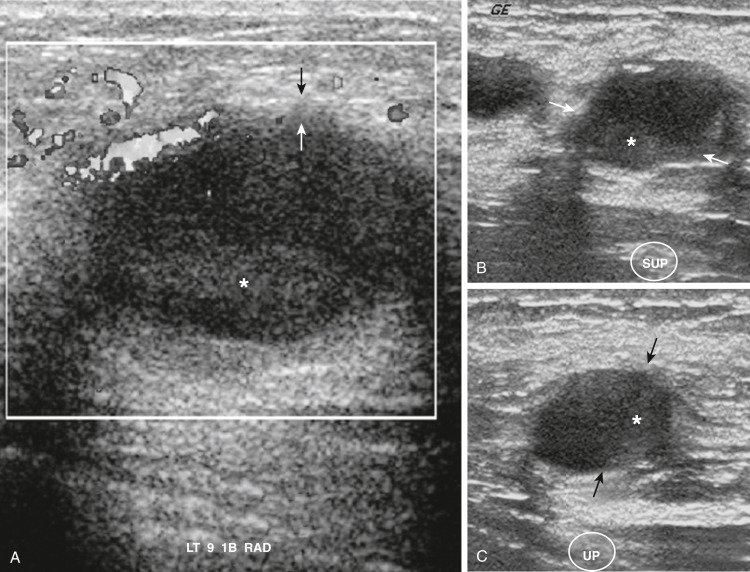
  • When severe, mastitis can lead to a breast abscess (5% to 11%) or septicemia.
  • Delayed diagnosis and treatment of lactational mastitis can lead to discontinuation of breastfeeding, breast tissue damage, or recurrence.
  • In younger, nonlactating women, infection often presents as periductal mastitis and is caused by inflamed milk ducts near the nipple.
  • Granulomatous mastitis (GM) is a rarer form of benign inflammation of the breast and also most commonly occurs in reproductive-age women. It generally affects the breast peripherally.
  • Mastitis also can occur in infancy when there is breast hypertrophy from maternal hormones, called neonatal mastitis (NM).
    1. Approximately 50% of neonates with mastitis will develop an abscess.
Prevalence

Lactational mastitis occurs in up to 33% of mothers

Predominant Sex & Age

Females of reproductive age

Risk Factors

  • Previous mastitis
  • Milk stasis and missed feedings, or extended periods between feedings such as when an infant begins to sleep through the night
  • History of oversupply
  • Cracked, fissured, or sore nipples
  • Primiparity and infant attachment difficulties
  • Cleft lip or palate or short frenulum in infant
  • Use of manual breast pump
  • Foreign material: Breast implants, nipple piercings
  • Rapid weaning
  • Smoking (periductal mastitis)
  • Obesity (periductal mastitis)
  • Conditions that impair immunity (peripheral or granulomatous mastitis): Diabetes, steroid use, rheumatoid arthritis
Physical Findings & Clinical Presentation

  • Warmth, redness, noncyclic tenderness in breast
  • Unilateral or bilateral
  • Malaise, myalgias, fevers, chills, nausea
  • Decreased milk output
  • Breast is hard and swollen in a wedge-shaped area
  • Lactational mastitis tends to be found in the breast periphery, whereas nonlactational mastitis tends to be peri- or sub-areolar
  • In PM, breast mass near nipple with retraction or discharge
    1. Can present simultaneously with abscess or even mammary duct fistula
  • In GM, enlarged axillary lymph nodes or sinus tract formation
Etiology

  • In lactational mastitis, infection occurs as a result of milk stasis and irritation of the milk ducts due to local immune response to milk proteins.
  • Bacterial infection of subcutaneous tissue due to breaks in skin.
  • Most commonly, Staphylococcus aureus; less common, S. epidermidis, group A beta-hemolytic streptococci, S. pneumoniae, Escherichia coli, Candida albicans, Mycobacterium tuberculosis. Up to 40% are polymicrobial.
  • GM results from inflammation with epithelioid histiocytes and multinucleated giant cells and can be caused by etiologies like tuberculosis, sarcoidosis, foreign body reaction, parasitic and mycotic infections, or idiopathic.
  • Periductal mastitis occurs following inflammation around nondilated subareolar ducts and often can progress to abscess formation. Peripheral abscesses can result from trauma, usually in the setting of comorbid conditions impairing immunity such as diabetes or use of immunosuppressive medications.
  • Neonatal mastitis caused by S. aureus or gram-negative enteric bacteria.

Diagnosis

Differential Diagnosis

  • Engorgement, plugged duct (Table 1)
  • Breast abscess
  • Inflammatory or other breast cancer (3% of women diagnosed with breast cancer are lactating)
  • Paget disease of breast
  • Mastitis as a symptom of hyperprolactinemia or galactorrhea
  • GM can be manifestation of systemic disease (sarcoidosis, Wegener granulomatosis, giant cell arteritis [GCA], polyarteritis nodosa, tuberculosis [TB], syphilis)

TABLE 1 Comparison of Findings of Engorgement, Plugged Duct, and Mastitis

CharacteristicsEngorgementPlugged DuctMastitis
OnsetGradual, immediatelyGradual, after feedingsSudden, after 10 days postpartum
SiteBilateralUnilateralUsually unilateral
Swelling and heatGeneralizedMay shift/little or no heatLocalized red, hot, and swollen
Body temperature<38.4° C; 101.1° F<38.4° C; 101.1° F>38.4° C; 101.1° F
Systemic symptomsFeels wellFeels wellFlulike symptoms

From Lawrence RA, Lawrence RM: Breastfeeding: a guide for the medical profession, ed 5, St Louis, 1999, Mosby.

Workup

  • History and clinical exam with thorough breast exam are generally sufficient for diagnosis.
    1. Be sure to address time, course, and duration of symptoms as well as breast history, including lactation, recent trauma, and prior treatment
    2. Physical exam should include special attention to inflammatory changes and their location, skin changes, assessment of nipple for skin changes and discharge, axillary or supraclavicular adenopathy, and presence or absence of breast mass
  • Recurrent mastitis should include workup for underlying breast disease.
Laboratory Tests

  • Simple lactational mastitis requires no milk culture or laboratory studies.
  • Obtain midstream sample of milk for culture and sensitivities in refractory mastitis or in methicillin-resistant S. aureus-suspected cases.
  • CBC and blood cultures in toxic-appearing patients.
  • In abscess formation, culture of drainage or aspirate fluid.
  • Inpatient intravenous antibiotics may be necessary in severe cases or recalcitrant to outpatient treatment.
  • Gram stain and culture indicated in infant mastitis.
Imaging Studies

  • Not necessary unless refractory mastitis or abscess suspected.
    1. An abscess generally presents as a hypoechoic mass on ultrasound.
  • In the context of a discrete mass on exam, age-appropriate breast imaging starting with ultrasound (US) (Fig. E1) is recommended to exclude carcinoma.
  • In PM, US should be performed.
  • In GM, mammogram and US-guided fine needle aspiration (FNA) are standard.

Figure E1 Inflamed or infected cyst.

A, Acutely inflamed or infected cysts demonstrate three findings: (1) Abnormal uniform isoechoic wall thickening (between arrows), (2) dependent debris (asterisk), and (3) hyperemia of the thickened wall. Supine (B) and upright (C) images show the debris (asterisk), resembling sludge within a gallbladder, shifting to the dependent part of the cyst when the position of the patient is changed from supine to upright or lateral decubitus positions. Note the change in the position of the interface between the nondependent fluid and the dependent debris or pus (between arrows).

From Rumack CM et al: Diagnostic ultrasound, ed 4, Philadelphia, 2011, Mosby.

Treatment

Nonpharmacologic Therapy

  • Mainstay of therapy is effective milk removal through continued breastfeeding or pumping. Patient should be encouraged to continue breastfeeding her infant(s) throughout her treatment unless otherwise indicated.
  • Consider referral to a certified lactation consultant to improve breastfeeding technique.
    1. Positioning the infant with its chin pointed towards the affected area can help to drain the affected area.
  • Warm compresses, increased fluid intake, good nutrition, and rest.
  • In abscess formation (10% of women who are treated for bacterial mastitis), surgical drainage or needle aspiration is necessary, followed by antibiotic therapy based on sensitivities of culture.
  • In PM, aspiration or incision and drainage of abscesses should be performed, followed by culture of aspirate to guide antibiotic selection. If applicable, smoking cessation should be encouraged.
Acute General Rx

  • NSAIDs and analgesics (e.g., acetaminophen, ibuprofen). There is insufficient evidence to support or refute the effectiveness of antibiotic therapy.
  • Patients should be asked about history of penicillin allergy and history of methicillin-resistant S. aureus infection, either in herself or members of her household. Common antibiotic regimens include:
  • No history of MRSA (methicillin-resistant S. aureus):
    1. Penicillinase-resistant antibiotic: Dicloxacillin 250 mg 4×/day for 7 days
    2. Cephalexin 500 mg 4×/day for 10 to 14 days
    3. Inpatient: Nafcillin or oxacillin 2 g intravenous (IV) q4h
    4. Erythromycin may be used in patients allergic to penicillin
  • Suspected MRSA or high-risk penicillin allergy:
    1. Trimethoprim/sulfamethoxazole 160 mg/ 800 mg 2×/day for 10 to 14 days; should not be used when breastfeeding healthy infants <2 mo or compromised infants
    2. Clindamycin 300 mg 4×/day for 10 to 14 days
    3. Inpatient: Vancomycin 1 g IV q12h
  • Women should be reassured that antibiotics and antiinflammatory medicines are safe for her infant(s).
  • If no clinical response to antibiotics, MRSA or abscess should be considered. Again, if abscess is suspected or symptoms do not resolve with empiric antibiotic treatment, imaging should be performed to exclude other pathology. A biopsy should be performed based on imaging results.
  • Antibiotic treatment in patients with abscesses should continue for up to 10 days following aspiration or incision and drainage, and antibiotic choice should be guided by culture results.
  • Oxytocin nasal spray if letdown reflex disturbed.
  • Consider treatment for candidal infection if bilateral symptoms and infant with thrush. Both infant and mother will require treatment.
    1. Topical clotrimazole for mother and oral nystatin for infant, with careful washing of all pacifiers and nipples
    2. If resistant to topical treatment, can consider oral fluconazole; however, data in breastfeeding are limited
    3. Onset of candida infection can follow antibiotic treatment for presumed mastitis
  • Infant mastitis typically is treated in an inpatient setting with parenteral antibiotics based on results of Gram stain.
  • Most PM cases are treated adequately with a combination of antibiotics including anaerobic coverage (supported by culture results if available), needle aspiration/incision, and drainage. In recurrent cases, surgical removal of diseased ducts may be needed, which may necessitate referral to an experienced breast surgeon.
  • If antibiotic and NSAID treatment for GM fails, immunosuppressive drugs (steroids, methotrexate) can be used. Surgical management is not recommended due to associated slow wound healing.
Chronic Rx

  • No evidence proving benefit of prophylactic antibiotics to prevent lactational mastitis
  • In GM, systemic corticosteroids or wide surgical resection
Disposition

  • Most women with mastitis can be treated with antibiotics on an outpatient basis.
  • Criteria for admission include:
    1. Signs of sepsis or hemodynamic instability
    2. Rapidly progressing infection
    3. Immunocompromised status
  • If admission is necessary, the infant should be admitted with her to allow for continued breastfeeding.
Complementary & Alternative Medicine

  • Complementary therapies not assessed in prospective studies: Belladonna, Phytolacca, Chamomilla, sulfur, Bellis perennis, mupirocin, fucidic acid ointment, antisecretory factor, nisin
  • Several strains of lactobacilli have shown promise as probiotic agents that might be useful in treating mastitis, including L. fermentum and L. salivarius. These results should be replicated before this approach is adopted widely
Referral

  • Refer to surgeon for severe PM or significant lactational abscess that does not resolve with conservative measures

Pearls & Considerations

Comments

  • Early recognition and treatment is important to prevent complications such as breast abscesses, sepsis, and early weening.
  • 25% of breastfeeding mothers with one episode of mastitis stop breastfeeding.
  • Patients may experience a temporary decrease in milk supply that should improve once she begins to recover and as long as she continues to breastfeed or adequately express.
  • Increasing incidence of MRSA mastitis.
  • Lactational mastitis is a risk factor for vertical transmission of infections (i.e., HIV-1, cytomegalovirus, measles, hepatitis B and C).
  • When reassessing refractory nonlactational mastitis, the most important consideration is the possibility of cancer.
  • Nonlactational mastitis can be a manifestation of systemic disease.
  • GM mimics breast cancer both clinically and radiologically (>50% of reported cases are initially mistaken for carcinoma). This includes FNA, which is sometimes interpreted as malignant.
Related Content

Lactational Mastitis (Patient Information)

Mastitis (Patient Information)

Breast Abscess (Related Key Topic)

Fibrocystic Breast Disease (Related Key Topic)

Suggested Readings

    1. American College of Obstetricians and Gynecologists : Diagnosis and management of benign breast disorders, Practice Bulletin No 164Obstet Gynecol. ;127:e141-e156, 2016.
    2. American College of Obstetricians and Gynecologists : Breastfeeding challenges. ACOG Committee Opinion No. 820Obstet Gynecol. ;137:e42-e53, 2021.
    3. Amir L.H. : Managing common breastfeeding problems in the communityBr Med J. ;348, 2014.
    4. Amir L.H. : ABM clinical protocol #4: mastitis, revised March 2014Breastfeed Med. ;9:239-243, 2014.
    5. Boakes E. : Breast infection: a review of diagnosis and management practicesEur J Breast Health. ;14(3):136-143, 2018.
    6. Crepinsek M.A. : Interventions for preventing mastitis after childbirthCochrane Database Syst Rev. ;10, 2012.
    7. Dixon J.M., Khan L.R. : Treatment of breast infectionBMJ. ;342, 2011.
    8. Efrat M. : Neonatal mastitis-Diagnosis and treatmentIsrael J of Med Sci. ;31(9):558-560, 1995.
    9. Jahanfar S. : Antibiotics for mastitis in breastfeeding womenCochrane Database Syst Rev. ;2, 2013.