section name header

Basic Information

AUTHORS: Matthew Taylor, MD and Ross W. Hilliard, MD, FACP

Definition

Immunoglobulin A vasculitis (IgAV), formerly known as Henoch-Schönlein purpura (HSP), is a systemic, small-vessel, IgA immune complex-mediated leukocytoclastic vasculitis characterized by a tetrad of palpable purpura, GI symptoms (colicky abdominal pain, GI bleed), renal disease, and arthritis/arthralgias.

Synonyms

Henoch-Schönlein purpura (HSP)

Anaphylactoid purpura

Allergic purpura

ICD-10CM CODE
D69.0Immunoglobulin A vasculitis
Epidemiology & Demographics
Incidence

Annual incidence of 3 to 27 cases/100,000 population in children. 2/100,000 in adults.1

Prevalence

Most common vasculitis seen in children and younger age groups; seen in White and Asian populations (primarily Indian and Pakistani ancestry in a study conducted in the West Midlands, UK, with suggested predominance in Japan and Korea)2,3; 3 to 4 times more commonly than in Black patients.

Predominant Sex & Age

1.2 to 2:1 male:female ratio. Seen mostly from ages 3 to 12 with peak incidence between 4 and 6 yr. This disorder can be seen in older adolescents and young adults; however, 90% of cases are in patients <10 yr.2,4,5

Peak Incidence

In children, late autumn, winter, and early spring are the seasons with peak incidence. Cases in children are rare during the summer months. Seasonal variation is not seen in adults.2

Risk Factors

There are no formally identified risk factors; however, rates of IgAV are higher in patients with familial Mediterranean fever and MEFV mutations.6 Viral precipitants have been theorized to trigger onset of the disease.

Genetics

Recent studies have identified genetic susceptibility factors, but familial recurrence is rare. There appears to be a strong genetic predisposition with human leukocyte antigen (HLA) class 2 region related to HLA-DRB101 allele.7 There also may be protective genetic associations of HLA genes that may reduce the risk of acquiring the disease and explain ethnic differences. The ACE, IL, and HLA-B35 genes are associated with worse renal phenotype.

Physical Findings & Clinical Presentation

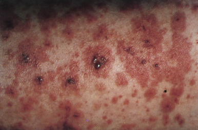
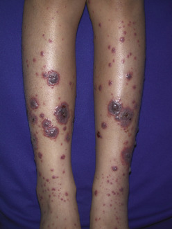
  • Palpable purpura (Fig. E1) of dependent areas, especially lower extremities (palms and soles) and areas subjected to pressure, such as the beltline in adults or buttocks in toddlers. Facial involvement is rare.
  • GI symptoms are seen in up to two thirds of patients. Common findings are abdominal pain, nausea, vomiting, diarrhea, cramping, hematochezia, and melena. Complications include GI bleeding (20% to 30%), bowel ischemia, intussusception (1% to 5%), and bowel perforation (<1%). Pancreatitis and acalculous cholecystitis are rare complications.
  • Subcutaneous edema in dependent and periorbital areas.
  • Arthralgias and arthritis in 60% to 85%. Typically, oligoarticular, affecting lower extremity and large joints. Periarticular swelling and tenderness also noted.
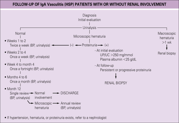
  • Renal involvement is seen in as many as 80% of older children, usually within the first month of illness. Fewer than 5% progress to end-stage renal failure, which is a major cause of morbidity. Renal manifestations may range from isolated hematuria, proteinuria, or severe crescentic glomerulonephritis.
  • Genitourinary involvement is seen between 2% and 38% of male children that can present with pain and edema of the scrotum, epididymitis, or orchitis that can mimic testicular torsion.8
  • Central nervous system (CNS) involvement is rare, but can manifest as confusion, weakness, visual deficits, or impaired consciousness.
Etiology

  • Presumptive etiology is exposure to a trigger antigen that causes antibody formation.
  • Antigen-antibody (immune) complex deposition, complement factor, and neutrophil infiltration then occurs in arteriole and capillary walls of skin, renal mesangium, and GI tract. IgA deposition is most common.
  • Abnormal IgA1 glycosylation is a leading cause of the elevated serum galactose-deficient IgA1 levels seen in the disease. Notably, this is a trait shared with IgA nephropathy indicating a similar pathogenesis or, perhaps, two variants of the same disease.
  • Antigen triggers include drugs, foods, immunizations, upper respiratory, or other viral illnesses. Group A β-hemolytic streptococcal infection is the most common precipitant in children, seen in up to one third of cases. Of childhood cases, 30% to 65% are preceded by an unspecified upper respiratory infection. Various precipitants have been described in case reports including parvovirus B19, Helicobacter pylori, intravesical bacille Calmette Guérin (BCG), and COVID-19.
  • Emerging data suggests that COVID-19 precipitation of IgA vasculitis is caused by rapid IgA activation and subsequent immune complex deposition; occurring as early as two days after initial symptom onset.9
  • Development of IgA vasculitis may be associated with exposure to medications, including antibiotics, antitumor necrosis factors, or chemotherapeutic agents or vaccines.1
  • All vaccine subtypes have been identified as a potential precipitant of drug-induced IgA vasculitis. It is hypothesized that vaccine-mediated immune responses mimic the disease-mediated immune responses that would otherwise trigger IgA vasculitis.10

Diagnosis

Differential Diagnosis

The diagnosis is made more difficult if the presenting symptoms exclude skin manifestations, wherein the differential will include alternative causes for abdominal pain, arthritis/arthralgia, and renal disease.

Purpuric Rash

  • Polyarteritis nodosa
  • Meningococcemia
  • Rocky Mountain spotted fever
  • Cryoglobulinemia thrombocytopenic purpura
  • Hypersensitivity vasculitis
  • Microscopic polyangiitis
Abdominal Pain

  • Acute abdomen
  • Intussusception
  • Inflammatory bowel disease
Renal Disease

  • Poststreptococcal glomerulonephritis
  • Granulomatosis with polyangiitis (Wegener granulomatosis)
Arthralgias

  • Connective tissue diseases (e.g., systemic lupus erythematosus)
  • Juvenile rheumatoid arthritis

Figure E1 Palpable purpura in a patient with IgA vasculitis.

From Hoffman R et al [eds]: Hematology: basic principles and practice, ed 4, Philadelphia, 2005, Saunders.

Figure E2 Extensive Palpable Purpura over the Lower Extremities in a 7-Yr-Old Girl with IgA Vasculitis

From Hochberg MC et al: Rheumatology, ed 5, St Louis, 2011, Mosby.

Workup

History, physical examination, laboratory testing, skin or renal biopsy

Laboratory Tests (Table E1

  • Laboratory abnormalities are not specific for IgA vasculitis but may help identify complications and rule out other diseases.
    1. Electrolytes, blood urea nitrogen, and creatinine
    2. Urinalysis
    3. Urine albumin: Urine creatinine or urine protein: Urine creatinine ratio to screen for renal involvement
    4. CBC
    5. Prothrombin time, fibrinogen, and fibrin degradation products
    6. Blood cultures
    7. Skin or renal biopsy
  • Leukocytosis and eosinophilia may be seen.
  • IgA levels are elevated in 50% of patients.
  • Glomerulonephritis may be present (microscopic hematuria, proteinuria, and red blood cell casts).
  • Pathologic examination may aid diagnosis.
    1. Characteristic skin biopsy findings include hemorrhage below the epidermis, as well as necrotic, thickened small vessels with IgA/C3 deposits. Notably, the absence of IgA immunofluorescence staining does not exclude the diagnosis of IgA vasculitis.
    2. There is no one characteristic renal lesion, although mesangial IgA deposition is often seen.
Imaging Studies

  • Imaging studies are generally not diagnostic of IgA vasculitis, although certain studies may be useful to narrow the differential diagnosis and/or screen for complications.
  • One important example of this is the use of ultrasound to screen for intussusception in those with IgA vasculitis and severe abdominal pain.

Treatment

Nonpharmacologic Therapy

Supportive care with pain management, adequate hydration, and nutrition is the primary intervention as up to 94% of cases in children and 87% cases in adults will resolve spontaneously.

Acute General Rx

  • Prednisone 1 to 2 mg/kg/day PO for 2 wk is typically given for severe arthritis and/or abdominal pain. An equivalent dose of methylprednisolone can be used with same efficacy.
    1. A double-blind, randomized, controlled trial (RCT) found that early treatment with prednisone reduced abdominal pain and joint symptoms but did not prevent development of renal disease. It was effective in the treatment of renal disease once it was established.4
  • Kidney disease: Improving Global Outcomes (KDIGO) guidelines12 recommend the same treatment for nephritis related to IgA vasculitis in children and adults.
    1. ACE inhibitor or ARB therapy is suggested for nephritis with persistent proteinuria and monitor urine protein excretion to assess for progression.13 If initial therapies are unsuccessful and glomerular filtration rate (GFR) >50 ml/min/1.73m2, treatment is similar to IgA nephritis where a prolonged (6-mo) glucocorticoid regimen is suggested along with RAAS inhibition and strict blood pressure control (<130/80 mm Hg).14
    2. Patients with crescentic glomerulonephritis with deteriorating renal function or nephrotic syndrome related to IgA vasculitis should be treated as if they have crescentic IgA nephritis; such treatment includes corticosteroids and cyclophosphamide.
    3. Children requiring kidney biopsy to guide management should have the procedure performed within 3 wk of proteinuria onset to help prevent the permanent kidney damage that often manifests within that timeframe.15
    4. In a 2018, study of 22 adult patients with IgA vasculitis, treatment with rituximab resulted in a 91% remission rate that appeared to be sustained for 6 mo post treatment.16
    5. Mycophenolate mofetil has been shown to be noninferior to glucocorticoid therapy in patients with nephrotic-range proteinuria.17
    6. An RCT of 24 children with nephrotic range proteinuria or crescentic HSPN on biopsy were treated with methylprednisolone or cyclosporine. All 11 patients treated with cyclosporine had resolution of proteinuria within 3 mo, compared to seven of the 13 patients treated with methylprednisolone. The six from the methylprednisolone group without resolution were subsequently treated with cyclosporine with five showing a response.18
    7. In a 2019 systematic review of 17 cases of patients with IgA vasculitis and severe or treatment-resistant purpura who were treated with dapsone, rash was resolved in each case. However, dapsone was associated with a high relapse rate (50%) and does not improve nonskin symptoms of IgA vasculitis.19
    8. Not all patients with nephritis or nephrotic-range proteinuria respond to these treatments; they eventually may require renal transplantation.
    9. Fig. E1 illustrates follow-up of IgA vasculitis patients with or without renal involvement.
  • Severe GI involvement (defined by intense pain, GI bleeding, and protein losing enteropathy) improved with intravenous immunoglobulin (IVIG) in eight pediatric patients with IgA vasculitis. IVIG was found to slow or stop effects on estimated GFR (eGFR) in patients with nephritis. A case series postulated an efficacious addition of plasma exchange to adults with severe renal and extrarenal complications.

BOX E1 Clinical Characteristics of Patients With Henoch-Schönlein Purpura/Immunoglobulin a Vasculitis

Skin (Involvement Is Present in All Patients)

  • Palpable purpura
  • Petechiae
  • Ecchymoses
  • Bullae
  • Ulceration
  • Maculopapular lesions
  • Urticarial lesions
  • Subcutaneous edema
Joints (up to 82%)

  • Arthritis or arthralgia
Gastrointestinal System (50%-75%)

  • Abdominal pain
  • Gastrointestinal hemorrhage
  • Bowel infarction
  • Bowel perforation
  • Duodenal obstruction
  • Intussusception
  • Acute pancreatitis
  • Hepatobiliary involvement
  • Protein-losing enteropathy
Renal (20%-60%)

  • Microscopic hematuria
  • Proteinuria
  • Macroscopic hematuria
  • Acute nephritic or nephritic syndrome
  • Hypertension
  • Acute renal failure
Urogenital

  • Orchitis
  • Scrotal edema
  • Ureteral stenosis
  • Testicular torsion
Neurologic

  • CNS vasculitis
  • Headache
  • Seizures
  • Visual abnormalities and verbal disability
  • Facial palsy
  • Peripheral neuropathy
Pulmonary

  • Diffuse alveolar hemorrhage
  • Intestinal infiltrates

From Hochberg MC: Rheumatology, ed 7, Philadelphia, 2019, Elsevier.

Chronic Rx

  • Even if renal involvement is not present after the initial episode, urinalysis and blood pressure should be checked every 2 to 4 wk for first 6 mo to detect development and progression of kidney disease.
  • Azathioprine may allow earlier corticosteroid discontinuation in patients with relapsing symptoms of IgA vasculitis.20
Disposition

  • Prognosis excellent, with spontaneous recovery within 4 wk in most patients.
  • Hospitalization is often required for significant GI or renal involvement.
  • Increased age of onset generally correlates with morbidity. While end-stage renal disease (ESRD) occurs in 10% to 30% of adult patients with IgA vasculitis at 15 years, chronic renal insufficiency is the most common long-term morbidity and affects adults more than children.
  • Recurrence occurs in approximately one third of patients, typically within first 4 to 6 mo after the initial episode, and most commonly affects patients with prior renal involvement. Recurrent episodes are generally less severe than the initial episode. For those with a relapsing course, immunosuppression may be required depending on the severity of symptoms.
Complementary & Alternative Medicine

No complementary or alternative treatment modalities proven to be effective for IgA vasculitis.

Referral

Rheumatology, nephrology, dermatology, or general surgery consultation based on clinical manifestations

Listed starting from the most frequent to the most rare.

Pearls & Considerations

Comments

  • Organ systems involved in IgA vasculitis are skin, joints, GI tract, and kidneys.
  • Palpable purpura, abdominal pain, arthritis, and renal involvement are common clinical manifestations.
  • GI symptoms and arthritis may precede rash. Skin and renal manifestations occur at the same time.
  • Most with spontaneous recovery within 4 wk of onset of symptoms.
  • End-stage renal disease occurs in only 5% of all patients with IgA vasculitis.
Patient/Family Education

Related Content

Henoch-Schönlein Purpura (Patient Information)

IgA Nephropathy (Related Key Topic)

TABLE E1 Laboratory Features of Henoch-Schönlein Purpura/Immunoglobulin a Vasculitis

InvestigationsPossible Findings
Initial investigations
Complete blood countMay reveal leukocytosis or anemia
Acute-phase reactions (ESR or CRP)Normal or elevated
UrinalysisNormal, hematuria, or proteinuria
Stool guaiac examinationNormal, positive
Secondary Investigations (Indicated According to Clinical Features and Abnormal Results of the Above)
Urine protein/creatinineNormal, increased, nephrotic range
Renal functional testsNormal, elevated creatinine, hypoalbuminemia
Plain abdominal supine radiography and chest radiographyNormal, air leak
Abdominal ultrasonographyThickened bowel wall, intussusception
Tests for differential diagnosisDiseases to be differentiated
Blood cultureSpecific infections
Imaging procedures and relevant biopsiesTo differentiate from PAN
ANCAANCA-associated vasculitis
Antinuclear antibodies, anti-double-stranded DNA, complement 3SLE, hypocomplementemic urticarial vasculitis

ANCA, Antineutrophil cytoplasmic antibody; CRP, C-reactive protein; DNA, deoxyribonucleic acid; ESR, erythrocyte sedimentation rate; PAN, polyarteritis nodosa; SLE, systemic lupus erythematosus.

From Hochberg MC: Rheumatology, ed 7, Philadelphia, 2019, Elsevier.

Figure E3 A practical approach for patients with renal involvement.

BP, Blood pressure; UC, urinary creatinine; UP, urinary protein.

From Hochberg MC: Rheumatology, ed 7, Philadelphia, 2019, Elsevier.

Related Content

  1. Piram M., Mahr A. : Epidemiology of immunoglobulin A vasculitis (Henoch-Schönlein): current state of knowledgeCurr Opin Rheumatol. ;25(2):171-178, 2013.
  2. Trapani S. : Henoch Schonlein purpura in childhood: epidemiological and clinical analysis of 150 cases over a 5-year period and review of literatureSemin Arthritis Rheum. ;35(3):143-153, 2005.
  3. Gardner-Medwin J.M. : Incidence of Henoch-Schönlein purpura, Kawasaki disease, and rare vasculitides in children of different ethnic originsLancet. ;360(9341):1197-1202, 2002.
  4. Oni L., Sampath S. : Childhood IgA vasculitis (Henoch Schonlein purpura): advances and knowledge gapsFront Pediatr. ;7, 2019.
  5. Hwang H.H. : Analysis of seasonal tendencies in pediatric Henoch-Schonlein purpura and comparison with outbreak of infectious diseasesMedicine. ;97, 2018.
  6. Bayram C. : Prevalence of MEFV gene mutations and their clinical correlations in Turkish children with Henoch-Schönlein purpuraActa Paediatr. ;100(5):745-749, 2011.
  7. González-Gay M.A. : IgA vasculitis: genetics and clinical and therapeutic managementCurr Rheumatol Rep. ;20(5), 2018.
  8. Mod S. : Acute scrotal swelling in Henoch-Schonlein purpura: case report and review of the literatureUrol Case Rep. ;6:9-11, 2016.
  9. Farooq H. : The pathogenesis of COVID-19-induced IgA nephropathy and IgA vasculitis: a systematic reviewJ Taibah Univ Med Sci. ;17(1):1-13, 2022.
  10. Rasmussen C. : Drug-induced IgA vasculitis in children and adults: revisiting drug causality using a dual pharmacovigilance-based approachAutoimmun Rev. ;20(1), 2021.
  11. Ozen S. : EULAR/PRINTO/PRES criteria for Henoch-Schonlein purpura, childhood polyarteritis nodosa, childhood Wegener granulomatosis and childhood Takayasu arteritis: Ankara 2008. Part II: final classification criteriaAnn Rheum Dis. ;68(5):798-806, 2010.
  12. Improving Global Outcomes (KDIGO) : KDIGO 2021 clinical practice guideline for the management of glomerular diseasesGlomerular Diseases Work Group, Kidney Int. ;100(4):S1-S276, 2021.
  13. Ozen S. : European consensus-based recommendations for diagnosis and treatment of immunoglobulin A vasculitis-the SHARE initiativeRheumatology. ;58(9):1607-1616, 2019.
  14. Cheung C.K. : An update on the current state of management and clinical trials for IgA nephropathyJ Clin Med. ;10(11), 2021.
  15. Mizerska-Wasiak M. : IgA vasculitis nephritis clinical course and kidney biopsy: national study in childrenPediatr Rheumatol Online J. ;19(1), 2021.
  16. Maritati F. : Rituximab for the treatment of adult-onset IgA vasculitis (Henoch-Schönlein)Arthritis Rheumatol. ;70:109-114, 2018.
  17. Du B. : Efficacy and safety of mycophenolate mofetil in patients with IgA nephropathy: an update meta-analysisBMC Nephrol. ;18, 2017.
  18. Jauhola O. : Cyclosporine A vs. methylprednisolone for Henoch-Schonlein nephritis: a randomized trialPediatr Nephrol. ;26:2159-2166, 2011.
  19. Lee K.H. : Treatment of refractory IgA vasculitis with dapsone: a systematic reviewClin Exp Pediatr. ;63(5):158-163, 2020.
  20. Fotis L. : Azathioprine therapy for steroid-resistant Henoch-Schönlein purpura: a report of 6 casesPediatr Rheumatol. ;14, 2016.