section name header

Basic Information

AUTHOR: Fred F. Ferri, MD

Definition

Graves disease is a hypermetabolic state caused by circulating immunoglobulin G (IgG) antibodies that bind to and activate the G-protein-coupled thyrotropin receptor. This activation stimulates follicular hypertrophy and hyperplasia, causing thyroid enlargement as well as increases in thyroid hormone production. It affects the thyroid, ocular muscles, and shin. It is characterized by thyrotoxicosis, diffuse goiter, and infiltrative ophthalmopathy (edema and inflammation of the extraocular muscles and an increase in orbital connective tissue and fat); infiltrative dermopathy characterized by lymphocytic infiltration of the dermis; accumulation of glycosaminoglycans; and occasionally edema.

Synonym

Thyrotoxicosis

ICD-10CM CODES
E05.00Thyrotoxicosis with diffuse goiter without thyrotoxic crisis or storm
E05.01Thyrotoxicosis with diffuse goiter with thyrotoxic crisis or storm
Epidemiology & Demographics
Incidence & Prevalence

Graves disease is the most common cause of hyperthyroidism. It affects 3% of women and 0.5% of men during their lifetime. There is a slight increased incidence among young African Americans. The annual incidence of Graves disease-associated ophthalmopathy is 16 cases/100,000 women and 3 cases/100,000 men. It is more common in Whites than Asians. Cigarette smoking is a risk factor.

Predominant Age

Peak incidence is between 30 and 60 yr.

Genetics

Patients often report a family history of Hashimoto thyroiditis, Graves disease, or other autoimmune conditions. Increased prevalence of HLA-B8 and HLA-DR3 in Whites with Graves disease. Concordance rate is 20% among monozygotic twins.

Physical Findings & Clinical Presentation

  • Diffusely enlarged thyroid. Thyroid bruit may be present. Cervical lymphadenopathy also may be present
  • Elevated systolic blood pressure with a widened pulse pressure
  • Tachycardia, palpitations, tremor, hyperreflexia
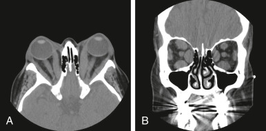
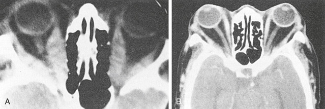
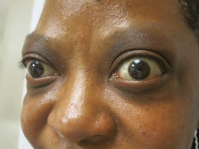
  • Exophthalmos (50% of patients) (Fig. E1, Fig. E2), lid retraction (lid lag), in which contraction of the levator palpebrae muscles of the eyelids show immobility of the upper eyelid with downward rotation of the eye
  • Nervousness, weight loss (weight gain in 10% of patients), heat intolerance, pruritus, muscle weakness, atrial fibrillation
  • Increased sweating, brittle nails, clubbing of fingers
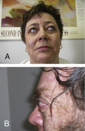
  • Localized infiltrative dermopathy (1% to 2% of patients) is most frequent over the anterolateral aspects of the legs, commonly over the pretibial area (pretibial myxedema) but can be found at other sites (especially after trauma). It is nonpitting and indurated. It is typically patchy with a peau d’orange appearance to the skin
  • Men may have gynecomastia, reduced libido, and erectile dysfunction. Women often have irregular menses

Figure E1 Proptosis seen in the setting of Graves disease.

A, Eye signs in Graves disease. B, Severe proptosis in Graves disease.

A and B, Courtesy Dr. Meir H. Kryger. From Kryger M et al: Principles and practice of sleep medicine, ed 6, Philadelphia, 2017, Elsevier.

Figure E2 Graves ophthalmopathy.

From Micheletti RG et al: Andrews’ diseases of the skin: clinical atlas, ed 2, Philadelphia 2023, Elsevier.

Etiology

Autoimmune etiology: Thyrotropin receptor antibodies (TRAb) mediated activation of thyroid-stimulating hormone receptor (TSHR). The activity of the thyroid gland is stimulated by the action of T cells, which induce specific B cells to synthesize antibodies against TSHRs in the follicular cell membrane.

Diagnosis

Differential Diagnosis

  • Anxiety disorder
  • Premenopausal state
  • Thyroiditis
  • Other causes of hyperthyroidism (e.g., toxic multinodular goiter, toxic adenoma)
  • Other: Metastatic neoplasm, diabetes mellitus, pheochromocytoma
Workup

  • The diagnosis is made clinically in most instances. The clinical assessment of the patient with Graves orbitopathy is summarized in Table 1.
  • The diagnostic workup includes a detailed medical history followed by laboratory and imaging studies and ECG. Patients often present with anxiety, heat intolerance, menstrual dysfunction, increased appetite, and weight loss. Elderly patients can have an atypical presentation (apathetic hyperparathyroidism). For additional information, refer to the topic “Hyperthyroidism.”

TABLE 1 Clinical Assessment of Patient With Graves Orbitopathy

Severity Measures (Using the Mnemonic NO SPECS)
NO SPECS ClassItemMethod
0. No signs or symptoms
1. Only signs, no symptomsLid apertureWith ruler in midline in mm
2. Soft tissue involvementEyelid and conjunctiva swelling and rednessInspection, color picturesa
3. ProptosisExophthalmosHertel in mm
4. Extraocular muscle involvementEye muscle motility
Diplopia
Impaired elevation, abduction
Subjective gradingb
5. Corneal involvementKeratitis, ulcerFluoresceine
6. Sight loss caused by optic nerve involvementDysthyroid optic neuropathy (DON)Visual acuity, color vision, visual fields, optic disc
Activity Measures (Using the Clinical Activity Score [CAS])
Inflammatory SignItemScore
PainSpontaneous retrobulbar pain
Pain on up gaze, side gaze, or down gaze
1
1
RednessRedness of the eyelids
Redness of the conjunctiva
1
1
SwellingSwelling of the eyelids
Swelling of the caruncle and/or plica
Chemosis
1
1
1
Maximum CAS Score (assessed momently)7
Impaired functionIncrease in proptosis 2 mm in 1-3 mo
Decrease of 8 degrees in eye muscle motility in any direction in 1-3 mo
Decrease in visual acuity of more than one line on the Snellen chart (using pinhole) in 1-3 mo
1
1
1
Maximum CAS Score (assessed over time)10

CAS, Clinical activity score.

a Color atlas in Dickinson AJ, Perros P: Controversies in the clinical evaluation of active thyroid-associated orbitopathy: use of a detailed protocol with comparative photographs for objective assessment, Clin Endocrinol (Oxf) 55:283-303, 2001.

b Intermittent diplopia = at awakening or when tired; inconstant diplopia = at extremes of gaze; constant diplopia = in primary or reading position.

From Melmed S et al: Williams textbook of endocrinology, ed 14, Philadelphia, 2020, Elsevier.

Laboratory Tests

  • Increased free thyroxine (T4) and free triiodothyronine (T3)
  • Decreased thyroid-stimulating hormone
  • Measurement of thyroid-stimulating antibodies (TSI) and TRAb
  • Table E2 summarizes assays of TSHR antibody nomenclature and indications
Imaging Studies

  • 24-h radioactive iodine uptake (RAIU): Increased homogeneous uptake.
  • Computed tomography (Fig. E3) or MRI of the orbits (Fig. E4) is useful if there is uncertainty about the cause of ophthalmopathy.

Figure E3 Axial (A) and Coronal (B) Computed Tomography Scans of a Patient with Graves Orbitopathy Demonstrating Generalized Enlargement of All Extraocular Muscles, Expansion of the Orbital Fat, and Marked Bilateral Proptosis

From Melmed S et al: Williams textbook of endocrinology, ed 14, Philadelphia, 2020, Elsevier.

Figure E4 Computed tomography scans of orbits in two patients with Graves orbitopathy.

A, Notice the obviously grossly swollen medial rectus extraocular muscles in both orbits and the resulting proptosis. B, The patient shows considerable proptosis with only minimal muscle enlargement, suggesting the presence of a large amount of retroorbital fat.

Courtesy Dr. Peter Som, New York.

TABLE E2 Assays of Thyroid-Stimulating Hormone Receptor Antibodies: Nomenclature and Indications

Nomenclature of TSHRAb Assays
TBII (TSH-binding inhibitory immunoglobulins)Measurement of inhibition of labeled TSH (or labeled thyroid-stimulating monoclonal antibody) binding to recombinant TSHR by serum antibodies
TSAb or TSI (thyroid-stimulating antibodies)Measurement of cAMP production by thyroid cell lines transfected with TSHR
TBAb (thyroid-blocking antibodies)Measurement of inhibition of cAMP production after TSH-mediated stimulation of thyroid cells or TSHR-transfected cells
Indications for Assay of TSHRAb
DiagnosisGraves hyperthyroidism
Graves orbitopathy and Graves dermopathy
Fetal and neonatal thyrotoxicosis
TreatmentChance of remission of hyperthyroidism at baseline, and during treatment with antithyroid drugs.

cAMP, Cyclic adenosine monophosphate; TSH, thyroid-stimulating hormone; TSHR, thyroid-stimulating hormone receptor; TSHRAb, thyroid-stimulating hormone receptor antibodies.

From Melmed S et al: Williams textbook of endocrinology, ed 14, Philadelphia, 2020, Elsevier.

Treatment

Nonpharmacologic Therapy

  • Patient education and discussion of therapeutic options.
  • Smoking cessation: Smoking is associated with an increased risk of progression of Graves ophthalmopathy.
Acute General Rx

  • Advantages and disadvantages of treatment options for Graves hyperthyroidism are summarized in Table E3.
  • Antithyroid drugs (thionamides, ATDs) to inhibit thyroid hormone synthesis or peripheral conversion of T4 to T3:
    1. Methimazole or propylthiouracil (PTU) are available. Methimazole is generally preferred because it has a longer half-life, allowing for once-daily dosing. PTU is preferred during pregnancy.
    2. Side effects: Skin rash (3% to 5%), arthralgias, myalgias, granulocytopenia (0.5%); rare side effects: Aplastic anemia, hepatic necrosis (PTU), cholestatic jaundice.
    3. Thionamide antithyroid drug therapy results in a remission in 40% to 50% of patients treated for 12 to 18 mo.
  • Radioactive iodine (RAI):
    1. Treatment of choice for patients >21 yr and younger patients who have not achieved remission after 1 yr of ATD therapy.
    2. Contraindicated during pregnancy and lactation.
    3. After radioactive therapy there may be an acute elevation of thyroid antibody titers and exacerbation of ocular symptoms in 15% to 20% of patients.
  • Surgery: Near-total thyroidectomy. Indications: Obstructing goiters despite RAI and ATD therapy, patients who refuse RAI and cannot be adequately managed with ATDs, and pregnant women inadequately managed with ATDs. Complications of surgery include hypoparathyroidism (4%) and vocal cord paralysis (1%).
  • Adjunctive therapy: Beta-adrenergic receptor blockers (e.g., atenolol 50 to 100 mg/day) to alleviate the beta-adrenergic symptoms of hyperthyroidism (tachycardia, tremor); contraindicated in patients with bronchospasm.
  • Graves ophthalmopathy: Methylcellulose eye drops to protect against excessive dryness, sunglasses to decrease photophobia, intraocular and systemic high-dose corticosteroids for severe exophthalmos. Worsening of ophthalmopathy after RAI therapy is often transient and can be prevented by the administration of prednisone. Other treatment options include antiinflammatory and immunosuppressive agents, radiation, and corrective surgical procedures. The administration of the antioxidant selenium (100 μg PO bid) has been recently reported as effective in improving quality of life, reducing ocular involvement, and slowing progression of the disease in patients with mild Graves orbitopathy. Its mechanism of action is believed to be an effect on the oxygen free radicals and cytokines that play a pathogenic role in Graves orbitopathy. Inhibition of the insulin-like growth factor I receptor (IGF-IR) is a new therapeutic strategy to combat the underlying autoimmune etiology of ophthalmopathy. Trials with teprotumumab, a human monoclonal antibody inhibitor of IGF-IR, in patients with active, moderate-to-severe ophthalmopathy have shown effectiveness in reducing proptosis.1
  • Dermopathy and acropachy: Topical corticosteroids are often used but are generally ineffective. Trials using rituximab infusion for dermopathy have shown striking improvement.

TABLE E3 Advantages and Disadvantages of Treatment Options for Graves Hyperthyroidism

Treatment OptionAdvantagesDisadvantages
Antithyroid drugsChance of permanent remission (35%)Side effects of ATDs
Long duration (12-18 mo)
High recurrence risk
Radioactive iodineSimplicity
Low recurrence risk
Risk of orbitopathy
Lifelong LT4 needed
Possible small increase in cancer risk
ThyroidectomyRapidity
Almost no recurrences
Low but unavoidable morbidity
Lifelong LT4 needed

ATDs, Antithyroid drugs; LT4, l -thyroxine.

From Melmed S et al: Williams textbook of endocrinology, ed 14, Philadelphia, 2020, Elsevier.

Chronic Rx

Patients undergoing treatment with ATDs should be seen every 1 to 3 mo until euthyroidism is achieved and every 3 to 4 mo while they are receiving ATDs.

Disposition

  • ATDs induce sustained remission in <60% of cases.
  • The incidence of hypothyroidism after RAI is >50% within the first yr and 2% per yr thereafter.
  • Complications of surgery include hypothyroidism (28% to 43% after 10 yr), hypoparathyroidism (4%), and vocal cord paralysis (1%).
  • Successful treatment of hyperthyroidism requires lifelong monitoring for the onset of hypothyroidism or the recurrence of thyrotoxicosis.
  • RAI therapy is followed by the appearance or worsening of ophthalmopathy more often than is therapy with methimazole, particularly in patients who are cigarette smokers. It can be prevented with the administration of prednisone 0.5 mg/kg body weight per day starting 2 to 3 days after RAI, continued for 1 mo, then tapered off over 2 mo.
  • Mild to moderate ophthalmopathy often improves spontaneously. Severe cases can be treated with high-dose glucocorticoids, orbital irradiation, or both. Orbital decompression may be used in patients with optic neuropathy and exophthalmos (see “Hyperthyroidism”).
Related Content

Graves Disease (Patient Information)

Hyperthyroidism (Related Key Topic)

Related Content

    1. Douglas R.S. : Teprotumumab for the treatment of active thyroid eye diseaseN Engl J Med. ;382(4):341-352, 2020.
    2. Bahn R.S. : Graves’ ophthalmopathyN Engl J Med. ;362:726-738, 2010.
    3. Burch H.B., Cooper D.S. : Management of Graves disease: a reviewJ Am Med Assoc. ;314(23):2544-2554, 2015.
    4. Marsocci C. : Selenium and the course of mild Graves’ orbitopathyN Engl J Med. ;364:1920-1931, 2011.
    5. Smith T.J., Hegedus L. : Graves’ diseaseN Engl J Med. ;375:1552-1565, 2016.