AUTHOR: Donny V. Huynh, MD


DefinitionInflammatory anemia, also known as anemia of chronic disease (ACD), refers to the impaired production of erythrocytes associated with chronic inflammatory states, such as cancer, chronic infection, or autoimmune diseases. Recent data have connected inflammatory anemia with severe, acute inflammation, such as critical illness, or with milder but persistent inflammatory signals that occur in obesity, aging, and kidney failure.1 It is a disorder of iron homeostasis (Table 1) promoted by hepcidin-25 in response to an inflammatory condition.
TABLE 1 Suspected Causes of Anemia of Chronic Disease
Shortened erythrocyte survival
Block in reuse of iron by erythrocyte
Direct inhibition of erythropoiesis
Relative deficiency of erythropoietin |
From Hoffman R et al: Hematology, basic principles and practice, ed 7, Philadelphia, 2018, Elsevier.
SynonymsAnemia of chronic disease
ACD
Anemia, inflammatory
| ICD-10CM CODES | | D63.8 | Anemia in chronic diseases classified elsewhere | | D63.0 | Anemia in neoplastic disease | | D64.8 | Anemia, unspecified |
|
Epidemiology & DemographicsPrevalence
- Second-most prevalent anemia after iron deficiency anemia:
- Around 11% of men and 10% of women ages 65 to 85 yr
- >20% of adults older than 85 yr
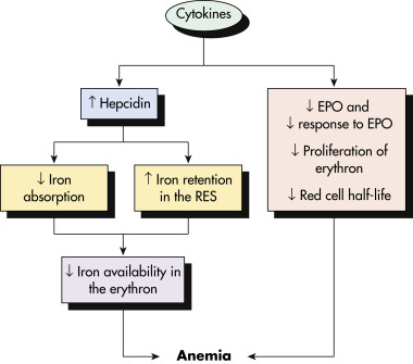
Figure 1 Pathophysiologic Factors Associated with the Development of Anemia of Chronic DiseaseEpo, Erythropoietin; Res, Reticuloendothelial System.

From Hoffman R et al: Hematology, basic principles and practice, ed 7, Philadelphia, 2018, Elsevier.
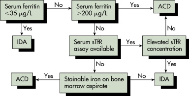
Pathophysiology (FIG. 1Iron is carried in the bloodstream shelled by a hollow protein called transferrin (<0.2% of total iron body content) or at the core of hemoglobin in red blood cells (RBCs; 60% of total iron body content). It is mainly stored (15% to 30% of total iron body content) inside the liver, spleen, and skeletal muscle as ferritin and in lysosomes as hemosiderin. The rest of the body iron content is trapped in skeletal muscle myoglobin and mitochondrial cytochromes. In clinical practice, ferritin is a surrogate for iron stores, and total iron binding capacity (TIBC) is a surrogate for transferrin and iron carrying capacity.
Cells involved in the response to inflammation cause the release of cytokines, such as interleukin 6 (IL-6), which stimulates hepatic release of hepcidin. Hepcidin is a circulating protein that blocks ferroportin, an iron channel responsible for the exit of iron from enterocytes (and thus gastrointestinal absorption) and macrophages (which accumulate iron from engulfed senescent blood cells). IL-1 and tumor necrosis factor (TNF)-alpha stimulate interferon-gamma release by marrow stromal cells, which in turn suppress the erythroid response to erythropoietin (EPO). In chronic kidney disease, ACD is a consequence of decreased production of EPO and decreased renal clearance of hepcidin. The low availability of serum iron causes iron deficiency in the bone marrow compartment and decreased reticulocyte levels.
Clinical Presentation
- Generalized symptoms include fatigue, shortness of breath, and weakness.
- It is important to consider other complaints if the underlying diagnosis is unknown, such as weight loss (malignancy, chronic infections, connective tissue diseases), anorexia, nausea, paresthesias, pleuritic chest pain, weight gain (chronic kidney disease [CKD]), diarrhea, bloody stools, abdominal pain, oral ulcers (IBD), and fevers (HIV, chronic infections).
- Physical findings may include pallor, lymphadenopathy, signs of connective tissue diseases (malar rash, sclerodactyly), palpable or visible masses, and localized findings for infection or malignancy.
Etiology
- Malignancy
- CKD (patients with CKD stage IV [glomerular filtration rate [GFR] <30 ml/min] should be screened for ACD)
- Congestive heart failure [CHF] (ACD is the main cause of anemia in CHF patients)
- Chronic infections
- Anemia of critical illness (develops within days)
- Connective tissue diseases

Treat the underlying disorder/disease.
Acute General Rx
- The treatment of inflammatory anemia is directed primarily at treatment of underlying conditions, which can improve symptoms and facilitate hemoglobin recovery.
- Packed RBC (PRBC) transfusion is usually reserved for severe anemia (with Hb level <7 g/dl or <8 g/dl in patients with cardiac disease), especially if complicated with ongoing bleeding.
Chronic Rx
- Erythropoiesis-stimulating agents (ESA) (epoetin alfa and darbepoetin alfa) are FDA approved for use in patients with anemia resulting from:
- Chronic kidney disease
- Chemotherapy
- A 1998 study, the Normal Hematocrit Cardiac Trial (NHCT), showed a nonsignificant increase in the combined endpoint death and nonfatal myocardial infarction in patients with goal hematocrit of 33% versus 27%. Subsequent studies (CHOIR, CREATE, and TREAT) showed that higher doses and higher hematocrit targets were associated with increased cardiovascular events.
- ESA dose should be individualized for each patient, and the lowest sufficient dose to reduce PRBC transfusions should be used.2 A hemoglobin target of ∼10 g% is widely accepted. Iron deficiency should be ruled out before ESA is started. After starting ESA therapy, ASH/ASCO guidelines recommend periodic monitoring of iron status. When there is no or suboptimal response to oral therapy, parenteral iron therapy should be considered before concluding that a patient is nonresponsive to iron therapy.
- The hepcidin-ferroportin axis is the target of development of novel agents of which the most promising are hypoxia-induced factor modulators. Hypoxia-inducible factor (HIF) is a transcription factor that promotes expression of erythropoietin. HIF is upregulated by inhibition of PHD. Small molecule inhibitors of prolyl hydroxylase domain dioxygenases (HIF-PHI [prolyl hydroxylase inhibitor]) stimulate the production of endogenous erythropoietin and improve iron metabolism.3 The clinical development of three oral agents targeting this axis-daprodustat, roxadustat, and vadadustat-has now completed randomized clinical development.
- Among patients with CKD undergoing dialysis, the oral hypoxia-inducible factor prolyl hydroxylase inhibitors (HIF-PHI) daprodustat was noninferior to ESAs regarding the change in the hemoglobin level from baseline and cardiovascular outcomes.
- Additionally, among patients with CKD and anemia who were not undergoing dialysis, daprodustat was noninferior to darbepoetin alfa with respect to the change in the hemoglobin level from baseline and with respect to cardiovascular outcomes.