section name header

Basic Information

AUTHORS: Hannah Fiske, MDand Harlan G. Rich, MD, FACP, AGAF

Definition

Achalasia is a motility disorder of the esophagus classically characterized by incomplete relaxation of the lower esophageal sphincter (LES) and aperistalsis of the esophageal smooth muscle resulting in functional obstruction of the esophagus.

Synonyms

  • Achalasia and cardiospasm
  • Achalasia (of cardia)
  • Aperistalsis of esophagus
  • Megaesophagus
  • Esophageal achalasia
  • Esophageal cardiospasm
ICD 10-CM CODES
K22.0Achalasia of cardia
Epidemiology & Demographics

  • Incidence has increased to approximately to 0.03 to 1.63 in 100,000 persons/yr due to studies using high-resolution esophageal manometry (HRM).1
  • Prevalence is around 10 per 100,000 persons.
  • Although the onset of symptoms may occur at any age, incidence is typically bimodal (between 20 and 40 yr, then after 60 yr) with greater incidence in the elderly.
  • Men and women are affected equally.
Physical Findings & Clinical Presentation

Symptoms:

  • Dysphagia (most commonly with both solids and liquids) without oropharyngeal transfer difficulties
  • Difficulty belching
  • Regurgitation or vomiting of undigested food
  • Chest pain and/or heartburn
  • Globus
  • Frequent hiccups
  • Symptoms of aspiration such as nocturnal cough; possible dyspnea and pneumonia
  • Weight loss
  • The Eckardt symptom score (assessing dysphagia, regurgitation, retrosternal pain, and weight loss) is a fairly reliable measure of achalasia severity and may be used to assess response to therapy
  • The Brief Esophageal Dysphagia Questionnaire (BEDQ) is more highly sensitive for manometric diagnosis of dysphagia; it is independent of GERD symptoms and has been shown to be a better symptom-generic evaluating tool, while the Eckardt score is a more achalasia-specific metric.2

Physical Exam:

  • While achalasia patients typically have a benign physical exam, focal lung examination abnormalities and wheezing are possible.
Etiology

  • Etiology is poorly understood.
  • Loss of intrinsic inhibitory neurons in the myenteric plexus and smooth muscle portion of the esophagus as well as depletion of networks of interstitial cells of Cajal of the LES result in the loss of inhibitory neurotransmitters nitric oxide and vasoactive intestinal polypeptide and unopposed excitatory cholinergic activity, leading to incomplete relaxation of the lower esophageal sphincter (LES) and loss of esophageal peristalsis.1,3
  • Loss of myenteric nerve fibers is associated with lymphocytic and eosinophilic infiltrates, capillaritis, plexitis, venulitis, nerve hypertrophy, and fibrosis.
  • There is evidence to suggest that esophageal achalasia may be autoimmune mediated, with an increase in immune cells, cytokines, chemokines, and autoimmune antibodies.4This disorder may be caused by autoimmune degeneration of the esophageal myenteric plexus in association with several human leucocyte antigen (HLA) class II DQ antigens. An eight amino-acid insertion in the cytoplasmic tail of HLA-DQβ1 has been identified as a strong achalasia risk factor. Antimyenteric plexus and other antineural autoantibodies have also been described. Patients with achalasia are more likely to have other autoimmune diseases.
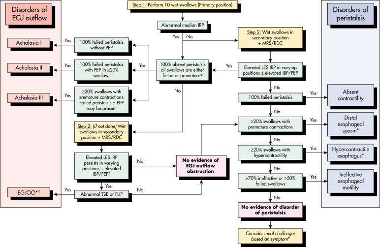
  • Abnormal immune reactions to neurotropic viruses, such as varicella zoster, measles, herpes simplex type 1, and human papilloma virus have been implicated. A host T-cell-mediated response may lead to neuronal injury; this has been suggested as the cause of types I and II achalasia (see “Imaging Studies” section).
  • Achalasia is also seen in Allgrove syndrome, a rare autosomal recessive disorder caused by gene mutation on chromosome 12q13 and defined by achalasia, alacrima, autonomic disturbance, and acetylcholine insensitivity. Neurons in this syndrome may be susceptible to oxidative injury.
  • Recent studies suggest that type III achalasia (see “Imaging Studies” section) is associated with myenteric inflammation but not neuronal loss; downregulation of nitric oxide synthase expression and increased cholinergic sensitivity are cytokine-mediated.
  • A type III achalasia pattern has been described in chronic daily opiate users.

Diagnosis

Differential Diagnosis

  • Primary achalasia:
    1. Idiopathic
  • Secondary achalasia:
    1. Chagas disease
    2. Vagal injury
    3. Prior surgery, including fundoplication; achalasia-like esophageal dilation has also been described after laparoscopic gastric banding
  • Pseudoachalasia (diseases that may mimic achalasia):
    1. Esophageal cancer
    2. Infiltrating gastric cancer
    3. Oat cell and bronchogenic lung cancer
    4. Lymphoma
    5. Amyloidosis
    6. Paraneoplastic syndrome
  • Angina
  • Bulimia
  • Anorexia nervosa
  • Gastric bezoar
  • Gastritis
  • Peptic ulcer disease
  • Postvagotomy dysmotility
  • Opioid-induced esophageal dysmotility
  • Esophageal disease (Table 1 ):
    1. Gastroesophageal reflux disease
    2. Sarcoidosis
    3. Amyloidosis
    4. Esophageal stricture
    5. Esophageal webs and rings
    6. Scleroderma
    7. Barrett esophagus
    8. Esophagitis
    9. Diffuse esophageal spasm

TABLE 1 Esophageal Motor Disorders

AchalasiaSclerodermaDistal Esophageal Spasm
SymptomsDysphagia
Regurgitation of nonacidic material
Gastroesophageal reflux disease
Dysphagia
Substernal chest pain (angina-like)
Dysphagia with pain
Radiographic appearanceDilated, fluid-filled esophagus
Distal bird-beak stricture
Aperistaltic esophagus
Free reflux
Peptic stricture
Simultaneous noncoordinated contractions
Manometric findingsHigh resting pressureLow resting pressureNormal pressure
Lower esophageal sphincterIncomplete or abnormal relaxation with swallow
BodyLow-amplitude, simultaneous contractions after swallowingLow-amplitude peristaltic contractions or no peristalsisSome peristalsis
Diffuse and simultaneous nonperistaltic contractions, occasionally high amplitude

From Andreoli TE et al: Andreoli and Carpenter’s Cecil essentials of medicine, ed 8, Philadelphia, 2010, Saunders.

Workup

  • Physical examination and laboratory analyses to rule out other causes (Fig. E1, Table 2 ) and assess complications
  • Imaging studies, manometry, and endoscopy (may be supportive or complementary)

TABLE 2 Chicago Classification v4.0 of Esophageal Motility Disorders5

DisorderDiagnostic Criteria
Disorders of EGJ Outflow
Type I achalasia (classic)Abnormal median IRP and 100% failed peristalsis
Type II achalasiaAbnormal median IRP, 100% failed peristalsis, and 20% of swallows with panesophageal pressurization
Type III achalasiaAbnormal median IRP and 20% of swallows with premature/spastic contractions and no evidence of peristalsis
EGJ outflow obstructionAbnormal median IRP (supine and upright), 20% elevated intrabolus pressure (supine), and sufficient evidence of peristalsis such that criteria for achalasia are not met; diagnosis requires clinical symptoms in addition to manometric criteria
Disorders of Peristalsis
DESNormal median IRP and 20% swallows with premature/spastic contractions with DCI >450 mm Hgseccm; diagnosis requires clinical symptoms in addition to manometric criteria
Hypercontractile (jackhammer) esophagusNormal median IRP and 20% hypercontractile swallows with DCI >8000 mm Hgseccm; diagnosis requires clinical symptoms in addition to manometric criteria
Absent contractilityNormal median IRP and 100% failed peristalsis/absent contractility (DCI <100 mm Hgseccm)
Ineffective esophageal motility (includes the definition of fragmented peristalsis)>70% ineffective swallows or with DCI <450 mm Hgseccm or 50% failed peristalsis

DES, Diffuse esophageal spasm; DCI, distal contractile interval; DL, distal latency; EGJ, esophagogastric junction; IRP, integrated relaxation pressure ULN, upper limits of normal.

FIG E1 Algorithm for applying the Chicago classification of esophageal motor disorders.

!!flowchart!!

CFV, Contractile front velocity; DCI, distal contractile interval; DL, distal latency; EGJ, esophagogastric junction; IBC, isobar contour; IRP, integrated relaxation pressure; PEP, panesophageal pressurization; ULN, upper limits of normal (see Table 2 for more details).

From Yadlapati R et al: What is new in Chicago Classification version 4.0, Neurogastroenterol Motil 33(1):e14053, 2021. https://doi.org/10.1111/nmo.14053.

Laboratory Tests

  • Assessment of nutritional status (evaluate for malnutrition, iron deficiency, vitamin/mineral deficiencies)
  • Complete blood count
  • ECG and/or stress test as appropriate to exclude cardiac etiologies of substernal chest pain
  • Serologic assays for Trypanosoma cruzi (Chagas disease) in appropriate individuals
Imaging Studies

Barium swallow with fluoroscopy (particularly a timed barium esophagram (TBE)) may demonstrate:

  • Uncoordinated or absent esophageal contractions (loss of peristalsis)
  • An acutely tapered contrast column (“bird’s beak”; Fig. E2)
  • Dilation of the distal esophagus (smooth muscle portion; Fig. E3)
  • Esophageal air-fluid level with evidence of poor esophageal emptying
  • Late-stage changes include tortuosity, angulation, dilated megaesophagus, retained food, and secretions

Manometry (Fig. E4) is considered the “gold standard” test to confirm the diagnosis. High-resolution manometry (HRM) or high-resolution esophageal pressure topography (HREPT) has defined subsets of patients with achalasia who may have different responses to medical or surgical therapies and prognoses. This technique uses the integrated relaxation pressure (IRP) >15 mm Hg to define better the failure of esophagogastric junction relaxation. HRM also utilizes the distal contraction integral (DCI) to define hypercontractile vs. weak swallows vs. failed peristalsis. The distal latency (DL) defines premature contractions.

  • In classic (Type I) achalasia, manometric abnormalities include 100% aperistalsis and incomplete or absent LES relaxation (with median IRP 15 mm Hg while supine or 12 mm Hg while upright) after swallow.
  • Type II achalasia shows panesophageal pressurization to greater than 30 mm Hg with 20% of test swallows, aperistalsis, and incomplete or absent LES relaxation.
  • Type III achalasia shows premature or spastic contractions of the distal esophagus with 20% of swallows (reduced DL, normal DCI), aperistalsis, and incomplete or absent LES relaxation.5
  • HREPT has also defined the achalasia variant esophagogastric junction outflow obstruction (EGJOO), where the IRP is 15 mm Hg, but peristalsis is present, excluding the diagnosis of achalasia. Some are associated with secondary causes; a small percentage may progress to achalasia; and, in many, the natural history is unclear, and response to therapy is variable. A clinical diagnosis of EGJOO requires both a manometric diagnosis of EGJOO as well as clinically relevant symptoms (dysphagia and/or noncardiac chest pain) supported by follow-up testing, including pharmacologic provocation, TBE, or functional luminal imaging probe (FLIP) technology.5If incidentally found on manometry, asymptomatic EGJOO does not require treatment.
  • Multiple rapid swallows and rapid drink challenges are provocative tests done as part of HREPT to distinguish achalasia from other motility disorders.5
  • Accuracy of manometric classification requires normal foregut anatomy, without prior surgical interventions and without anatomical variants, including hernias, which could alter manometric catheter measurements.5
  • Direct visualization by endoscopy (Fig. E5), including careful visualization of the esophagogastric junction and cardia, should be performed prior to manometry to exclude other causes of dysphagia, including EGJOO, strictures, secondary causes of achalasia (including infiltrating cancers), and pseudoachalasia. It is important that patients on long-term opioids be weaned off prior to manometric testing to exclude opioid-induced esophageal dysmotility; opioids have been linked to shortened DL, elevated DCI, and elevated IRP.

FLIP is a new technique that can measure compliance and distensibility across the esophagogastric junction and may demonstrate both impaired LES relaxation and response to achalasia therapy.6

FIG E2 Classic esophagram of a patient with achalasia.

The esophagus is dilated with a “bird’s beak” tapering of the distal esophagus. Retained secretions form the heterogeneous air-fluid level seen at the top of the barium column.

From Flint PW et al: Cummings otolaryngology, head and neck surgery, ed 7, Philadelphia 2021, Elsevier.

FIG E3 Esophagram of late-stage achalasia.

The esophagus now has a sigmoidlike tortuosity with a large amount of retained debris.

From Flint PW et al: Cummings otolaryngology, head and neck surgery, ed 7, Philadelphia 2021, Elsevier.

FIG E4 Manometric findings in achalasia.

Aperistalsis is manifested by isobaric contractions without propagation. The lower esophageal sphincter pressure, which is elevated, shows minimal relaxation with swallowing.

From Flint PW et al: Cummings otolaryngology, head and neck surgery, ed 7, Philadelphia 2021, Elsevier.

FIG E5 Endoscopic view of the distal esophagus in a patient with achalasia.

The esophagus is dilated with retained fluid and debris.

From Flint PW et al: Cummings otolaryngology, head and neck surgery, ed 7, Philadelphia 2021, Elsevier.

Treatment

Nonpharmacologic Therapy

  • Achalasia is treatable but incurable. The goals of therapy are to decrease LES pressure to relieve the functional obstruction, improve symptoms, and prevent progression to a dilated esophagus, also referred to as a megaesophagus. Current treatments are unable to improve esophageal peristalsis.
  • Pneumatic dilation (PD) uses a fixed-diameter and high-pressure dilating balloon across the LES under fluoroscopic visualization to disrupt the muscle fibers and may benefit 50% to 93% of patients. Most protocols use a graded dilation approach, starting with a 30-mm balloon and repeating if required with a 35-mm or 40-mm balloon. Studies show this may be more effective in females, older patients, and HRM type 2 patients. Esophageal rupture or perforation is a rare complication (2% to 4%). It may be managed conservatively in stable patients with a small perforation but may require surgery with larger tears or mediastinal contamination. Some patients will experience heartburn after, usually responsive to proton-pump inhibitor therapy.
  • Surgical: Laparoscopic, or less commonly open, Heller esophagomyotomy (HM) is effective (90%). Approximately 35% of patients undergoing surgery will develop reflux disease. As a result, some surgeons will perform an anterior Dor or posterior Toupet antireflux repair (fundoplication) as part of the surgical procedure. Studies suggest this may be more effective in men and younger patients. An observational study has suggested that those with prior endoscopic treatment before myotomy may not do as well as those who have a primary myotomy.
  • A large European study suggested that patients may expect similar medium-term outcomes from myotomy and balloon dilation. A meta-analysis suggests better long-term durability of myotomy, while balloon dilation may be more cost-effective. About 20% to 30% of patients undergoing either therapy may require retreatment within 5 to 7 yr.
  • Endoscopic submucosal myotomy (peroral endoscopic myotomy [POEM]) has a high success rate comparable to laparoscopic HM (particularly in HRM type 3 patients in whom a longer myotomy, tailored to the length of the diseased segment, improves outcome), decreased hospital length of stay, few adverse events (which are similar to those associated with Heller myotomy and include pneumomediastinum, pneumothorax, pneumoperitoneum, pleural effusion, pneumonia, and bleeding), and exceedingly rare mortality. Since no antireflux procedure is performed, there is a modest risk (up to 53%) of developing pathologic reflux. POEM should only be performed at high-volume centers. A randomized multicenter study showed a higher treatment success rate for POEM at 2 yr when compared with pneumatic dilation.7,8
  • FLIP and TBE may help determine response to therapy.6
  • Esophagectomy has been performed in patients with end-stage achalasia with a dilated, often sigmoid-shaped or megaesophagus, who have failed myotomy or pneumatic dilation. Esophagectomy is coupled with either gastric pull-up, colon interposition, or jejunal interposition, where the stomach, colon, or small bowel are used to functionally replace the esophagus.
  • Self-expanding metal stents (SEMS) have been studied as a temporary treatment in a few studies with low-quality evidence and are not currently recommended. Patients with SEMS often reported chest pain and regurgitation.
  • Transoral incisionless fundoplication (TIF) may be an endoscopic treatment alternative for postprocedural GERD, specifically after POEM.9
Acute General Rx

  • Medications may be useful for short-term symptom relief and in patients with refractory chest pain. They should only be considered in patients unable to receive, or who are scheduled for, more definitive procedures. LES pressure may be lowered temporarily by up to 50% through sublingual use of long-acting nitrates (e.g., isosorbide dinitrate 5 to 20 mg) or calcium channel blockers (e.g., nifedipine 10 to 30 mg). Side effects are common, and duration of relief tends to be short. Sildenafil was shown to be effective in a few small, short-term studies based on manometric results but is generally not recommended given minimal clinical improvement and negative side effect profile. Anticholinergics such as atropine and theophylline are less commonly used.
  • Botulinum toxin (BT) injection will benefit up to 85% of patients by inhibiting acetylcholine release from cholinergic nerve endings and blocking the unopposed cholinergic stimulation of the LES, without impacting the myogenic tone. Up to half will require repeat injections by 6 mo. A few studies have suggested that repeated injections may have diminished efficacy and can lead to fibrosis, complicating subsequent attempts at surgical therapy. Studies have identified age >40, type II or III achalasia, and EGJOO to have more favorable response to BT injections. BT is recommended as first-line therapy for patients who may not be candidates for surgical options.
  • Many patients will require proton pump inhibitor therapy for gastroesophageal reflux after effective disruption of the LES.
  • For patients with weight loss secondary to achalasia, studies regarding dietary modification as adjunctive treatment are underway to help improve weight after intervention.
  • Future therapies, including neural cell transplantation and electrical stimulation, are currently under investigation in animal models, but likely are years away from implementation.

Pearls & Considerations

Comments

  • Medication has a limited role in treatment.
  • Botulinum toxin is transiently effective in improving symptoms and should be considered primarily in patients too elderly or ill to be considered for surgery.
  • Pneumatic dilation, surgical myotomy, and POEM provide more durable long-term responses and are the treatment of choice for most patients.
  • Patients with achalasia may be at long-term risk of squamous cell carcinoma of the esophagus and non-reflux-associated esophagitis. Treated patients may be at long-term risk for reflux esophagitis, Barrett esophagus, and adenocarcinoma. Endoscopic surveillance is not routinely recommended in these patients.
Related Content

Achalasia (Patient Information)

Dysphagia (Related Key Topic)

Related Content

    1. Mari A. : Diagnosis and management of achalasia: updates of the last two yearsJ Clin Med. ;10(16), 2021.doi:10.3390/jcm10163607
    2. Chen S. : The number of interstitial cells of cajal differs among different subtypes of achalasia and is related to patients’ prognosis Published 2021 Aug 24 Clin Transl Gastroenterol. ;12(8), 2021.doi:10.14309/ctg.0000000000000388
    3. Cisternas D. : The Brief Esophageal Dysphagia Questionnaire shows better discriminative capacity for clinical and manometric findings than the Eckardt score: results from a multicenter study, [published online ahead of print, 2021 Aug 2]Neurogastroenterol Motil. , 2021.doi:10.1111/nmo.14228
    4. Wu X.Y. : The etiology of achalasia: an immune-dominant diseaseJ Dig Dis. ;22(3):126-135, 2021.doi:10.1111/1751-2980.12973. 33583137
    5. Yadlapati R. : What is new in Chicago Classification version 4.0?Neurogastroenterol Motil. ;33(1), 2021.doi:10.1111/nmo.14053
    6. Holmstrom A.L. : Comparison of preoperative, intraoperative, and follow-up functional luminal imaging probe measurements in patients undergoing myotomy for achalasiaGastrointest Endosc. ;94(3):509-514, 2021.doi:10.1016/j.gie.2021.02.031
    7. Richter J.E. : Tailoring therapy for achalasiaGastroenterol Hepatol. ;16:249-257, 2020.
    8. Campagna R.A.J. : Outcomes of 100 patients more than 4 years after POEM for achalasiaAnn Surg. ;273(6):1135-1140, 2021.
    9. Brewer Gutierrez O.I. : Is transoral incisionless fundoplication (TIF) an answer to post-peroral endoscopic myotomy gastroesophageal reflux? A multicenter retrospective study [published online ahead of print, 2021 May 28] Endoscopy. ;210, 2021.doi:10.1055/a-1446-8953