Author: Kapil S. Meleveedu, MD
Acute lymphoblastic leukemia (ALL) is a malignancy of precursor B or T lymphocytes (lymphoblasts) characterized by uncontrolled proliferation of malignant lymphocytic cells with replacement of normal bone marrow elements and bone marrow failure. Lymphoblastic lymphoma (LBL) is diagnosed when the disease presents in extramedullary sites (most commonly as mediastinal mass in T-cell disease) and <20% of the bone marrow is involved.
|
Expected overall 5 yr relative survival is 71% but substantially better in children and approaches 95%.
TABLE E1 Clinical Presentation of Acute Lymphoblastic Leukemia
Symptoms/Signs | Etiology | Management |
---|---|---|
Fever | Disease or infection | Always conduct fever workup and provide broad antimicrobial coverage until infectious etiology is ruled out |
Fatigue, pallor | Anemia (ALL infiltrating bone marrow) | Packed red blood cell transfusion (slow if anemia is severe, avoid in hyperleukocytosis) |
Petechiae, bruising, bleeding | Thrombocytopenia (ALL infiltrating bone marrow) | Transfuse with platelets |
Pain | Leukemia infiltrating bones/joints, or expanding marrow cavity | Establish diagnosis and start chemotherapy |
Respiratory distress/superior vena cava syndrome | Mediastinal mass | Avoid sedation in presence of tracheal compression. Establish diagnosis as soon as possible and start chemotherapy |
ALL, Acute lymphoblastic leukemia.
From Hoffman R et al: Hematology, basic principles and practice, ed 8, Philadelphia, 2023, Elsevier.
Disorders associated with lymphocytosis (lymphocytes >5000/mcl):
TABLE E3 WHO Classification of Precursor B-cell and T-cell Lymphoid Neoplasms
NK, Natural killer; WHO, World Health Organization.
TABLE E4 More Common Recurrent Cytogenetic Abnormalities in Acute Lymphoblastic Leukemia (ALL)/Lymphoma
Abnormality | Clinical Relevance | ||
---|---|---|---|
B cell ALL | |||
t(9;22)(q34;q11.2); BCR-ABL1 | Incidence approximately 3% in children, 25% in adults, rising with age; requires therapy with ABL1 tyrosine kinase inhibitors. | ||
t(v*;11q23) KMT2A rearranged | Most common variant is t(4;11); often presents with very high white blood cell count; confers worse prognosis; rare in adults; common in infant leukemia. Hematopoietic stem cell transplant is often required. | ||
t(12;21)(p13;q22); ETV6-RUNX1 | Common in children (20%-30%); rare in adults; confers improved prognosis with intensive asparaginase. | ||
Hyperdiploidy (>50 chromosomes) | Seen in about 25% of children, less in adults; confers favorable prognosis. | ||
Hypodiploidy (<44 chromosomes) | Uncommon; confers worse prognosis. | ||
t(1;19)(q23;p13.3); TCF3-PBX1 | Incidence approximately 5%; intermediate/favorable in children, intermediate/poor in adults. Benefits from high dose methotrexate. | ||
BCR-ABL1-like | Shares similar gene expression profile as BCR-ABL1 but lacks the t(9;22) fusion. Adverse prognosis. Might benefit from tyrosine kinase inhibitors and/or JAK2 inhibitors. | ||
T-cell ALL | More common in adults 60%-70%. | ||
NOTCH1 mutation |
v*, Variable gene partners. Many of these disorders also have distinct immunophenotypes by flow cytometry. Additional molecular abnormalities of recently defined relevance include mutations of IKZF1, which encodes a lymphoid transcription factor IKAROS, is associated with high relapse rates and gene expression profile similar to BCR-ABL1 translocated disease. Gene expression profiling has identified a subgroup of "Philadelphia chromosome-like" ALL with a gene expression similar to BCR-ABL1 translocation associated disease, which confers worse prognosis, but which may identify new opportunities for targeted therapies.
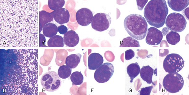
Figure E1 Morphologic Features of Acute Lymphoblastic Leukemia in the Blood and Bone Marrow Aspirate
A, In acute lymphoblastic leukemia (ALL), the peripheral blood count can be low, normal, or high, although frequently it is high and composed of mostly blasts. B, A similar range in cellularity is true regarding the bone marrow. Typically, lymphoblasts are small to intermediate in size and have round nuclei with dispersed chromatin and indistinct nucleoli. C, They have scant pale blue cytoplasm. A small lymphocyte (C, middle, left) is useful for comparison. D, In many cases, the lymphoblasts are monotonous, but in some cases, the lymphoblast morphology is varied with some large cells, some cells with abundant cytoplasm, and other cells with prominent nucleoli. In the older French-American-British classification, these two patterns were referred to as "ALL-L1" and "ALL-L2," respectively, although they have been shown not to have any clinical significance. Other cytologic variants of lymphoblasts are shown in E-H. These include (E) lymphocytelike blasts, (F) blasts with azurophilic granules, (G) blasts with "hand-mirror" morphology, and (H) blasts with vacuoles. The small lymphocytelike blasts can be difficult to distinguish from normal mature lymphocytes or chronic lymphocytic leukemia cells in the blood, making flow immunophenotyping important in the distinction. Lymphoblasts with granules can be misleading because they can be mistaken for myeloblast or monoblasts. The hand-mirror cells appear to be an artifact because they can be seen only focally on a smear. Vacuoles in the blasts can make the blasts difficult to distinguish from Burkitt cells. However, flow cytometry can easily distinguish the immature ALL blasts from the mature B cells seen in a leukemic presentation of Burkitt lymphoma.
(From Hoffman R et al: Hematology, basic principles and practice, ed 8, Philadelphia, 2023, Elsevier.)
TABLE E2 Initial Evaluation of a Patient with Acute Lymphoblastic Leukemia
ASCT, Allogeneic stem cell transplantation; BM, bone marrow; CBC, complete blood count; CT, computed tomography; HLA, human leukocyte antigen; LDH, lactate dehydrogenase; LFT, liver function test.
From Hoffman R et al: Hematology, basic principles and practice, ed 8, Philadelphia, 2023, Elsevier.
TABLE E6 Risk Factors for Treatment Failure in Recent ALL Trials
t(v*;11q23) MLL rearranged Hypodiploidy Minimal residual disease after remission or consolidation* Philadelphia chromosome-like genomic signature (in Ph-ALL) Early precursor T (ETP) ALL (absent CD1a, CD8, weak CD5, myeloid or stem cell antigen expression) |
ALL, Acute lymphoblastic leukemia.
* Measured variously after induction or consolidation therapy.
Standardized testing for this is still in development, but it may have important treatment implications. Note also that many historic risk factors (e.g., T-cell vs B-cell disease) have not been independent risk factors in current trials.
From Roberts KG et al: Targetable kinase-activating lesions in Ph-like acute lymphoblastic leukemia, N Engl J Med 371:1005-1015, 2014.
TABLE E7 Prognostic Factors in Acute Lymphoblastic Leukemia
Factor | Prognosis | Clinical Application |
---|---|---|
Age | ||
<1 yr | KMT2Ar (70%-80% infants) poor outcome; KMT2AWT same outcome as older children | KMT2AWT do well on standard ALL therapy. Potential role for proteasome inhibitors, histone deacetylase inhibitors, hypomethylating agents, BCL2 targeted agents, immunotherapy for KMT2Ar |
1-9 yr | Lower (standard) risk | ALL biology may change risk |
>9 yr | Higher risk | ALL biology may change risk |
WBC | ||
<50 × 109/L | Lower (standard) risk | ALL biology may change risk |
≥50 × 109/L | Higher risk | ALL biology may change risk |
CNS | ||
CNS3 | Higher risk of CNS and bone marrow relapse | Therapy intensification, +/ cXRT |
CNS2 traumatic lumbar puncture with blasts | Higher risk of CNS relapse | CNS directed therapy intensification |
Testicular | Higher risk | Therapy intensification, radiation for persistent testicular leukemia |
Immunophenotype | ||
T cell | Higher risk | Poor outcome abolished with current therapy; nelarabine an active agent |
pre-B (cIgM+) | Standard risk | Poor outcome abolished with current therapy |
Early pre-B | Standard risk | Genetics may change risk |
Early T-cell precursor | Adverse prognosis | Poor outcome abolished with current therapy |
Ploidy | ||
>50 (DI >1.16) (or trisomies of chromosomes 4 and 10) | Low risk | Good response to antimetabolites, may have delayed MRD clearance, but do well provided MRD <0.1% at end induction |
<44 | Higher risk | Therapy intensification, high prevalence of germline TP53 mutations |
Genetic Alterations | ||
t(9;22)/BCR-ABL1 | Higher risk | ABL TKI |
t(4;11)/KMT2A-AF4 | Higher risk | Potential role for proteasome inhibitors, histone deacetylase inhibitors, hypomethylating agents, BCL2 targeted agents, immunotherapy |
t(1;19)/E2A-PBX1 | Higher risk of CNS relapse | Improved outcome with current therapy |
t(12;21)/ETV6-RUNX1 | Low risk | |
IKZF1 | Poor prognosis present in 80% Ph+ and also in Ph-like ALL | Potential role for tyrosine kinase, JAK inhibitors |
ABL-class fusions | High risk | Potential benefits from ABL-targeting TKI |
CRLF2 rearrangements | In half of Ph-like cases, associated with Hispanic/Latino, poor outcome | Potential role for JAK inhibitors |
Other JAK activating lesions (JAK1/2 fusions and activating mutations, EPOR rearrangements, IL7R lesions, SH2B3 lesions) | Poor prognosis | Potential role for JAK inhibitors |
DUX4 rearranged | Favorable prognosis. Commonly accompanied by ERG dysregulation. Frequent IKZF1 deletions, but these not associated with poor prognosis. | |
MEF2D fusions | Possible poor prognosis, older age | Potential role for HDAC inhibition |
ZNF384 fusions | Younger age, immunophenotype with weak CD10 and expression of myeloid markers; stem cell like gene expression signature; TCF3-ZNF384 fusions associated with poor prognosis | |
NT5C3 mutations | Associated with 6MP/6TG resistance and relapse | Possible role for NT5C3 inhibitors, inosine-5-monophosphate dehydrogenase inhibition |
CREBBP | Associated with drug resistance and relapse | Potential benefit from histone deacetylase inhibitors |
MRD | ||
Day 15 <0.01% | Excellent outcome | No benefit from 2nd delayed intensification |
Day 8 PB <1% | Associated with excellent outcome in NCI SR patients with favorable cytogenetics, end induction MRD <0.01% | Do well with relatively low-intensity therapy |
Day 8 PB ≥1% | Associated with higher risk for NCI SR patients with neutral cytogenetics even if end induction BM MRD <0.01% | Benefit from intensified postinduction therapy |
Slow early responders | Higher MRD=higher risk of relapse. Prognostic MRD threshold may be genotype specific (0.01% for most, but 0.1% for some favorable cytogenetic subsets) | Benefit from intensified postinduction therapy |
End of consolidation (or after 4 mo of therapy) >0.01% | Dismal outcome | Transplantation in first CR; CD19 targeting CART being explored |
ALL, Acute lymphoblastic leukemia; BM, bone marrow; CART, chimeric antigen receptor T-cell therapy; CNS, central nervous system; CR, complete remission; DI, DNA index; HDAC, histone deacetylase; JAK, Janus kinase; KMT2Ar, KMT2A-rearranged; KMT2AWT, KMT2A wild-type; MRD, minimal residual disease; NCI SR, National Cancer Institute standard risk; TKI, tyrosine kinase inhibitor; WBC, white blood cell.
Hoffman R et al: Hematology, basic principles and practice, ed 8, Philadelphia, 2023, Elsevier.
TABLE E8 Markers for Poor Prognosis in Adult Acute Lymphoblastic Leukemia
Established Risk Factors | |||
Age | >60 yr | ||
Presenting WBC count | >30,000/μL (B-cell ALL); >100,000/μL (T-cell ALL) | ||
Immunophenotype | Pro-B cell; early T cella | ||
Cytogenetics | t(4;11)(q21;q23) and other MLL rearrangements | ||
t(9;22)(q34;q11.2) - Philadelphia chromosome | |||
Hypodiploidy (<44 chromosomes) | |||
Complex (>5 abnormalities) | |||
Therapy response | Time to complete remission >4 wk | ||
MRD | ≥0.01% at 3-6 mo after initiation of therapyb | ||
Emerging Risk Factors | |||
Immunophenotype | CD20 | ||
Molecular | BAALC | ||
FUS | |||
ERG | |||
IKZF1c | |||
Ph-like ALL |
ALL, Acute lymphoblastic leukemia; ETP, early T-cell precursor; MRD, minimal residual disease; Ph, Philadelphia chromosome; WBC, white blood cell.
a Initial report characterizing ETP ALL showed a poor outcome. However, subsequent studies have shown variable association with response to therapy.
b Different studies have used different time points for MRD assessment.
c Focal deletions in IKZF1 are present in up to 70% of Ph-like ALL. However, IKZF1 deletions are associated with adverse outcome irrespective of association with Ph-like phenotype.
From Hoffman R et al: Hematology, basic principles and practice, ed 8, Philadelphia, 2023, Elsevier.
Laboratory Tumor Lysis Syndromea | |||
| |||
Clinical Tumor Lysis Syndrome | |||
a Laboratory tumor lysis syndrome present if two or more abnormalities are present within 3 days before or 7 days after therapy.
b Corrected calcium is measured calcium (mg/dl) + 0.8 × (4 - measured albumin g/dl).
From Arber DA et al: The 2016 revision to the World Health Organization classification of myeloid neoplasms and acute leukemia, Blood 127:2391-2405, 2016.