Author: Husam Abu-Nejim, MD and Vishnu Kadiyala, MD and Aravind Rao Kokkirala, MD, FACC
Heart failure (HF) is a complex clinical syndrome that can result from any structural or functional cardiac disorder that impairs the ability of the ventricle to fill with or eject blood. The cardinal manifestations of HF are dyspnea, fatigue, and fluid retention. The pathophysiology of HF (Fig. 1) is related to progressive activation of the neuroendocrine system to compensate for decreased effective circulating volume (Table 1), leading to total body volume overload and circulatory insufficiency.1 These events culminate in the development of pulmonary congestion as well as peripheral edema. Specifically, the renin-angiotensin-aldosterone system (RAAS) is implicated; once activated, it can lead to volume expansion (sodium retention) and cardiac fibrosis (mediated through angiotensin II). Another recognized mechanism is disordered adrenergic stimulation as a key component of progression of disease. The term congestive heart failure (CHF) usually denotes a volume-overloaded status as a result of HF. Given that not all patients have volume overload at the time of the evaluation, congestive heart failure should be distinguished from the broader term heart failure.1
TABLE 1 Compensatory Mechanisms in Heart Failure
Compensatory Response | Stimuli | Beneficial Effects | Adverse Effects | Potential Pharmacologic Interventions |
---|---|---|---|---|
Renin-angiotensin system activation | Maintain vital organ perfusion through vasoconstriction and sodium retention | |||
Adrenergic activation | ↓CO/BP | β-Adrenergic blocking agents | ||
Renal salt and water retention |
| |||
↑Natriuretic peptide secretion | Volume expansion (atrial stretch) | None known | Natriuretic peptides |
ACE, Angiotensin-converting enzyme; ARB, angiotensin receptor blocker; BP, blood pressure; CO, cardiac output; LV, left ventricular; LVEDP, left ventricular end-diastolic pressure.
From Sellke FW et al: Sabiston & Spencer surgery of the chest, ed 9, Philadelphia, 2016, Elsevier.
Figure 1 Schematic of the pathophysiology of acute heart failure.
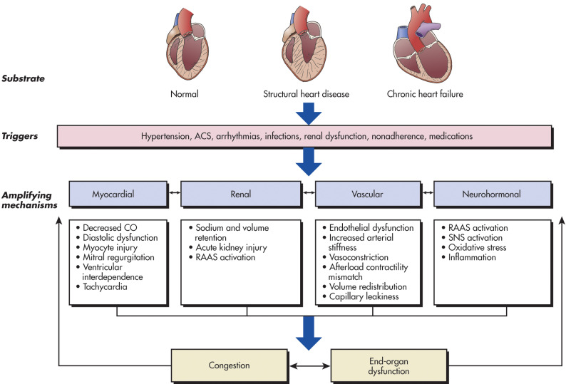
ACS, Acute coronary syndrome; CO, cardiac output; RAAS, renin-angiotensin-aldosterone; SNS, sympathetic nervous system.
(From Libby P et al: Braunwalds heart disease: a textbook of cardiovascular medicine, ed 12, Philadelphia, 2022, Elsevier.)
The American College of Cardiology/American Heart Association (ACC/AHA) describes the following four stages of HF.2 This staging model was designed to emphasize the evolution and progression of HF over a continuum and the preventability of HF in at-risk patients.
In addition to the ACC/AHA stages described above, the New York Heart Association (NYHA) defines four functional classes of HF designed to describe the symptoms of stage C and D HF.3 The functional classes are intended to assess the symptoms of HF and may fluctuate with therapy. It should be noted that current guidelines employ the functional classes to aid in determination of appropriate treatment.
Table 2 compares the ACC/AHA and the NYHA classification. Table 3 describes a simplified classification and common clinical characteristics of patients with acute HF.
TABLE 3 Simplified Classification and Common Clinical Characteristics of Patients with Acute Heart Failure
Clinical Classification | Symptom Onset | Triggers | Signs and Symptoms | Clinical Assessment | Course |
---|---|---|---|---|---|
Decompensated heart failure | Usually gradual | Noncompliance, ischemia, infections | Peripheral edema, orthopnea, dyspnea on exertion | Variable, high rehospitalization rate | |
Acute hypertensive heart failure | Usually sudden | Hypertension, atrial arrhythmias, ACS | Dyspnea (often severe), tachypnea, tachycardia, rales common | ||
Cardiogenic shock | Variable | Progression of advanced HF or major myocardial insult (e.g., large AMI, acute myocarditis) | End-organ hypoperfusion; oliguria, confusion, cool extremities |
ACS, Acute coronary syndrome; AMI, acute myocardial infarction; CXR, chest x-ray film; LV, left ventricular; RV, right ventricular; SBP, systolic blood pressure.
From Zipes DP: Braunwalds heart disease: a textbook of cardiovascular medicine, ed 11, Philadelphia, 2019, Elsevier.
TABLE 2 American College of Cardiology/American Heart Association (ACC/AHA) Stages of Heart Failure (HF) Compared to the New York Heart Association (NYHA) Functional Classification
ACC/AHA Stages of Heart Failure | NYHA Functional Classification | ||
---|---|---|---|
A | At high risk for HF but without structural heart disease or symptoms of heart failure. | None | |
B | Structural heart disease but without signs or symptoms of heart failure. | I | No limitation of physical activity. Ordinary physical activity does not cause symptoms of heart failure. |
C | Structural heart disease with prior or current symptoms of heart failure. | I | No limitation of physical activity. Ordinary physical activity does not cause symptoms of heart failure. |
II | Slight limitation of physical activity. Comfortable at rest, but ordinary physical activity results in symptoms of heart failure. | ||
III | Marked limitation of physical activity. Comfortable at rest, but less than ordinary activity causes symptoms of heart failure. | ||
D | Refractory heart failure requiring specialized interventions. | IV | Unable to carry on any physical activity without symptoms of heart failure, or symptoms of heart failure at rest. |
HF, Heart failure.
From Libby P et al: Braunwalds heart disease: a textbook of cardiovascular medicine, ed 12, Philadelphia, 2022, Elsevier.
|
Several conditions are associated with an increased risk of developing HF. If these are identified and treated appropriately, it may be possible to delay, if not prevent, the onset of HF and also reduce rates of decompensations.4
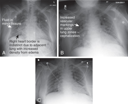
The clinical and physical examination findings should be given the highest priority when determining the diagnosis of HF (Fig. 2). These signs and symptoms are dependent on the severity of disease, precipitant factors, comorbid conditions, and whether the HF symptoms are predominantly right-sided or left-sided. Clues in the patients history when evaluating HF are summarized in Table 4.
TABLE 6 Physical Findings of Heart Failure
a Indicative of more severe disease.
From Libby P et al: Braunwalds heart disease; a textbook of cardiovascular medicine, ed 12, Philadelphia, 2022, Elsevier.
TABLE 5 Common Presenting Symptoms and Signs of Decompensated Heart Failure
Symptoms | Signs | ||
---|---|---|---|
Predominantly Related to Volume Overload | |||
Dyspnea (exertional, paroxysmal nocturnal dyspnea, orthopnea, or at rest); cough; wheezing | Rales, pleural effusion | ||
Foot and leg discomfort | Peripheral edema (legs, sacral) | ||
Abdominal discomfort/bloating; early satiety or anorexia | Ascites/increased abdominal girth; right upper quadrant pain or discomfort; hepatomegaly/splenomegaly; scleral icterus | ||
Increased weight | |||
Elevated jugular venous pressure, abdominojugular reflux | |||
Increasing S3, accentuated P2 | |||
Predominantly Related to Hypoperfusion | |||
Fatigue | Cool extremities | ||
Altered mental status, daytime drowsiness, confusion, or difficulty concentrating | Pallor, dusky skin discoloration, Hypotension | ||
Dizziness, pre-syncope, or syncope | Pulse pressure (narrow)/proportional pulse pressure (low) | ||
Pulsus alternans | |||
Other Signs and Symptoms of AHF | |||
Depression | Orthostatic hypotension (hypovolemia) | ||
Sleep disturbances | S4 | ||
Palpitations | Systolic and diastolic cardiac murmurs |
AHF, Acute heart failure.
From Libby P et al: Braunwalds heart disease: a textbook of cardiovascular medicine, ed 12, Philadelphia, 2022, Elsevier.
TABLE 4 Using the Medical History to Assess the Heart Failure Patient
From Libby P et al: Braunwalds heart disease: a textbook of cardiovascular medicine, ed 12, Philadelphia, 2022, Elsevier.
Figure 2 Flow chart for the evaluation of patients with heart failure (HF).
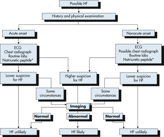
The diagnosis of HF is made using a combination of clinical judgment and initial and subsequent testing. Following thorough history and physical examination together with initial diagnostic testing, imaging (such as with echocardiography [ECG]) may still be necessary in ambiguous cases to definitively identify or exclude the diagnosis.
(From Libby P et al: Braunwalds heart disease, a textbook of cardiovascular medicine, ed 12, Philadelphia, 2022, Elsevier.)
Acute precipitants of HF decompensation include nonadherence with salt restriction or medications (most common cause), any acute systemic illness, infection, arrhythmias (e.g., atrial fibrillation), ischemia or infarction, uncontrolled hypertension, new medications (e.g., negative inotropic agents such as calcium channel blockers/antiarrhythmic agents), NSAIDs, renal dysfunction, toxins (e.g., ethanol and anthracyclines), surgery, or valvular catastrophe.10
The dichotomy of whether HF occurs in the setting of preserved or reduced LV systolic function plays an important role in treatment strategies. Patients with HFpEF may have significant abnormalities in active relaxation and passive stiffness of the LV as well as valvular disease. HfrEF denotes poor pump function.10
TABLE 7 Mechanisms/Factors Contributing to the Pathophysiology of Heart Failure with Preserved Ejection Fraction
Cardiovascular | |||
LV Structure | |||
LV Function | |||
| |||
Cardiomyocyte | |||
| |||
Extracellular Matrix | |||
| |||
Extracardiac | |||
ADP,Adenosine diphosphate; ATP, adenosine triphosphate; LA, left atrium; LV, left ventricle; RV, right ventricle; SPARC, secreted protein, acidic and rich in cysteine [osteonectin]; TGF, transforming growth vector.
From Mann DL et al: Braunwalds heart disease, ed 10, Philadelphia, 2015, Elsevier.
Figure 4 Congestive heart failure.
Mild left ventricular hypertrophy with restricted filling, ejection fraction >55%, and no pericardial effusion. This 63-yr-old man with coronary artery disease, chronic renal insufficiency, and diastolic heart failure (ejection fraction >55%) presented multiple times for dyspnea (A, B, and C, first through third clinical presentations). Each of these three radiographs shows signs of moderate pulmonary edema. The diaphragms and costophrenic angles are clear, suggesting no pleural effusion. The right heart border in all three images is indistinct because of interstitial edema in these locations. Portions of the left heart border are also indistinct. The upper lung fields have a hazy appearance indicating mild edema. Fluid is visible in the minor fissure on all three images. Does the similarity of these radiographs mean that edema is not the cause of the patients dyspnea? No, he simply presented with pulmonary edema on all three occasions.
(From Broder JS: Diagnostic imaging for the emergency physician, Philadelphia, 2011, Saunders.)
Figure E3 Indications for the use of biomarkers in heart failure.
*Other biomarkers of injury or fibrosis include soluble ST2 receptor, galectin-3, and high-sensitivity troponin. ACC, American College of Cardiology; AHA, American Heart Association; ADHF, acute decompensated heart failure; BNP, B-type natriuretic peptide; COR, class of recommendation; ED, emergency department; HF, heart failure; NT-proBNP, N-terminal pro-B-type natriuretic peptide; NYHA, New York Heart Association; pts, patients.
(From Yancy CW et al: 2017 ACC/AHA/HFSA focused update of the 2013 ACCF/AHA guideline for the management of heart failure: a report of the American College of Cardiology/American Heart Association Task Force on Clinical Practice Guidelines and the Heart Failure Society of America, J Am Coll Cardiol 2017;70[6]:776.)
TABLE 9 ACC/AHA/HFSA Guidelines for Use of Biomarkers in Heart Failure
Class | Level of Evidence | |
---|---|---|
Biomarkers for Prevention of HF | ||
IIa | For patients at risk of developing HF, natriuretic peptide biomarker-based screening can be useful to prevent the development of left ventricular dysfunction (systolic or diastolic) or new-onset HF. | B-R |
Biomarkers for Diagnosis | ||
I | In patients presenting with dyspnea (acute or chronic), measurement of natriuretic peptide biomarkers is useful to support a diagnosis or exclusion of HF. | A |
Biomarkers for Prognosis or Added Risk Stratification | ||
I | Measurement of BNP or NT-proBNP is useful for establishing prognosis or disease severity in chronic HF. | A |
I | Measurement of baseline levels of natriuretic peptide biomarkers and/or cardiac troponin on admission to the hospital is useful to establish a prognosis in acutely decompensated HF. | A |
IIa | During an HF hospitalization, a predischarge natriuretic peptide level can be useful to establish a postdischarge prognosis. | B-NR |
IIb | In patients with chronic HF, measurement of other clinically available tests, such as biomarkers of myocardial injury or fibrosis, may be considered for additive risk stratification. | B-NR |
ACC, American College of Cardiology; AHA, American Heart Association; BNP, B-type natriuretic peptide; HF, heart failure; HFSA, Heart Failure Society of America; NT-proBNP, N-terminal pro-B-type natriuretic peptide.
From Zipes DP: Braunwalds heart disease: a textbook of cardiovascular medicine, ed 11, Philadelphia, 2019, Elsevier.
TABLE E8 ACC/AHA Guidelines for Initial and Serial Evaluation of Heart Failure
Class | Level of Evidence | |
---|---|---|
Indication: History, Physical Examination, and Risk Scoring | ||
I | A thorough history and physical examination should be obtained/performed in patients presenting with HF to identify cardiac and noncardiac disorders or behaviors that might cause or accelerate the development or progression of HF. | C |
In patients with idiopathic DCM, a three-generational family history should be obtained to aid in establishing the diagnosis of familial DCM. | C | |
Volume status and vital signs should be assessed at each patient encounter. This includes serial assessment of weight, as well as estimates of jugular venous pressure and the presence of peripheral edema or orthopnea. | B | |
IIa | Validated multivariable risk scores can be useful to estimate subsequent risk of mortality in ambulatory or hospitalized patients with HF. | C |
Indication: Diagnostic Tests and Biomarkers (also see below) | ||
I | Initial laboratory evaluation of patients presenting with HF should include complete blood count, urinalysis, serum electrolytes (including calcium and magnesium), blood urea nitrogen, serum creatinine, glucose, fasting lipid profile, liver function tests, and thyroid-stimulating hormone. | C |
Serial monitoring, when indicated, should include serum electrolytes and renal function. | C | |
A 12-lead ECG should be performed initially on all patients presenting with HF. | C | |
In ambulatory patients with dyspnea, measurement of BNP or N-terminal pro-B-type natriuretic peptide (NT-proBNP) is useful to support clinical decision making regarding the diagnosis of HF, especially in the setting of clinical uncertainty, and measurement of BNP or NT-proBNP is useful for establishing prognosis or disease severity in chronic HF. | A | |
IIa | Screening for hemochromatosis or HIV is reasonable in select patients who present with HF. | C |
Diagnostic tests for rheumatologic diseases, amyloidosis, or pheochromocytoma are reasonable in patients presenting with HF in whom there is a clinical suspicion of these diseases. | C | |
BNP-guided or NT-proBNPguided HF therapy can be useful to achieve optimal dosing of GDMT in select, clinically euvolemic patients followed in a well-structured HF disease management program. | B | |
IIb | The usefulness of serial measurement of BNP or NT-proBNP to reduce hospitalization or mortality in patients with HF is not well established. The measurement of other clinically available tests, such as biomarkers of myocardial injury or "fibrosis," may be considered for additive risk stratification in patients with chronic HF. | B |
Indication: Noninvasive Cardiac Imaging | ||
I | Patients with suspected or new-onset HF, or those presenting with acute decompensated HF, should undergo a chest radiograph to assess heart size and pulmonary congestion and to detect alternative cardiac, pulmonary, and other diseases that may cause or contribute to the patients symptoms. | C |
A two-dimensional echocardiogram with Doppler should be performed during initial evaluation of patients presenting with HF to assess ventricular function, size, wall thickness, wall motion, and valve function. | C | |
Repeat measurement of EF and measurement of the severity of structural remodeling are useful to provide information in patients with HF who have had a significant change in clinical status; who have experienced or recovered from a clinical event; who have received treatment, including GDMT, that might have had a significant effect on cardiac function; or who may be candidates for device therapy. | C | |
IIa | Noninvasive imaging to detect myocardial ischemia and viability is reasonable in patients presenting with de novo HF who have known CAD and no angina, unless the patient is not eligible for revascularization of any kind. | C |
Viability assessment is reasonable in select situations when planning revascularization in HF patients with CAD. | B | |
Radionuclide ventriculography or MRI can be useful to assess LVEF and volume when echocardiography is inadequate. | C | |
MRI is reasonable when assessing myocardial infiltrative processes or scar burden. | B | |
III: No benefit | Routine repeat measurement of LV function assessment in the absence of clinical status change or treatment interventions should not be performed. | B |
Indication: Invasive Evaluation | ||
I | Invasive hemodynamic monitoring with a pulmonary artery catheter should be performed to guide therapy in patients who have respiratory distress or clinical evidence of impaired perfusion in whom the adequacy or excess of intracardiac filling pressures cannot be determined from clinical assessment. | C |
IIa | Invasive hemodynamic monitoring can be useful for carefully selected patients with acute HF who have persistent symptoms despite empiric adjustment of standard therapies and (a) whose fluid status, perfusion, or systemic or pulmonary vascular resistance is uncertain; (b) whose systolic pressure remains low, or is associated with symptoms, despite initial therapy; (c) whose renal function is worsening with therapy; (d) who require parenteral vasoactive agents; or (e) who may need consideration for mechanical circulatory support or transplantation. | C |
When ischemia may be contributing to HF, coronary arteriography is reasonable for patients eligible for revascularization. | C | |
Endomyocardial biopsy can be useful in patients presenting with HF when a specific diagnosis is suspected that would influence therapy. | C | |
III: No benefit | Routine use of invasive hemodynamic monitoring is not recommended in normotensive patients with acute decompensated HF and congestion with symptomatic response to diuretics and vasodilators. | B |
III: Harm | Endomyocardial biopsy should not be performed in the routine evaluation of patients with HF. | C |
ACC, American College of Cardiology; AHA, American Heart Association; BNP, B-type natriuretic peptide; CAD, coronary artery disease; DCM, dilated cardiomyopathy; ECG, electrocardiogram; EF, ejection fraction; GDMT, guideline-directed medical therapy; HIV, human immunodeficiency virus; LV, left ventricle; LVEF, left ventricular ejection fraction; MRI, magnetic resonance imaging.
TABLE E11 ACC/AHA Guidelines for Treatment of Asymptomatic Left Ventricular Systolic Dysfunction (Stage B)
Class | Indication | Level of Evidence |
---|---|---|
I | In all patients with a recent or remote history of MI or ACS and reduced EF, ACE inhibitors should be used to prevent symptomatic HF and reduce mortality. In patients intolerant of ACE inhibitors, ARBs are appropriate unless contraindicated. | A |
In all patients with a recent or remote history of MI or ACS and reduced EF, evidence-based β-blockers should be used to reduce mortality. β-Blockade and ACE inhibition should be used in all patients with a recent or remote history of MI regardless of EF or presence of HF. | B | |
In all patients with a recent or remote history of MI or ACS, statins should be used to prevent symptomatic HF and cardiovascular events. | A | |
Blood pressure should be controlled in accordance with clinical practice guidelines for hypertension to prevent symptomatic HF. | A | |
ACE inhibitors should be used in all patients with a reduced EF to prevent symptomatic HF. | A | |
β-Blockers should be used in all patients with a reduced EF to prevent symptomatic HF. | C | |
IIa | To prevent sudden death, placement of an ICD is reasonable in patients with asymptomatic ischemic cardiomyopathy who are at least 40 days post-MI, have an LVEF of 30% or less, are on appropriate medical therapy, and have reasonable expectation of survival with a good functional status for more than 1 yr. | B |
III: Harm | Nondihydropyridine calcium channel blockers with negative inotropic effects may be harmful in asymptomatic patients with low LVEF and no symptoms of HF after MI. | B |
ACC, American College of Cardiology; ACE, angiotensin-converting enzyme; ACS, acute coronary syndrome; AHA, American Heart Association; ARB, angiotensin receptor antagonist; EF, ejection fraction; HF, heart failure; ICD, implantable cardioverter-defibrillator; LVEF, left ventricular ejection fraction; MI, myocardial infarction.
From Zipes DP: Braunwalds heart disease: a textbook of cardiovascular medicine, ed 11, Philadelphia, 2019, Elsevier.
TABLE E10 ACC/AHA Guidelines for Treating Patients at High Risk of Developing Heart Failure (Stage A)
Class | Indication | Level of Evidence |
---|---|---|
I | Hypertension and lipid disorders should be controlled in accordance with contemporary guidelines to lower the risk of HF. | A |
I | In patients at increased risk, stage A, the optimal blood pressure in those with hypertension should be less than 130/80 mm Hg. | B-R |
I | Other conditions that may lead to or contribute to HF, such as obesity, diabetes mellitus, tobacco use, and known cardiotoxic agents, should be controlled or avoided. | C |
II | For patients at risk of developing HF, natriuretic peptide biomarker-based screening followed by team-based care, including a cardiovascular specialist optimizing GDMT, can be useful to prevent the development of left ventricular dysfunction (systolic or diastolic) or new-onset HF. | B-R |
ACC, American College of Cardiology; AHA, American Heart Association; GDMT, guideline-directed medical therapy; HF, heart failure.
From Zipes DP: Braunwalds heart disease: a textbook of cardiovascular medicine, ed 11, Philadelphia, 2019, Elsevier.
Figure 5 Treatment Algorithm Stage C and D Heart Failure with a Reduced Ejection Fraction
For all medical therapies, dosing should be optimized and serial assessment exercised. (Key: *See text for important treatment directions. Hydral-Nitrates green box: The combination of ISDN/HYD with ARNI has not been robustly tested. BP response should be carefully monitored. See 2013 ACC/AH heart failure guidelines. §Participation in investigational studies is also appropriate for stage C, NYHA class II and III HF. ACEI, Angiotensin-converting enzyme inhibitor; ARB, angiotensin receptor blocker; ARNI, angiotensin receptor neprilysin inhibitor; BP, blood pressure; bpm, beats per minute; C/I, contraindication; CrCl, creatinine clearance; CRT-D, cardiac resynchronization therapy-device; Dx, diagnosis; GDMT, guideline-directed management and therapy; HF, heart failure; HFrEF, heart failure with reduced ejection fraction; ICD, implantable cardioverter-defibrillator; ISDN/HYD, isosorbide dinitrate hydral-nitrates; K+, potassium; LBBB, left bundle branch block; LVAD, left ventricular assist device; LVEF, left ventricular ejection fraction; MI, myocardial infarction; NSR, normal sinus rhythm; NYHA, New York Heart Association.
(From Yancy CW et al: 2017 ACC/AHA/HFSA focused update of the 2013 ACCF/AHA guideline for the management of heart failure: a report of the American College of Cardiology/American Heart Association Task Force on Clinical Practice Guidelines and the Heart Failure Society of America, J Am Coll Cardiol 2017;70[6]:776.)
TABLE E13 ACC/AHA Recommendations for the Hospitalized Patient with Heart Failure (HF)
Class | Indication | Level of Evidencea |
---|---|---|
I | Thorough history and physical examination to evaluate for adequacy of systemic perfusion, volume status, contribution of precipitating factors and/or comorbidities, and whether HF is associated with preserved ejection fraction. | C |
Concentrations of B-type natriuretic peptide (BNP) or N-terminal pro-B-type natriuretic peptide (NT-proBNP) to evaluate dyspnea if the contribution of heart failure is not known. | A | |
Acute coronary syndrome should be promptly identified by electrocardiogram and cardiac troponin testing, and treated, as appropriate to the overall condition and prognosis of patient. | C | |
Oxygen therapy should be administered to relieve symptoms related to hypoxemia. | C | |
Improve systemic perfusion in patients who present with rapid decompensation and hypoperfusion associated with decreasing urine output and other manifestations of shock. | C | |
Treatment of significant fluid overload with intravenous loop diuretics. The diuretic dose should be titrated to relieve symptoms and to reduce extracellular fluid volume excess. | B, C | |
Monitor the effects of therapy with careful measurement of fluid intake and output; vital signs; body weight, and symptoms of systemic perfusion and congestion. | C | |
Intensify the diuretic regiment when the diuresis is inadequate to relieve congestion. | C | |
Intravenous inotropic or vasopressor drugs should be administered to maintain systemic perfusion and preserve end-organ performance in patients with clinical evidence of hypotension associated with hypoperfusion and elevated cardiac filling pressures. | C | |
Invasive hemodynamic monitoring to guide therapy in patients who are in respiratory distress or with clinical evidence of impaired perfusion if filling pressures cannot be determined from clinical assessment. | C | |
Medications should be reconciled and adjusted as appropriate on admission to and discharge from the hospital. | C | |
Maintenance treatment with oral therapies known to improve outcomes (ACE inhibitors or ARBs and β-blocker therapy) in the absence of hemodynamic instability or contraindications. | C | |
Initiation of treatment with oral therapies known to improve outcomes (ACE inhibitors or ARBs and β-blocker therapy) in stable patients prior to hospital discharge. | B | |
During transition from intravenous to oral diuretic therapy, patient should be monitored carefully for supine and upright hypotension, worsening renal function, and HF signs/symptoms. | C | |
Comprehensive written discharge instructions for patients and their caregivers is strongly recommended. | C | |
Post-discharge systems of care, if available, should be used to facilitate the transition to effective outpatient care. | B | |
IIa | Urgent cardiac catheterization and revascularization in patients with acute HF with known or suspected acute myocardial ischemia due to occlusive coronary disease when there are signs and symptoms of inadequate systemic perfusion and revascularization is likely to prolong meaningful survival. | C |
Intravenous nitroglycerin, nitroprusside, or nesiritide for patients with evidence of severely symptomatic fluid overload in the absence of systemic hypotension. | C | |
Ultrafiltration for patients with refractory congestion not responding to medical therapy. | B | |
IIb | Intravenous inotropic drugs (dopamine, dobutamine, or milrinone) for patients presenting with documented severe systolic dysfunction, low blood pressure, and evidence of low cardiac output, with or without congestion, to maintain systemic perfusion and preserve end-organ performance. | C |
III | Use of parenteral inotropes in normotensive patients with acute decompensated HF without evidence of decreased organ perfusion. | B |
Routine use of invasive hemodynamic monitoring in normotensive patients with acute decompensated HF and congestion with symptomatic response to diuretics and vasodilators. | B |
ACC, American College of Cardiology; ACE, angiotensin-converting enzyme; AHA, American Heart Association; ARB, angiotensin-receptor blocker.
a See guidelines text for definition of level of evidence categories.
From Zipes DP: Braunwalds heart disease: a textbook of cardiovascular medicine, ed 11, Philadelphia, 2019, Elsevier.
TABLE E12 ACC/AHA Guidelines for Treatment of Symptomatic Left Ventricular Systolic Dysfunction (Stage C)
Class | Indication | Level of Evidence |
---|---|---|
Nonpharmacologic Interventions | ||
I | Patients with HF should receive specific education to facilitate HF self-care. | B |
Exercise training (or regular physical activity) is recommended as safe and effective for patients with HF who are able to participate to improve functional status. | A | |
IIa | Cardiac rehabilitation can be useful in clinically stable patients with HF to improve functional capacity, exercise duration, HRQOL, and mortality. | B |
Sodium restriction is reasonable for patients with symptomatic HF to reduce congestive symptoms. | C | |
Continuous positive airway pressure (CPAP) can be beneficial to increase LVEF and improve functional status in patients with HF and sleep apnea. | B | |
Pharmacologic Interventions | ||
I | Measures listed as class I recommendations for patients in stages A and B are recommended where appropriate. | A, B, C |
GDMT should be the mainstay of pharmacologic therapy for HFrEF. | A | |
Diuretics | ||
I | Diuretics are recommended in patients with HFrEF who have evidence of fluid retention, unless contraindicated, to ameliorate symptoms. | C |
Angiotensin-Converting Enzyme Inhibitors/Adrenergic Receptor Blockers | ||
I | ACE inhibitors are recommended in patients with HFrEF and current or previous symptoms, unless contraindicated, to reduce morbidity and mortality. | A |
ARBs are recommended in patients with HFrEF with current or previous symptoms who are ACE inhibitor-intolerant, unless contraindicated, to reduce morbidity and mortality. | A | |
IIa | ARBs are a reasonable choice to reduce morbidity and mortality as alternatives to ACE inhibitors for first-line therapy in patients with HFrEF, especially in those already taking ARBs for other indications, unless contraindicated. | A |
IIb | Addition of an ARB may be considered in persistently symptomatic patients with HFrEF who are already being treated with an ACE inhibitor and a β-blocker in whom an aldosterone antagonist is not indicated or tolerated. | A |
III: Harm | Routinely combining an ACE inhibitor, an ARB, and an aldosterone antagonist. | C |
β-Blockers | ||
I | Use of one of the three β-blockers proven to reduce mortality (i.e., bisoprolol, carvedilol, and sustained-release metoprolol succinate) is recommended for all patients with current or previous symptoms of HFrEF, unless contraindicated, to reduce morbidity and mortality. | A |
Aldosterone Receptor Antagonists | ||
I | Aldosterone receptor antagonists (or mineralocorticoid receptor antagonists) are recommended in patients with NYHA class II-IV and LVEF of ≤35%, unless contraindicated, to reduce morbidity and mortality. | A |
I | Aldosterone receptor antagonists are recommended to reduce morbidity and mortality after an acute MI in patients with LVEF of ≤40% who develop symptoms of HF or who have a history of diabetes mellitus, unless contraindicated. | B |
III: Harm | Inappropriate use of aldosterone receptor antagonists is potentially harmful because of life-threatening hyperkalemia or renal insufficiency when serum creatinine is >2.5 mg/dl in men or >2.0 mg/dl in women (or estimated glomerular filtration rate <30 ml/min/1.73 m2), and/or potassium >5.0 mEq/liter. | B |
SGLT-2 Inhibitors | ||
I | In patients with symptomatic chronic HFrEF, SGLT2 inhibitors are recommended to reduce hospitalization for HF and cardiovascular mortality, irrespective of the presence of type 2 diabetes. | A |
IIa | In patients with HFmrEF (LVEF 41%-49%), SGLT2 inhibitors can be beneficial in decreasing HF hospitalizations and cardiovascular mortality. | B |
Hydralazine and Isosorbide Dinitrate | ||
I | The combination of hydralazine and isosorbide dinitrate is recommended to reduce morbidity and mortality for patients self-described as African Americans with NYHA class III-IV HFrEF receiving optimal therapy with ACE inhibitors and β-blockers, unless contraindicated. | A |
IIa | A combination of hydralazine and isosorbide dinitrate can be useful to reduce morbidity or mortality in patients with current or previous symptomatic HFrEF who cannot be given an ACE inhibitor or ARB because of drug intolerance, hypotension, or renal insufficiency, unless contraindicated. | B |
Digoxin | ||
IIa | Digoxin can be beneficial in patients with HFrEF, unless contraindicated, to decrease hospitalizations for HF. | B |
Anticoagulation | ||
I | Patients with chronic HF with permanent/persistent/paroxysmal atrial fibrillation and an additional risk factor for cardioembolic stroke (history of hypertension, diabetes mellitus, previous stroke or transient ischemic attack, or ≥75 yr of age) should receive chronic anticoagulant therapy. | A |
I | The selection of an anticoagulant agent (warfarin, dabigatran, apixaban, or rivaroxaban) for permanent/persistent/paroxysmal atrial fibrillation should be individualized on the basis of risk factors, cost, tolerability, patient preference, potential for drug interactions, and other clinical characteristics, including time in the international normalized ratio therapeutic range if the patient has been taking warfarin. | C |
IIa | Chronic anticoagulation is reasonable for patients with chronic HF who have permanent/persistent/paroxysmal atrial fibrillation but no additional risk factor for cardioembolic stroke. | B |
III: No benefit | Anticoagulation is not recommended in patients with chronic HFrEF without atrial fibrillation, a previous thromboembolic event, or a cardioembolic source. | B |
Statins | ||
III: No benefit | Statins are not beneficial as adjunctive therapy when prescribed solely for HF. | A |
Omega-3 Fatty Acids | ||
IIa | Omega-3 PUFA supplementation is reasonable to use as adjunctive therapy in patients with NYHA class II-IV symptoms and HFrEF or HFpEF, unless contraindicated, to reduce mortality and cardiovascular hospitalizations. | B |
Drugs of Unproven Value or That May Cause Harm | ||
III: No benefit | Nutritional supplements as treatment for HF are not recommended in patients with current or previous symptoms of HFrEF. | B |
Hormonal therapies other than to correct deficiencies are not recommended for patients with current or previous symptoms of HFrEF. | C | |
III: Harm | Drugs known to adversely affect the clinical status of patients with current or previous symptoms of HFrEF are potentially harmful and should be avoided or withdrawn whenever possible (e.g., most antiarrhythmic drugs, most calcium channel blocking drugs [except amlodipine], NSAIDs, or thiazolidinediones). | B |
Long-term use of infused positive inotropic drugs is potentially harmful for patients with HFrEF, except as palliation for patients with end-stage disease who cannot be stabilized with standard medical treatment (see recommendations for stage D). | C | |
Calcium Channel Blockers | ||
III: No benefit | Calcium channel blocking drugs are not recommended for routine therapy in patients with HFrEF. | A |
ACC, American College of Cardiology; ACE, angiotensin-converting-enzyme; AHA, American Heart Association; ARB, angiotensin-receptor blocker; GDMT, guideline-directed medical therapy; HF, heart failure; HFpEF, heart failure with preserved ejection fraction; HFrEF, heart failure with reduced ejection fraction; HRQOL, health-related quality of life; LVEF, left ventricular ejection fraction; MI, myocardial infarction; NYHA, New York Heart Association; NSAID, nonsteroidal antiinflammatory drug; PUFA, polyunsaturated fatty acid.
Adapted from Mann DL et al: Braunwalds heart disease, ed 10, Philadelphia, 2015, Elsevier.
TABLE 17 Therapeutic Approaches for Volume Management in Acute Heart Failure (AHF)
Severity of Volume Overload | Diuretic | Dose (mg) | Comments |
---|---|---|---|
Moderate | Furosemide, or | 20-40, or up to 2.5 times oral dose | IV administration preferable in symptomatic patients |
Bumetanide, or | 0.5-1.0 | Titrate dose according to clinical response. | |
Torsemide | 10-20 | Monitor Na+, K+, creatinine, BP | |
Severe | Furosemide, or | Intravenously | |
Bumetanide, or | 1-4/0.5-2 mg/h infusion (max, 2-4 mg/h, limit 2-4 h) | Bumetanide and torsemide have higher oral bioavailability than furosemide, but IV administration preferable in AHF. | |
Torsemide | 20-100/5-20 mg/h | ||
Ultrafiltration | 200-500 ml/h | Adjust ultrafiltration rate to clinical response; monitor for hypotension; consider hematocrit sensor. | |
Refractory to loop diuretics | Add HCTZ, or | 25-50 twice daily | Combination with loop diuretic may be better than very high dose of loop diuretics alone. |
Metolazone, or | 2.5-10 once daily | Metolazone more potent if creatinine clearance <30 ml/min | |
Chlorothiazide, or | |||
Spironolactone | 25-50 once daily | Spironolactone best choice if patient not in renal failure and normal or low serum K+, although may not be very potent | |
In case of alkalosis | Acetazolamide | 0.5 | Intravenously |
Refractory to loop diuretics and thiazides | Add dopamine (renal vasodilation), or | ||
dobutamine or milrinone (inotropic agent) | |||
Ultrafiltration, or hemodialysis if coexisting renal failure |
BP, Blood pressure; IV, intravenous; HCTZ, hydrochlorothiazide; PO, by mouth.
From Libby P et al: Braunwalds heart disease: a textbook of cardiovascular medicine, ed 12, Philadelphia, 2022, Elsevier.
TABLE E16 ACC/AHA/HFSA Guidelines for Treatment of Patients with Stage C Heart Failure and Preserved Left Ventricular Ejection Fraction (HFpEF)
Class | Indication | Level of Evidence |
---|---|---|
I | Systolic and diastolic blood pressure should be controlled in accordance with published clinical practice guidelines to prevent morbidity. | B |
Diuretics should be used for relief of symptoms due to volume overload. | C | |
IIa | Coronary revascularization is reasonable in patients with coronary artery disease in whom symptoms (angina) or demonstrable myocardial ischemia is judged to be having an adverse effect on symptomatic heart failure. | C |
Management of atrial fibrillation according to published clinical practice guidelines is reasonable to improve symptomatic heart failure. | C | |
The use of β-blocking agents, ACE inhibitors, and ARBs in patients with hypertension is reasonable to control blood pressure. | C | |
IIb | In appropriately selected patients with HFpEF (with EF ≥45%, elevated BNP levels or heart failure admission within 1 yr, estimated glomerular filtration rate >30 ml/min, creatinine <2.5 mg/dl, potassium <5.0 mEq/L), aldosterone receptor antagonists might be considered to decrease hospitalizations. | B-R |
IIb | The use of ARBs might be considered to decrease hospitalizations. | B |
III: No benefit | The routine use of nitrates or phosphodiesterase-5 inhibitors to increase activity or quality of life in patients with HFpEF is ineffective. | B-R |
III: No benefit | Routine use of nutritional supplements is not recommended. | C |
ACC, American College of Cardiology; ACE, angiotensin-converting enzyme; AHA, American Heart Association; ARB, angiotensin receptor blocker; BNP, B-type (brain) natriuretic peptide; HFSA, Heart Failure Society of America.
From Zipes DP: Braunwalds heart disease: a textbook of cardiovascular medicine, ed 11, Philadelphia, 2019, Elsevier.
TABLE 14 Intravenous Vasoactive Agents for the Treatment of Acute Heart Failure
Intravenous Medication | Initial Dose | Effective Dose Rangea | Comments |
---|---|---|---|
Vasodilators | |||
Nitroglycerin; glyceryl trinitrate | 20 μg/min | 40-400 μg/min | Hypotension, headache; Tolerance with continuous use after 24 h |
Isosorbide dinitrate | 1 mg/h | 2-10 mg/h | Hypotension, headache; Tolerance with continuous use within 24 h |
Nitroprusside | 0.3 μg/kg/min | 0.3-5 μg/kg/min (usually <4 μg/kg/min) | Caution in patients with active myocardial ischemia; Hypotension; cyanide side effects (nausea, dysphoria); thiocyanate toxicity; light sensitive |
Nesiritideb | 2 μg/kg bolus with 0.010-0.030 μg/kg/min infusionc | 0.010-0.030 μg/kg/min | |
Inotropes | |||
Dobutamine | 1-2 μg/kg/min | 2-20 μg/kg/min | For inotropy and vasodilation; Hypotension, tachycardia, arrhythmias; ? mortality |
Dopamine | 1-2 μg/kg/min | 2-4 μg/kg/min | For inotropy and vasodilation; Hypotension, tachycardia, arrhythmias; ? mortality |
4-5 μg/kg/min | 5-20 μg/kg/min | For inotropy and vasoconstriction; Tachycardia, arrhythmias; ?mortality | |
Milrinone | 25-75 μg/kg bolus over 10-0 minc followed by infusion | 0.10-0.75 μg/kg/min | For vasodilation and inotropy; Hypotension, tachycardia, arrhythmias; Renal excretion; ?mortality |
Enoximoneb | 0.25-0.75 mg/kg | 1.25-7.5 μg/kg/min | For vasodilation and inotropy; Hypotension, tachycardia, arrhythmias; ? mortality |
Levosimendanb | 12-24 μg/kg bolus over 10 mina followed by infusion | 0.5-2.0 μg/kg/min | For vasodilation and inotropy; active metabolite present for approximately 84 h; Hypotension, tachycardia, arrhythmias; ?mortality |
Epinephrine | 0.05-0.5 μg/kg/min | For vasoconstriction and inotropy; Tachycardia, arrhythmias, end-organ hypoperfusion; ?mortality | |
Norepinephrine | 0.2-1.0 μg/kg/min | For vasoconstriction and inotropy; Tachycardia, arrhythmias, end-organ hypoperfusion; ?mortality |
Use higher dose range for chronic diuretic use, renal insufficiency, and severe volume overload. Diuretic naïve patients should receive lower doses, initially.
a In general, titration of medication is accomplished by doubling of dose with careful monitoring for adverse effects.
b Not approved for use in all countries.
c Some clinicians do not administer a bolus dose, so as to decrease the risk of hypotension. Bolus not recommended in patients with hypotension.
From Libby P et al: Braunwalds heart disease, a textbook of cardiovascular medicine, ed 12, Philadelphia, 2022, Elsevier.
TABLE 15 Diuretics for Treating Fluid Retention in Chronic Heart Failure
Drug | Initial Daily Dose(S) | Maximum Total Daily Dose | Duration of Action |
---|---|---|---|
Loop Diureticsa | |||
Bumetanide | 0.5-1.0 mg once or twice | 10 mg | 4-6 h |
Furosemide | 20-40 mg once or twice | 600 mg | 6-8 h |
Torsemide | 10-20 mg once | 200 mg | 12-16 h |
Ethacrynic acid | 25-50 mg once or twice | 200 mg | 6 h |
Thiazide Diureticsb | |||
Chlorothiazide | 250-500 mg once or twice | 1000 mg | 6-12 h |
Chlorthalidone | 25 mg once | 100 mg | 24-72 h |
Hydrochlorothiazide | 25 mg once or twice | 200 mg | 6-12 h |
Indapamide | 2.5 mg once | 5 mg | 36 h |
Metolazone | 2.5-5.0 mg once | 5 mg | 12-24 h |
Potassium-Sparing Diuretics | |||
Amiloride | 5.0 mg once | 20 mg | 24 h |
Triamterene | 50-100 mg twice | 300 mg | 7-9 h |
AVP Antagonists | |||
Satavaptan | 25 mg once | 50 mg once | NS |
Tolvaptan | 15 mg once | 60 mg once | NS |
Lixivaptan | 25 mg once | 250 mg twice | NS |
Conivaptan (IV) | 20 mg IV loading dose followed by | 100 mg once | 7-9 h |
20 mg continuous IV infusion/day | 40 mg IV | ||
Sequential Nephron Blockade | |||
Metolazone | 2.5-10 mg once plus loop diuretic | ||
Hydrochlorothiazide | 25-100 mg once or twice plus loop diuretic | ||
Chlorothiazide (IV) | 500-1000 mg once plus loop diuretic |
Unless indicated, all doses are for oral diuretics.
IV, Intravenous; mg, milligrams; NS, not specified.
a Equivalent doses: 40 mg furosemide = 1 mg bumetanide = 20 mg torsemide = 50 mg of ethacrynic acid.
b Do not use if estimated glomerular filtration is less than 30 ml/min or with cytochrome 3A4 inhibitors.
Modified from Hunt SA, et al. ACC/AHA 2005 guideline update for the diagnosis and management of chronic heart failure in the adult: a report of the American College of Cardiology/American Heart Association Task Force on Practice Guidelines, J Am Coll Cardiol 46:e1-e82, 2005. In Libby P et al: Braunwalds heart disease: a textbook of cardiovascular medicine, ed 12, Philadelphia, 2022, Elsevier.
The goals of HF therapy are clinical improvement followed by stabilizing, slowing, or even reversing deterioration in myocardial function, and ultimately a reduction in risk of morbidity (including hospitalization rates) and mortality.2
TABLE E19 ACCF/AHA Guidelines for Indications for Implantable Cardioverter-Defibrillators (ICDs)
Class | Indication | Level of Evidence |
---|---|---|
I | ICD therapy is recommended for primary prevention of SCD in selected patients with HFrEF at least 40 days post-MI with LVEF less than 35% and NYHA Class II or III symptoms receiving chronic GDMT, who are expected to live more than 1 yr. | A |
I | ICD therapy is recommended for primary prevention of SCD in selected patients with HFrEF at least 40 days post-MI with LVEF less than 30% and NYHA Class I symptoms receiving GDMT, who are expected to live more than 1 yr. | B |
IIa | To prevent SCD, placement of ICD is reasonable in patients with asymptomatic ischemic cardiomyopathy who are at least 40 days post-MI, have an LVEF of 30% or less, are on appropriate medical therapy, and have reasonable expectation of survival with a good functional status for more than 1 yr. | B |
IIb | ICD therapy to prevent SCD in patients with nonischemic cardiomyopathy who are at least 40 days post-MI, have LVEF less than 35%, with NYHA Class II or III symptoms while undergoing chronic optimal medical therapy, and have reasonable expectation of survival for more than 1 yr with good functional status. | B |
Usefulness of implantation of ICD is of uncertain benefit to prolong meaningful survival in patients with high risk of non-SCD, as predicted by frequent hospitalizations, advanced frailty, or comorbidities such as systemic malignancy or severe renal dysfunction. | B |
HFrEF, Heart failure with reduced ejection fraction; MI, myocardial infarction; SCD, sudden cardiac death; see Table E18 for other abbreviations.
From Zipes DP: Braunwalds heart disease: a textbook of cardiovascular medicine, ed 11, Philadelphia, 2019, Elsevier.
TABLE E18 ACCF/AHA Guidelines for Cardiac Resynchronization Therapy (CRT)
Class | Indication | Level of Evidence |
---|---|---|
I | CRT is indicated for patients who have LVEF of 35% or less, sinus rhythm, LBBB with QRS duration of 150 msec or greater, and NYHA Class II, III, or ambulatory IV symptoms on GDMT. | |
IIa | CRT can be useful for patients who have LVEF of 35% or less, sinus rhythm, non-LBBB pattern with QRS duration of 150 msec or greater, and NYHA Class III/ambulatory Class IV symptoms on GDMT. | A |
CRT can be useful for patients who have LVEF of 35% or less, sinus rhythm, LBBB with QRS duration of 120-149 msec, and NYHA Class II, III, or ambulatory IV symptoms on GDMT. | B | |
CRT can be useful in patients with atrial fibrillation and LVEF of 35% or less on GDMT if (a) the patient requires ventricular pacing or otherwise meets CRT criteria and (b) atrioventricular nodal ablation or pharmacologic rate control will allow near-100% ventricular pacing with CRT. | B | |
CRT can be useful for patients on GDMT who have LVEF of 35% or less and are undergoing placement of new or replacement device with anticipated requirement for significant (>40%) ventricular pacing. | C | |
IIb | CRT may be considered for patients who have LVEF of 35% or less, sinus rhythm, non-LBBB pattern with QRS duration of 120-149 msec, and NYHA Class III/ambulatory Class IV on GDMT. | B |
CRT may be considered for patients who have LVEF of 35% or less, sinus rhythm, non-LBBB pattern with QRS duration of 150 msec or greater, and NYHA Class II symptoms on GDMT. | B | |
CRT may be considered for patients who have LVEF of 30% or less, ischemic etiology of HF, sinus rhythm, LBBB with QRS duration of 150 msec or greater, and NYHA Class I symptoms on GDMT. | C | |
III: No benefit | CRT is not recommended for patients with NYHA Class I or II symptoms and non-LBBB pattern with QRS duration less than 150 msec. | |
CRT is not indicated for patients whose comorbidities and/or frailty limit survival with good functional capacity to less than 1 yr. |
ACCF, The American College of Cardiology Foundation; AHA, American Heart Association; GDMT, guideline-directed medical therapy; LBBB, left bundle-branch block; LVEF, left ventricular ejection fraction; NYHA, New York Heart Association.
From Libby P et al: Braunwalds heart disease: a textbook of cardiovascular medicine, ed 12, Philadelphia, 2022, Elsevier.
TABLE E22 Surgery for Management of Heart Failure (HF): Guideline Recommendations
2016 ESC Guidelines for the Diagnosis and Treatment of Acute and Chronic Heart Failure | |||
Recommendations for Myocardial Revascularization in Patients With Chronic HF | |||
| |||
2014 ACC/AHA/AATS/PCNA/SCAI/STS Focused Update of the Guideline for the Diagnosis and Management of Patients With Stable Ischemic Heart Diseasea | |||
| |||
2014 AHA/ACC Guideline for the Management of Patients With Valvular Heart Diseaseb | |||
Indications for Aortic Valve Surgery in Aortic Stenosis (as) With Left Ventricular Dysfunction | |||
| |||
Indications for Aortic Valve Surgery in Aortic Regurgitation (AR) With LV Dysfunction | |||
Indications for Mitral Valve Surgery in Functional Mitral Regurgitation (MR) | |||
| |||
ESC/EACTS Guidelines on the Management of Valvular Heart Disease (Version 2012)c | |||
Indications for Mitral Valve Surgery in Chronic Secondary Mitral Regurgitation (MR) | |||
|
AATS, American Association for Thoracic Surgery; ACC, American College of Cardiology; AHA, American Heart Association; AS, aortic stenosis; AVR, aortic valve replacement; CABG, coronary artery bypass grafting; CAD, coronary artery disease; CRT, cardiac resynchronization therapy; EACTS, European Association for Cardio-Thoracic Surgery; ESC, European Society of Cardiology; HFrEF, heart failure with reduced ejection fraction; LAD, left anterior descending coronary artery; LM, left main coronary artery; LV, left ventricle; LVEF, left ventricular ejection fraction; NYHA, New York Heart Association; PCI, percutaneous coronary intervention; PCNA, Preventive Cardiovascular Nurses Association; SCAI, Society for Cardiovascular Angiography and Interventions; STS, Society of Thoracic Surgeons; TAVR, transcatheter aortic valve replacement.
a Fihn SD et al: 2014 ACC/AHA/AATS/PCNA/SCAI/STS focused update of the guideline for the diagnosis and management of patients with stable ischemic heart disease: a report of the American College of Cardiology/American Heart Association Task Force on Practice Guidelines, and the American Association for Thoracic Surgery, Preventive Cardiovascular Nurses Association, Society for Cardiovascular Angiography and Interventions, and Society of Thoracic Surgeons, Circulation 130:1749-1767, 2014.
b Nishimura RA et al: 2014 AHA/ACC guideline for the management of patients with valvular heart disease, J Thorac Cardiovasc Surg 148:E1-E132, 2014.
c Vahanian A et al: Guidelines on the management of valvular heart disease (version 2012), Eur Heart J 33:2451-2496, 2012.
From Zipes DP: Braunwalds heart disease: a textbook of cardiovascular medicine, ed 11, Philadelphia, 2019, Elsevier.
TABLE E21 ACC/AHA Guidelines for Surgical/Percutaneous/Transcatheter Interventional Treatments of Heart Failure
Class | Indication | Level of Evidence |
---|---|---|
I | Coronary artery revascularization via CABG or percutaneous intervention is indicated for patients (HFpEF and HFrEF) on GDMT with angina and suitable coronary artery anatomy, especially for a left main artery stenosis (>50%) or left main-equivalent disease. | C |
IIa | CABG to improve survival is reasonable in patients with mild to moderate LV systolic dysfunction (EF 35%-50%) and significant (≥70% diameter stenosis) multivessel CAD or proximal left anterior descending coronary artery stenosis when viable myocardium is present in the region of intended revascularization. | B |
CABG or medical therapy is reasonable to improve morbidity and cardiovascular mortality for patients with severe LV dysfunction (EF <35%), HF, and significant CAD. | B | |
Surgical aortic valve replacement is reasonable for patients with critical aortic stenosis and a predicted surgical mortality of no greater than 10%. | B | |
Transcatheter aortic valve replacement after careful candidate consideration is reasonable for patients with critical aortic stenosis who are deemed inoperable. | B | |
IIb | CABG may be considered with the intent of improving survival in patients with ischemic heart disease with severe LV systolic dysfunction (EF <35%) and operable coronary anatomy whether or not viable myocardium is present. | B |
Transcatheter mitral valve repair or mitral valve surgery for functional mitral insufficiency is of uncertain benefit and should only be considered after careful candidate selection and with a background of GDMT. | B | |
Surgical reverse remodeling or LV aneurysmectomy may be considered in carefully selected patients with HFrEF for specific indications, including intractable HF and ventricular arrhythmias. | B |
ACC, American College of Cardiology; AHA, American Heart Association; CABG, coronary artery bypass grafting; CAD, coronary artery disease; EF, ejection fraction; GDMT, guideline-directed medical therapy; HF, heart failure; HFpEF, heart failure with preserved ejection fraction; HFrEF, HF with reduced ejection fraction; LV, left ventricle.
From Zipes DP: Braunwalds heart disease, a textbook of cardiovascular medicine, ed 11, Philadelphia, 2019, Elsevier.
TABLE E20 ACC/AHA Guidelines for Treatment of Patients with End-Stage Heart Failure (Stage D)
Class | Indication | Level of Evidence |
---|---|---|
Nonpharmacologic Interventions | ||
IIa | Fluid restriction (1.5-2 L/day) is reasonable in stage D, especially in patients with hyponatremia. | B |
Inotropic Support | ||
I | Until definitive therapy (e.g., coronary revascularization, MCS, heart transplantation) or resolution of the acute precipitating problem, patients with cardiogenic shock should receive temporary IV inotropic support to maintain systemic perfusion and preserve end-organ performance. | C |
IIa | Continuous IV inotropic support is reasonable as "bridge therapy" in patients with stage D refractory to GDMT and device therapy who are eligible for and awaiting MCS or cardiac transplantation. | B |
IIb | Short-term, continuous IV inotropic support may be reasonable in those hospitalized patients presenting with documented severe systolic dysfunction who present with low blood pressure and significantly depressed cardiac output, to maintain systemic perfusion and preserve end-organ performance. | B |
Long-term, continuous IV inotropic support may be considered as palliative therapy for symptom control in select patients with stage D despite optimal GDMT and device therapy who are not eligible for either MCS or cardiac transplantation. | B | |
III: Harm | Long-term use of either continuous or intermittent, IV parenteral positive inotropic agents, in the absence of specific indications or for reasons other than palliative care, is potentially harmful in the patient with HF. | B |
Use of parenteral inotropic agents in hospitalized patients without documented severe systolic dysfunction, low blood pressure, or impaired perfusion, and evidence of significantly depressed cardiac output, with or without congestion, is potentially harmful. | B | |
Mechanical Circulatory Support (MCS) | ||
IIa | MCS is beneficial in carefully selected patients with stage D HFrEF in whom definitive management (e.g., cardiac transplantation) or cardiac recovery is anticipated or planned. | B |
Nondurable MCS, including the use of percutaneous and extracorporeal ventricular assist devices (VADs), is reasonable as a "bridge to recovery" or "bridge to decision" for carefully selected patients with HFrEF with acute, profound hemodynamic compromise. | B | |
Durable MCS is reasonable to prolong survival for carefully selected patients with stage D HFrEF. | B | |
Cardiac Transplantation | ||
I | Evaluation for cardiac transplantation is indicated for carefully selected patients with stage D HF despite GDMT, device, and surgical management. | C |
ACC, American College of Cardiology; AHA, American Heart Association; GDMT, Guideline-directed medical therapy; HF, heart failure; HFrEF, heart failure with reduced ejection fraction; IV, intravenous.
From Zipes DP: Braunwalds heart disease: a textbook of cardiovascular medicine, ed 11, Philadelphia, 2019, Elsevier.
Figure E6 Algorithm for evaluation of the potential heart transplant recipient.
BMI, Body mass index; FVC, forced vital capacity; FEV, forced expiratory volume; HIV, human immunodeficiency virus; VAD, ventricular assist device.
(From Zipes DP: Braunwalds heart disease: a textbook of cardiovascular medicine, ed 11, Philadelphia, 2019, Elsevier.)