section name header

Basic Information

Author: Junior Uduman, MD, MS

Definition

Acute kidney injury (AKI) is defined as a rapid impairment in kidney function that results in oliguria and retention of nitrogenous products in the blood normally excreted by the kidneys, that is, azotemia. The decline in kidney function can result in volume overload and dysregulation of acid-base status and electrolytes. Current consensus criteria for diagnosis of AKI requires an increase in serum creatinine of 0.3 mg/dl within 48 h or 1.5 times baseline serum creatinine over 7 days, and/or a decline in urine in output to <0.5 ml/kg/h for 6 to 12 h. AKI is further graded by severity as described in Table 1.1

TABLE 1 Consensus Acute Kidney Injury Definitions and Classification Systems

Serum CreatinineUrine Output
RIFLE Criteria
RiskIncrease in SCr to 1.5 times baseline or decrease in GFR by >25% within 7 days<0.5 ml/kg/h for >6 h
InjuryIncrease in SCr to >2 times baseline or decrease in GFR by >50% within 7 days<0.5 ml/kg/h for >12 h
FailureIncrease in SCr to >3 times baseline or decrease in GFR by >75% within 7 days or increase in SCr to 4 mg/dl with an acute rise of 0.5 mg/dl<0.3 ml/kg/h for >24 h or anuria for 12 h
LossComplete loss of kidney function requiring dialysis for >4 wk
ESRDComplete loss of kidney function requiring dialysis for >3 mo
AKIN Criteria
Stage 1Increase in SCr by 0.3 mg/dl or increase in SCr to 1.5 times baseline within 48 h<0.5 ml/kg/h for 6 h
Stage 2Increase in SCr to >2 times baseline within 48 h<0.5 ml/kg/h for 12 h
Stage 3Increase in SCr to >3 times baseline within 48 h or increase in SCr to 4 mg/dl with a rise of 0.5 mg/dl within 24 h or initiation of dialysis<0.3 ml/kg/h for 24 h or anuria for 12 h
KDIGO Criteria
Stage 1Increase in SCr by 0.3 mg/dl within 48 h or increase in SCr to 1.5 times baseline within 7 days<0.5 ml/kg/h for 6 h
Stage 2Increase in SCr to >2 times baseline within 7 days<0.5 ml/kg/h for 12 h
Stage 3Increase in SCr to >3 times baseline within 7 days or increase in SCr to 4 mg/dl or initiation of dialysis<0.3 ml/kg/h for 24 h or anuria for 12 h

AKIN, Acute kidney injury network; ESRD, end-stage renal disease; GFR, glomerular filtration rate; KDIGO, kidney disease improving global outcomes; RIFLE, risk, injury, failure, loss, end-stage kidney disease; SCr, serum creatinine.

From Newman M et al: Perioperative medicine, ed 2, Philadelphia, 2022, Elsevier.

Synonyms

  • AKI
  • Acute renal failure (ARF)
  • Acute kidney failure
ICD-10 CM CODES
N17.0Acute kidney failure with tubular necrosis
N17.1Acute kidney failure with acute cortical necrosis
N17.2Acute kidney failure with medullary necrosis
N17.8Other acute kidney failure
N17.9Acute kidney failure, unspecified
N99.0Postprocedural (acute) (chronic) kidney failure
O90.4Postpartum acute kidney failure
Epidemiology & Demographics

  • An estimated 20% of hospitalized patients and 60% of intensive care unit patients develop AKI.
  • AKI occurs in 20% of patients with moderate sepsis and in >50% of patients with septic shock and positive blood cultures.
  • More than 40% of hospital-associated AKI is iatrogenic.
  • AKI in hospitalized patients is associated with increased hospital length-of-stay and cost.
  • Most common cause of AKI in hospitalized patients is from intrinsic kidney failure due to acute tubular necrosis (ATN).
  • Key risk factors for AKI include older age, preexisting chronic kidney disease, diabetes mellitus, and/or preexisting proteinuria.
Physical Findings & Clinical Presentation

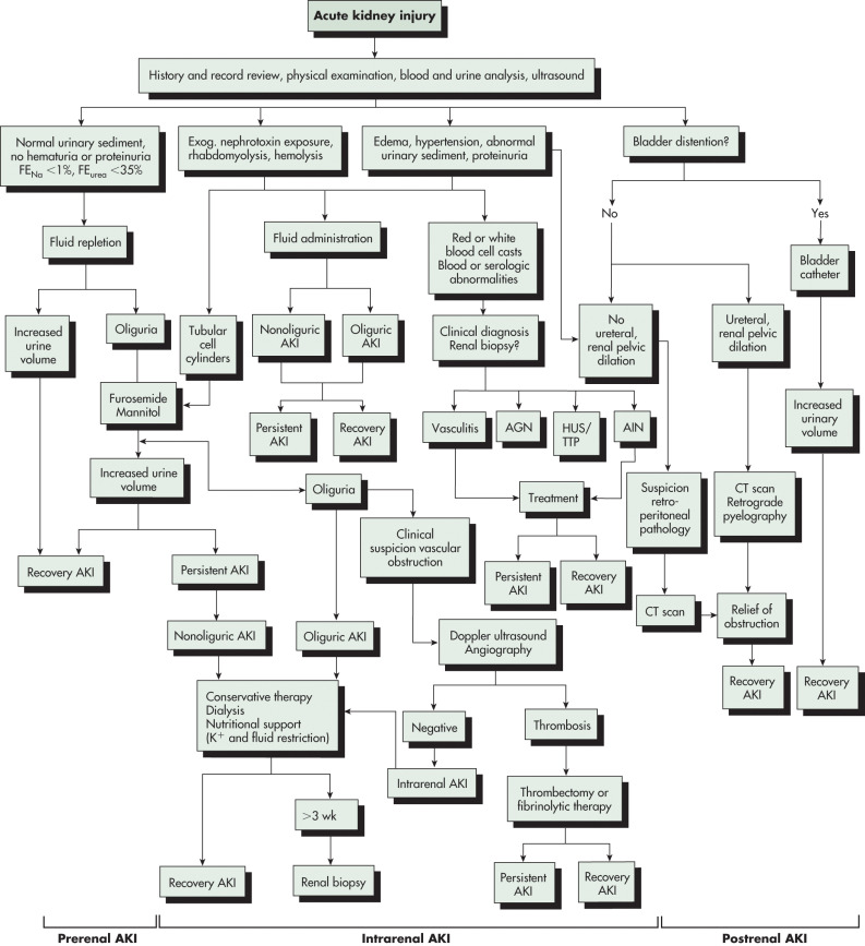
  • The clinical presentation of AKI depends on the presence of any preexisting conditions, the underlying conditions, the precipitating event(s) that caused the AKI and the severity of AKI (Fig E1).
  • Early or mild AKI is frequently asymptomatic.
  • Frequent presenting symptoms include weakness, anorexia, generalized malaise, and nausea.
  • Patients may develop oliguric or nonoliguric kidney injury.
  • Oliguria is defined as <400 to 500 ml of urine per 24 h. Anuria is frequently seen in ATN or bilateral obstructive uropathy.
  • Physical examination should focus on evaluation of volume status, eliciting systemic signs of AKI, and findings supportive of kidney injury etiology.
  • Clinical signs and symptoms are numerous; some key findings are highlighted:
    1. Peripheral edema from volume overload, heart failure, liver failure, or nephrotic syndrome
    2. Pulmonary edema
    3. Cardiac dysrhythmias
    4. Neurologic findings include altered mental status, delirium, lethargy, myoclonus, seizures, and asterixis
    5. Pruritus, uremic odor
    6. Flank pain
    7. Painless hematuria may be seen with glomerulonephritis (GN), whereas painful hematuria is more consistent with obstructive uropathy
    8. Pericardial effusion and/or pericardial rub
    9. Fever, skin rash, and arthralgia can be seen with systemic vasculitis
    10. Classic triad of fever, rash, and eosinophilia in the setting of AKI strongly implicates allergic interstitial nephritis (AIN). However, the simultaneous appearance of all three manifestations occurs in only 30% of cases. When AIN is considered the cause of AKI, a careful review of medications is required

Figure E1 The Clinical Presentation of Acute Kidney Injury (AKI) Depends on the Presence of any Preexisting Conditions, the Precipitating Event (s) that Caused the AKI and the Severity of the AKI

!!flowchart!!

(From Cameron P et al: Textbook of adult emergency medicine, ed 5, London, 2019, Elsevier.)

Etiology

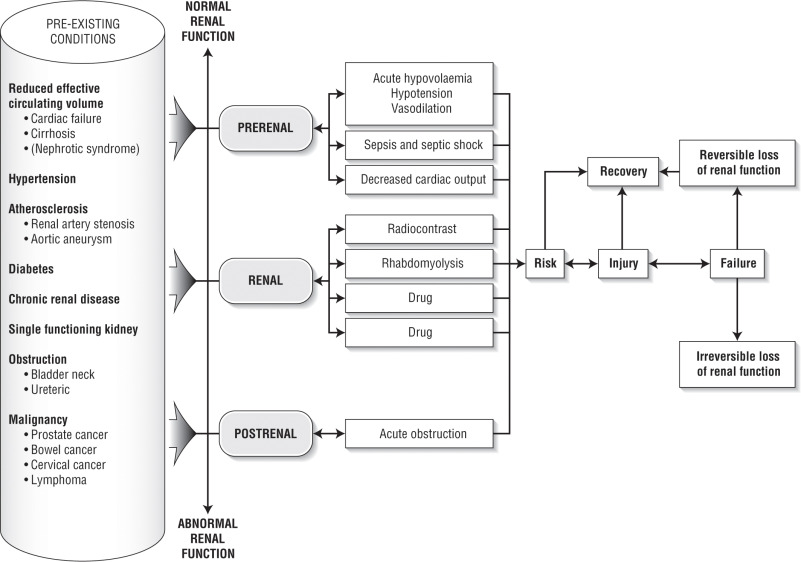
  • Prerenal: Inadequate renal perfusion caused by hypovolemia, congestive heart failure (impaired cardiac output), cirrhosis (fluid third-spacing), sepsis (vasodilation), abdominal compartment syndrome, or other. Sixty percent of community-acquired cases of AKI are due to prerenal conditions.
  • Postrenal: Bladder outlet obstruction (prostatic enlargement, urethral fibrosis), ureteral obstruction (stones, bladder masses, retroperitoneal fibrosis, ureteral fibrosis), or renal vein occlusion. With two functioning kidneys, bilateral obstruction is usually required to produce significant AKI. Postrenal causes of AKI account for 5% to 15% of community-acquired AKI.
  • Intrinsic renal: ATN, AIN, and GN. Common causes of ATN include ischemia (e.g., hypotension or shock, postcardiac bypass, or aorta surgery), rhabdomyolysis, sepsis, drug toxicity (e.g., aminoglycosides, amphotericin, cisplatin), and iodinated radiocontrast-induced nephropathy. Contrast-induced nephropathy is the third most common cause of new-onset AKI in hospitalized patients. However, most of these cases have multiple confounding factors and are better characterized as contrast-associated nephropathy. AIN can develop after exposure to medications, most commonly NSAIDs, antibiotics, and proton pump inhibitors. Microvascular diseases that cause AKI include thrombotic microangiopathies (e.g., thrombotic thrombocytopenic purpura, classic and atypical hemolytic-uremic syndrome, and preeclampsia) and cholesterol emboli.
  • Causes of AKI are listed in Table 2.
  • Nearly one third of AKI cases may be prevented or mitigated.

TABLE 2 Etiologies of Acute Kidney Injury

Prerenal Causes (Decreased Renal Blood Flow)Intrinsic Renal CausesPostrenal Causes
  • Hypovolemia
  • Renal losses (diuretics, osmotic agents, polyuria)
  • Gastrointestinal losses (vomiting, diarrhea)
  • Cutaneous losses (burns, exfoliative syndromes)
  • Hemorrhage
  • Pancreatitis
  • Decreased Cardiac Output
  • Congestive heart failure
  • Pulmonary embolism
  • Acute myocardial infarction
  • Severe valvular heart disease
  • Abdominal compartment syndrome
  • Renal artery obstruction (stenosis, embolism, thrombosis, dissection)
  • Systemic Vasodilation
  • Sepsis
  • Anaphylaxis
  • Anesthetics
  • Drug overdose
  • Afferent Arteriolar Vasoconstriction
  • Hypercalcemia
  • Drugs (NSAIDs, amphotericin B, calcineurin inhibitors, orepinephrine, radiocontrast agents, aminoglycosides)
  • Hepatorenal syndrome
  • Efferent arteriolar vasodilation (angiotensin converting enzyme inhibitors, aldosterone receptor blockers)
  • Trauma
  • Vascular: Large and Small Vessels
  • Renal vein obstruction (thrombosis, ventilation with high-level PEEP, abdominal compartment syndrome)
  • Microangiopathy (thrombotic thrombocytopenic purpura, hemolytic-uremic syndrome, disseminated intravascular coagulation, preeclampsia)
  • Malignant hypertension
  • Scleroderma renal crisis
  • Transplant rejection
  • Atheroembolic disease
  • Glomerular
  • Antiglomerular basement membrane disease (Goodpasture syndrome)
  • Antineutrophil cytoplasmic antibody-associated glomerulonephritis (Wegener granulomatosis)
  • Immune complex glomerulonephritis, systemic lupus erythematosus, postinfectious cryoglobulinemia, primary membranoproliferative glomerulonephritis
  • Tubular
  • Ischemic
  • Cytotoxic
  • Heme pigment (rhabdomyolysis, intravascular hemolysis)
  • Crystals (tumor lysis syndrome, seizures, ethylene glycol poisoning, vitamin C megadose, acyclovir, indinavir, methotrexate)
  • Drugs (aminoglycosides, lithium, amphotericin B, pentamidine, cisplatin, ifosfamide, radiocontrast agents), synthetic cannabinoid use
  • Interstitial
  • Drugs (penicillins, cephalosporins, NSAIDs, proton pump inhibitors, allopurinol, rifampin, indinavir, mesalamine, sulfonamides, trimethoprim)
  • Infection (pyelonephritis, viral infection)
  • Systemic Disease
  • Sjögren syndrome, sarcoidosis, systemic lupus erythematosus, lymphoma, leukemia, interstitial nephritis, uveitis
  • Calculus
  • Ureteral Obstruction
  • Tumor (intrinsic or extrinsic)
  • Fibrosis
  • Ligation during pelvic surgery
  • Bladder Neck Obstruction
  • Benign prostatic hypertrophy
  • Prostate cancer
  • Neurogenic bladder
  • Tricyclic antidepressants
  • Ganglionic blockers
  • Bladder tumor
  • Calculus
  • Hemorrhage/clot
  • Urethral Obstruction
  • Strictures
  • Tumor
  • Phimosis
  • Renal calcinosis
  • Obstructed urinary catheter, ureteral stent, or ileal conduit
  • Pelvic trauma, retroperitoneal hematoma

NSAIDs, Nonsteroidal antiinflammatory drugs; PEEP, positive end-expiratory pressure.

Modified from Cameron JL, Cameron AM: Current surgical therapy, ed 10, Philadelphia, 2011, Saunders.

Diagnosis

Table 3 summarizes useful clinical features, urinary findings, and confirmatory tests in the differential diagnosis of AKI.

TABLE 3 Useful Clinical Features, Urinary Findings, and Confirmatory Tests in the Differential Diagnosis of Acute Kidney Injury

Cause of Acute Kidney InjurySome Suggestive Clinical FeaturesTypical Urinalysis ResultsSome Confirmatory Tests
Prerenal azotemiaEvidence of true volume depletion (thirst, postural or absolute hypotension and tachycardia, low jugular venous pressure, dry mucous membranes and axillas, weight loss, fluid output greater than input) or decreased effective circulatory volume (e.g., heart failure, liver failure), treatment with NSAID, diuretic, or ACE inhibitor/ARB
  • Hyaline casts
  • FENa <1%
  • UNa <10 mmol/L
  • SG >1.018
Occasionally requires invasive hemodynamic monitoring; rapid resolution of AKI with restoration of renal perfusion
Diseases Involving Large Renal Vessels
Renal artery thrombosisHistory of atrial fibrillation or recent myocardial infarction, nausea, vomiting, flank or abdominal pain
  • Mild proteinuria
  • Occasionally RBCs
Elevated LDH level with normal transaminase levels, renal arteriogram, MAG3 renal scan, MRA*
AtheroembolismUsually age >50 yr, recent manipulation of aorta, retinal plaques, subcutaneous nodules, palpable purpura, livedo reticularis
  • Often normal
  • Eosinophiluria
  • Rarely casts
Eosinophilia, hypocomplementemia, skin biopsy, renal biopsy
Renal vein thrombosisEvidence of nephrotic syndrome or pulmonary embolism, flank painProteinuria, hematuriaInferior venacavogram, Doppler flow studies, MRV*
Diseases of Small Renal Vessels and Glomeruli
Glomerulonephritis or vasculitisCompatible clinical history (e.g., recent infection), sinusitis, lung hemorrhage, rash or skin ulcers, arthralgias, hypertension, edemaRBC or granular casts, RBCs, white blood cells, proteinuriaLow complement levels; positive antineutrophil cytoplasmic antibodies, antiglomerular basement membrane antibodies, antistreptolysin O antibodies, anti-DNase, cryoglobulins; renal biopsy
HUS/TTPCompatible clinical history (e.g., recent gastrointestinal infection, cyclosporine, anovulants), pallor, ecchymoses, neurologic findingsMay be normal, RBCs, mild proteinuria, rarely RBC or granular castsAnemia, thrombocytopenia, schistocytes on peripheral blood smear, low haptoglobin level, increased LDH, renal biopsy
Malignant hypertensionSevere hypertension with headaches, cardiac failure, retinopathy, neurologic dysfunction, papilledemaMay be normal, RBCs, mild proteinuria, rarely RBC castsLVH by echocardiography or ECG, resolution of AKI with BP control
Ischemic or Nephrotoxic Acute Tubular Necrosis
IschemiaRecent hemorrhage, hypotension, surgery often in combination with vasoactive medication (e.g., ACE inhibitor, NSAID)
  • Muddy-brown granular or tubular epithelial cell casts, FENa >1%,
  • UNa >20 mmol/L
  • SG 1.010
Clinical assessment and urinalysis usually inform diagnosis
Exogenous toxinRecent contrast medium-enhanced procedure; nephrotoxic medications; certain chemotherapeutic agents often with coexistent volume depletion, sepsis, or chronic kidney disease
  • Muddy-brown granular or tubular epithelial cell casts FENa >1%,
  • UNa >20 mmol/L
  • SG 1.010
Clinical assessment and urinalysis usually inform diagnosis
Endogenous toxinHistory suggestive of rhabdomyolysis (coma, seizures, drug abuse, trauma)Urine supernatant tests positive for heme in absence of RBCsHyperkalemia, hyperphosphatemia, hypocalcemia, increased CK, myoglobin
History suggestive of hemolysis (recent blood transfusion)Urine supernatant pink and tests positive for heme in absence of RBCsHyperkalemia, hyperphosphatemia, hypocalcemia, hyperuricemia, and free circulating hemoglobin
History suggestive of tumor lysis (recent chemotherapy), myeloma (bone pain), or ethylene glycol ingestionUrate crystals, dipstick-negative proteinuria, oxalate crystals, respectivelyHyperuricemia, hyperkalemia, hyperphosphatemia (for tumor lysis); circulating or urinary monoclonal protein (for myeloma); toxicology screen, acidosis, osmolal gap (for ethylene glycol)
Diseases of the Tubulointerstitium
Allergic interstitial nephritisRecent ingestion of drug and fever, rash, loin pain, or arthralgiasWhite blood cell casts, white blood cells (frequently eosinophiluria), RBCs, rarely RBC casts, proteinuria (occasionally nephritic)Systemic eosinophilia, renal biopsy
Acute bilateral pyelonephritisFever, flank pain and tenderness, toxic stateLeukocytes, occasionally white blood cell casts, RBCs, bacteriaUrine and blood cultures
Postrenal AKIAbdominal and flank pain, palpable bladderFrequently normal, hematuria if stones, prostatic hypertrophyPlain abdominal radiography, renal ultrasonography, postvoid residual bladder volume, computed tomography, retrograde or antegrade pyelography

ACE, Angiotensin-converting enzyme; AKI, acute kidney injury; ARB, angiotensin receptor blocker; BP, blood pressure; CK, creatine kinase; DNase, deoxyribonuclease; ECG, electrocardiography; FENa, fractional excretion of sodium; HUS, hemolytic uremic syndrome; LDH, lactate dehydrogenase; LVH, left ventricular hypertrophy; MAG3, mercaptoacetyltriglycine; MRA, magnetic resonance angiography; MRV, magnetic resonance venography; NSAID, nonsteroidal antiinflammatory drug; RBC, red blood cell; SG, specific gravity; TTP, thrombotic thrombocytopenic purpura; UNa, urinary sodium concentration.

* Contrast-enhanced MRA and MRV should be used with extreme caution in patients with AKI.

From Skorecki K et al: Brenner & Rector’s the kidney, ed 10, Philadelphia, 2016, Elsevier.

Differential Diagnosis

Refer to "Etiology." Diagnostic tests to distinguish prerenal and renal AKI are described in Table 4. A diagnostic approach to patients with suspected AKI is shown in Fig. 2.

TABLE 4 Diagnostic Tests to Distinguish Between Prerenal and Renal Acute Kidney Injury

IndexPrerenal CausesRenal Causes
FENa<1%>2%
Urine sodium<10 mmol/L>40 mmol/L
Urine/plasma osmolality>1.51-1.5
Renal failure index<1>2
BUN-to-serum creatinine ratio>20<10

BUN, Blood urea nitrogen; FENa, fractional excretion of sodium. Calculation of FENa: (Urine sodium × Plasma creatinine)/(Plasma sodium × Serum creatinine) ×100. Renal failure index: (Urine sodium × Urine creatinine)/Plasma creatinine.

From Cameron JL, Cameron AM: Current surgical therapy, ed 10, Philadelphia, 2011, Saunders.

Figure 2 Diagnostic approach to patients with suspected acute kidney injury (AKI).

!!flowchart!!

AGN, Acute glomerulonephritis; AIN, acute interstitial nephritis; CT, computed tomography; Exog, exogenous; HUS/TTP, hemolytic-uremic syndrome/thrombotic thrombocytopenic purpura.

(From Floege J et al: Comprehensive clinical nephrology, ed 4, Philadelphia, 2010, Saunders.)

Laboratory Tests

  • Elevated serum creatinine.
  • Standard estimating equations for glomerular filtration rate (GFR) require steady-state creatinine levels and are not recommended to estimate GFR during AKI.
  • Elevated blood urea nitrogen (BUN): BUN-to-creatinine ratio is commonly >20:1 in prerenal azotemia, postrenal azotemia, and acute GN.
  • BUN-to-creatinine ratio is <20:1 in acute interstitial nephritis and ATN.
  • Hyperkalemia, hyperphosphatemia, and metabolic acidosis are common.
  • Hypocalcemia with hyponatremia or hypernatremia may occur, depending on etiology.
  • Urinalysis is the initial diagnostic evaluation. Prerenal and postrenal AKI are typically characterized by a normal urinalysis. Conversely, abnormal findings should prompt further workup for specific intrinsic causes of AKI that may require intervention. Hematuria and proteinuria imply GN; heavy (>3+) proteinuria is associated with leukocyturia and may signify AIN. Microscopic examination of urine sediment may facilitate diagnosis: Granular casts in ATN, dysmorphic red blood cells or red blood cell casts in acute GN, and white blood cell casts in AIN.
  • In oliguric patients, obtain urine sodium and creatinine concentrations for determination of fractional excretion of sodium: FENa = 100% × (UNa × PCr)/(PNa × UCr). FENa <1% occurs in prerenal AKI and >1% occurs in intrinsic AKI. FENa may be falsely elevated in patients taking diuretics or falsely low in several intrinsic renal conditions, including acute GN, contrast-induced nephropathy, and rhabdomyolysis. (UNa, urinary sodium; UCr, urinary creatinine; PCr, plasma creatinine; PNa, plasma sodium).
  • Fractional excretion of urea (FEUrea) can be used to assess renal dysfunction in AKI. FEUrea is more useful than FENa during diuretic therapy. FEUrea is calculated as follows: FEUrea = 100% × (UUrea × PCr)/(PUrea × UCr). If FEUrea <35% prerenal AKI is likely, and if FEUrea >50% intrinsic AKI is likely. (UUrea, urinary urea; PUrea, plasma urea).
  • Urinary osmolality range of 250 mOsm/kg H2O to 300 mOsm/kg H2O in ATN (isosthenuria), <400 mOsm/kg H2O in postrenal azotemia, and >500 mOsm/kg H2O in prerenal azotemia and acute GN.
  • In suspected GN (e.g., hematuria plus proteinuria), additional serologic testing may be warranted. Abnormal liver function tests and elevated inflammatory markers are nonspecific. Immune complex deposition disorders (e.g., infectious GN, lupus nephritis, cryoglobulinemic vasculitis) are characterized by decreased complement levels (C3, C4). Specific testing includes antinuclear antibodies (lupus), antineutrophil cytoplasmic antibodies (ANCA-associated vasculitis), antiglomerular basement membrane antibodies (Goodpasture disease), and cryoglobulins. Kidney biopsy is frequently required for diagnostic confirmation.
  • Creatinine phosphokinase level is indicated if rhabdomyolysis is suspected; positive blood reaction on a urine dipstick with typically few or no red blood cells by microscopy may indicate myoglobinuria from rhabdomyolysis.
  • Serum free light chain analysis, serum and urine protein electrophoresis, and serum and urine immunofixation electrophoresis for suspected multiple myeloma or other plasma cell dyscrasias. Myeloma can cause AKI via a variety of mechanisms, including tubular precipitation of filtered light chains (cast nephropathy), hypercalcemia, and amyloidosis, among others.
  • Kidney biopsy may be indicated in patients with intrinsic kidney failure when considering specific therapy. The major indications for kidney biopsy include the following: Differential diagnosis of nephrotic syndrome, distinguishing lupus vasculitis from other vasculitides, distinguishing lupus membranous nephropathy from idiopathic membranous nephropathy, confirmation of hereditary nephropathies based on ultrastructure, diagnosis of rapidly progressive GN, distinguishing AIN from ATN, and separation of primary glomerulonephritides. In addition to establishing a diagnosis, biopsy may determine renal prognosis and guide direction of management. Severe interstitial fibrosis is associated with poor renal outcomes.
  • Biomarkers of kidney injury have been explored for earlier diagnosis of AKI and to separate intrinsic from prerenal causes. Candidate markers include neutrophil gelatinase-associated lipocalin (NGAL), kidney-injury molecule 1 (KIM-1), and the product of tissue inhibitor of metalloprotein-ase-2 and insulin-like growth-factor binding protein-7 (TIMP2*IGFBP7).
  • TIMP2*IGFBP7 is U.S. FDA approved for risk prediction of AKI. However, there remains little published experience with this test, and the clinical role remains undefined.
Imaging Studies

  • ECG for arrhythmia detection, especially in hyperkalemia: Peaked T waves in precordial leads, widening QRS interval, and/or bradycardia with AV node blockade.
  • Chest radiograph to detect signs of congestive heart failure and pulmonary renal syndromes that frequently present with alveolar hemorrhage (antiglomerular basement membrane or ANCA-associated vasculitis).
  • Bladder scan to assess post-void residual urine when urinary obstruction is suspected.
  • Kidney ultrasonography to determine kidney sizes (distinguishes acute from chronic kidney disease), presence of obstruction, and renal vascular status (Doppler study).

Treatment

Management of AKI depends on the underlying etiology. Some conditions (e.g., GN) require specific therapy. The general focus of treatment for established AKI is supportive care and limiting additional injury. Fig. 3 illustrates an algorithm for management of AKI.

Figure 3 Algorithm for management of acute kidney injury (AKI).

!!flowchart!!

AIN, Acute interstitial nephritis; ATN, acute tubular necrosis; BPH, benign prostatic hypertrophy; GN, glomerulonephritis; HRS, hepatorenal syndrome.

(From Ronco C et al: Critical care nephrology, ed 3, Philadelphia, 2019, Elsevier.)

Nonpharmacologic Therapy

  • Withdraw nephrotoxic medications.
  • Evaluate volume status and correct hypovolemia. Goal of therapy is to increase cardiac output and improve tissue perfusion.
  • Avoid excessive fluid administration in patients who are nonvolume responsive.1,5,6
  • Dietary modification: (1) Energy prescription (29 to 36 Kcal/kg/day), (2) potassium restriction (60 mmol/day), (3) sodium restriction (90 mmol/day), (4) phosphorus restriction (<800 mg/day), and (5) high biologic value protein (1.2 g/kg/day) depending on requirement for dialysis.7
  • Daily weight to monitor for fluid retention, in addition to intake and output measurement.
  • Modification of drug dosages or schedules of renally excreted medications. Dosing should consider the trajectory of renal function, during evolving and recovering AKI, and may require additional adjustments in patients who require dialysis.8
Acute General Rx

  • Correct electrolyte abnormalities and metabolic acidosis.
  • Administer loop diuretics for volume overload.
  • Administer vasopressors for circulatory shock or vasodilators, when appropriate, to optimize cardiac output in congestive heart failure.

Specific treatment is variable and depends on cause of AKI.

  • Prerenal: Intravenous (IV) volume expansion with isotonic solutions in hypovolemic patients or those with shock. Balanced crystalloid solutions are preferable.
  • Intrinsic kidney failure: Discontinue all potential nephrotoxins and treat condition(s) causing kidney failure. In severe AIN cases, consider a trial of corticosteroids. For acute noninfectious GN, high-dose pulse corticosteroids are first-line therapy, typically in conjunction with other immunomodulatory therapy and/or plasma exchange, depending on the clinical scenario.
  • Postrenal: Eliminate cause of obstruction. Immediate bladder catheter insertion for suspected bladder outlet obstruction. This maneuver should precede the kidney ultrasonogram during the diagnostic workup. Percutaneous nephrostomy tubes or ureteral stents may be required for upper urinary tract obstruction.
  • Hyperkalemia-related ECG changes: IV calcium if electrocardiographic changes are present; IV insulin and/or glucose to shift potassium into cells; and IV bicarbonate therapy when metabolic acidosis is present to shift potassium into cells. These three treatments are temporary, and definitive therapy requires potassium removal via the GI tract by cation exchangers or via the urinary tract by diuretics or dialytic therapy.

Dialysis in AKI:

  • General indications for initiation of dialysis during AKI:
    1. Uremic symptoms (e.g., encephalopathy, pericarditis, seizures).
    2. Extracellular fluid volume overload refractory to medical management.
    3. Severe acid-base derangement(s) refractory to medical management.
    4. Significant electrolyte derangement(s) (e.g., hyperkalemia, hyponatremia) refractory to medical management.
  • Among critically ill patients, kidney replacement therapy may be required despite an absence of other indications. Optimal timing of initiation of dialysis remains controversial and is not solely dependent on previous metabolic parameters. Multiple recent clinical trials have not shown that early dialysis in AKI is beneficial.
  • Intermittent hemodialysis and continuous renal replacement therapy (CRRT) have similar outcomes for patients with AKI. However, CRRT is associated with greater hemodynamic stability and fluid removal compared to conventional intermittent hemodialysis. CRRT is employed in critically ill patients with hemodynamic instability.
Adjunctive Rx

  • Monitoring of renal function parameters and electrolytes.
  • Renally excreted drugs are adjusted according to creatinine clearance or GFR to prevent further kidney damage or other medication-related toxicities.
  • Prevent further renal insult with appropriate volume expansion, particularly before contrast administration, and avoid nephrotoxic agents. Volume expansion with isotonic solutions is more effective than hydration with hypotonic solutions. Isotonic saline or bicarbonate-containing solutions are effective.
  • Renal function recovery (ability to discontinue dialysis) varies from 50% to 75% in AKI survivors. Preexisting chronic kidney disease, longer duration of dialysis dependence, congestive heart failure, and older age are negative prognostic factors for renal function recovery.
  • Overall mortality rate in AKI is nearly 25% and approaches 50% to 60% in patients who require acute dialysis.
  • The combination of AKI and sepsis is associated with a mortality rate as high as 70%.

Pearls & Considerations

However studies associating AKI with rapid subsequent loss of kidney function have been shown to have methodological limitations and after pre-AKI, eGFR, proteinuria, and other covariables are accounted for, the association between mild to moderate AKI and worsening subsequent loss of kidney function in patients with CKD has been found to be small.13

Related Content

Suggested Readings

  1. Guadry S : Extracorporeal kidney-replacement therapy for acute kidney injuryN Engl J Med. 386(10):964-975, 2022.
  2. Prowle JR : Fluid management for the prevention and attenuation of acute kidney injuryNat Rev Nephrol. 10:37-47, 2014.

Related Content

    1. Au TH : The prevention of contrast-induced nephropathyAnn Pharmacother. 48:1332-1342, 2014.
    2. Chawla LS : Acute kidney injury and chronic kidney disease as interconnected syndromesN Engl J Med. 371:58-66, 2014.
    3. Godin M : Clinical approach to the patient with AKI and sepsisSemin Nephrol. 35:12-22, 2015.
    4. Gotfried J : Finding the cause of acute kidney injury: which index of fractional excretion is better?Cleve Clin J Med. 79(2):121-126, 2012.
    5. Grams ME : Fluid balance, diuretic use, and mortality in acute kidney injuryClin J Am Soc Nephrol. 6(5):966-973, 2011.
    6. Kaddourah A : Epidemiology of acute kidney injury in critically ill children and young adultsN Engl J Med. 376:11-20, 2017.
    7. Kane-Gill SL, Goldstein SL : Drug-induced acute kidney injury: a focus on risk assessment for preventionCrit Care Clin. 31:675-684, 2015.
    8. Improving Global Outcomes (KDIGO) Acute Kidney Injury Work Group. KDIGO Clinical Practice Guideline for Acute Kidney InjuryKidney inter., Suppl.. 2:1-138, 2012.
    9. Levey AS, James MT : In the clinic: acute kidney injuryAnn Intern Med. 167(9):ITC66-ITC80, 2017.
    10. Li Y : Nutritional support for acute kidney injuryCochrane Database Syst Rev. 1:CD005426, 2010.
    11. Meersch M : Prevention of cardiac surgery-associated AKI by implementing the KDIGO guidelines in high risk patients identified by biomarkers: the PrevAKI randomized controlled trialIntensive Care Med. 43(11):1551-1561, 2017.
    12. Mehran R : Contrast-associated acute kidney injuryN Engl J Med. 380(22):2146-2155, 2019.
    13. Muiru AN : Risk for chronic kidney disease progression after acute kidney injury: findings from the Chronic Renal Insufficiency Cohort StudyAnn Int Med. 176:961-968, 2023.