Author: Miriam Nathan, MD and Tara C. Bouton, MD, MPH, TM

The intracellular protozoan parasite Cryptosporidium parvum is associated with gastrointestinal diseases, mostly diarrhea and biliary tree disease, especially in patients with AIDS or other immunocompromised hosts. It is also associated with sporadic infections and waterborne outbreaks in immunocompetent hosts. Cryptosporidiosis is a notifiable disease in the U.S.
Other species, including C. hominis, C. felis, C. muris, C. canis, C. suis, and C. meleagridis, are now described to be pathogens as well.
- •Approximately 2% of diarrheal illness in industrial countries, 5% to 10% in developing countries.
- •Immunocompromised patients, especially those with HIV/AIDS, are particularly susceptible to infection. 10% to 20% of HIV patients in the U.S. may excrete cysts.
- •Cryptosporidiosis is a leading cause of all waterborne outbreaks in the U.S. An estimated 823,000 cryptosporidiosis cases occur annually, although fewer than 2% are reported. In 2019, more than 13,000 cases of cryptosporidiosis were reported in the country. The highest incidence was reported in the Midwest.1
Worldwide, especially developing countries; associated with poor hygiene as a waterborne pathogen.
- •Person-to-person (day care, family members)
- •Animal-to-person (pets, farm animals). Fig. E1 describes the life cycle of Cryptosporidium
- •Environmental (water-associated outbreaks, including travel associated with swimming in or drinking contaminated water or eating contaminated food)
- •May be a significant pathogen causing diarrhea in patients with AIDS
Figure E1 Life cycle of Cryptosporidium.
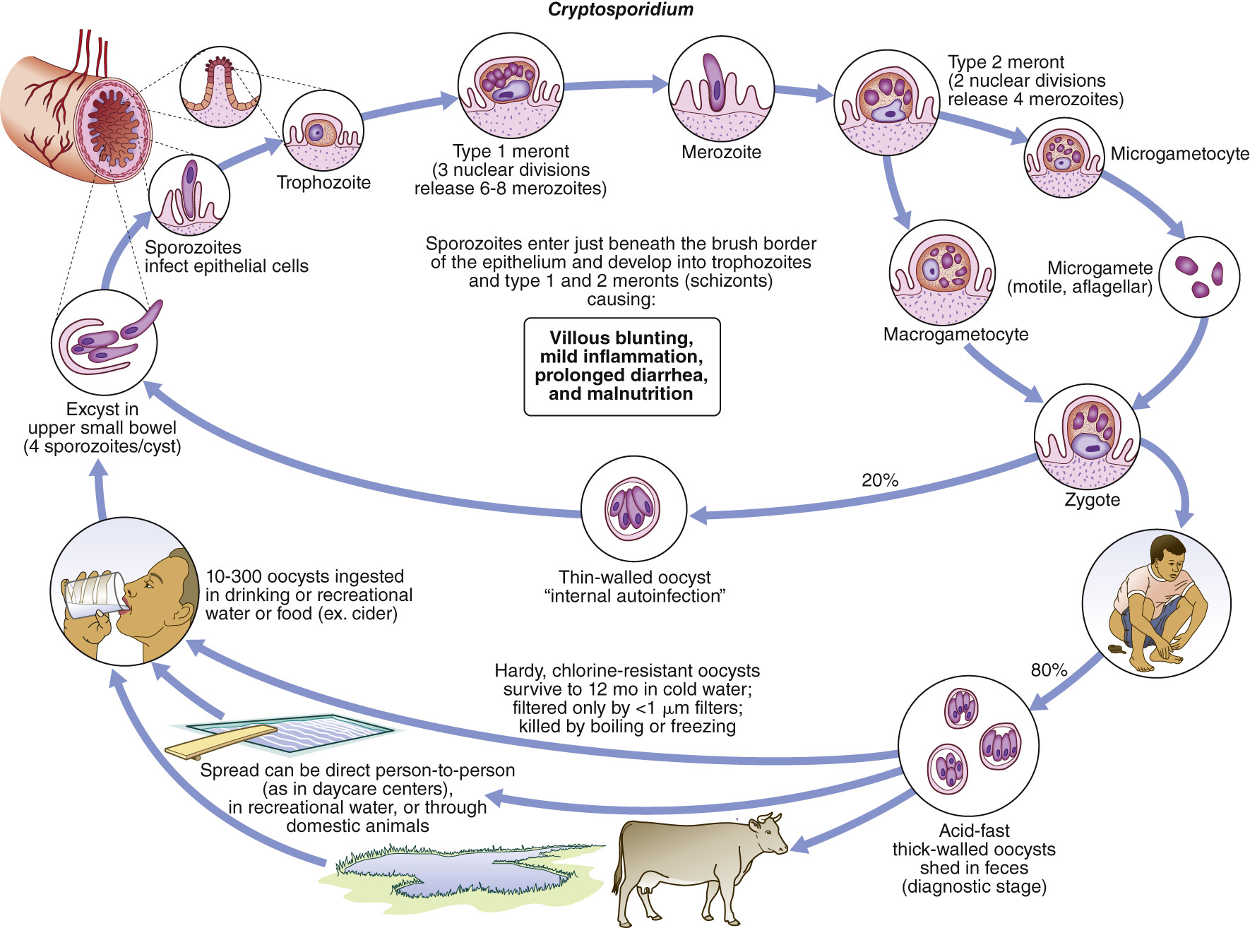
Oocysts are excreted in the feces. After ingestion, the sporozoites are released from the oocysts and attach to and invade intestinal epithelial cells. The cells engulf the parasites into a parasitophorous vacuole, where they enlarge to form the trophozoites; undergo asexual multiplication, forming type 1 meronts; and release the motile merozoites. The type II meronts differentiate into microgamonts and macrogamonts. The microgametes fertilize the macrogametes to form the zygote. The zygotes develop into the oocysts. Two different types of oocysts are produced: the thick-walled, which is commonly excreted from the host, and the thin-walled, which is primarily involved in autoinfection. Oocysts are infective on excretion, thus permitting direct and immediate fecal-oral transmission.
(From Guerrant RL et al [eds]: Tropical infectious diseases, ed 3, Philadelphia, 2011, Saunders; In Bennett JE et al: Mandell, Douglas, and Bennett’s principles and practice of infectious diseases, ed 9, Philadelphia, 2020, Elsevier.)
- •Spectrum of illness ranging from asymptomatic to severe enteritis (Table E1). Typical cases in immunocompetent hosts result in self-limited diarrhea, whereas immunocompromised hosts are characterized by profuse, watery, nonbloody diarrhea that may lead to dehydration and weight loss
- •More frequent in children
- •Usually limited to the gastrointestinal tract; however, in individuals with AIDS, the disease may be fulminant and life-threatening (CD4 counts <50)
- •Diarrhea, severe abdominal pain (2 to 28 days)
- •Impaired digestion, dehydration
- •Fever, malaise, fatigue, nausea, vomiting
- •Pneumonia if aspirated
TABLE E1 Clinical Manifestation of Cryptosporidiosis
| Host | Clinical Manifestations | Comments |
| Normal Host | Acute watery diarrhea | Relapses common Persistent diarrhea common |
| Children in Developing Countries | Acute watery diarrhea Persistent diarrhea | Diarrhea more severe in children with malnutrition Persistent diarrhea affecting nutritional status, growth, and intellectual function |
| Immunocompromised Host | Acute watery diarrhea Relapsing diarrhea Persistent or chronic diarrhea Cholera-like illness Extraintestinal involvement | Transient, self-limited, similar to disease in normal host Very common Usually found in patients with low CD4 count or malnutrition Voluminous watery diarrhea, only with very low CD4 count Respiratory tract, biliary tract, and pancreas |
From Cherry JD et al: Feigin and Cherry’s pediatric infectious diseases, ed 8, Philadelphia, 2019, Elsevier.