Author: Gerald Hagler, MD, MBA and Emily Mylhousen, MD

Neurofibromatosis (NF) is an autosomal-dominant, neurocutaneous disorder causing a tumor predisposition predominantly in the form of neurofibromas, optic gliomas, Lisch nodules, café au lait spots, axillary and inguinal freckling, and osseous lesions. Traditionally NF has been divided into three major subtypes: NF type 1 (NF1), NF type 2 (NF2), and schwannomatosis (SWN). However, nomenclature and diagnostic criteria for neurofibromatosis were recently updated (Box E1) to incorporate clinical and genetic discoveries made since the previous consensus conference in the late 1990s.
BOX E1 Revised Diagnostic Criteria for Neurofibromatosis Type 1 (NF1)
A: The diagnostic criteria for NF1 are met in an individual who does not have a parent diagnosed with NF1 if two or more of the following are present:
B: A child of a parent who meets the diagnostic criteria specified in A merits a diagnosis of NF1 if one or more of the criteria in A are present. |
From Legius E et al: Revised diagnostic criteria for neurofibromatosis type 1 and Legius syndrome: an international consensus recommendation, Genet Med 23,1506-1513, 2021.
| ICD-10CM CODES | |||
| Q85.00 | Neurofibromatosis, unspecified | ||
| Q85.01 | Neurofibromatosis, type 1 | ||
| Q85.02 | Neurofibromatosis, type 2 | ||
| Q85.03 | Schwannomatosis | ||
- •NF1 and NF2-related SWN are autosomal dominant; however, approximately 50% of cases are de novo mutations with no family history.
- •NF1 has an incidence of 1/3000 live births and prevalence of 1/5000 persons.
- •NF2-related SWN has an incidence of 1/25,000 live births and prevalence of 1/60,000 persons.
- •These two disorders affect approximately 100,000 people in the US.
- •Affects males and females equally.1
- •NF1 may be associated with optic gliomas, astrocytomas, spinal neurofibromas, pheochromocytomas, and chronic myeloid leukemia.
- •NF2-related SWN may be associated with acoustic neuromas, meningiomas, spinal schwannomas, and gliomas.2
- •The incidence of non-NF2-related SWN is 1/40,000 live births. The disease is mostly sporadic in nature; however, approximately 20% of cases appear to be inherited.1
- •Common features of NF1 include:
- 1.Café-au-lait macules (100% of children by age 2 yr)
- a.Hyperpigmented skin lesions (Fig. E1) occurring anywhere on the body except the face, palms, and soles
- b.Appear early in life and increase in size and number during puberty
- c.Are focal or diffuse
- 2.Axillary and inguinal freckling (70%)
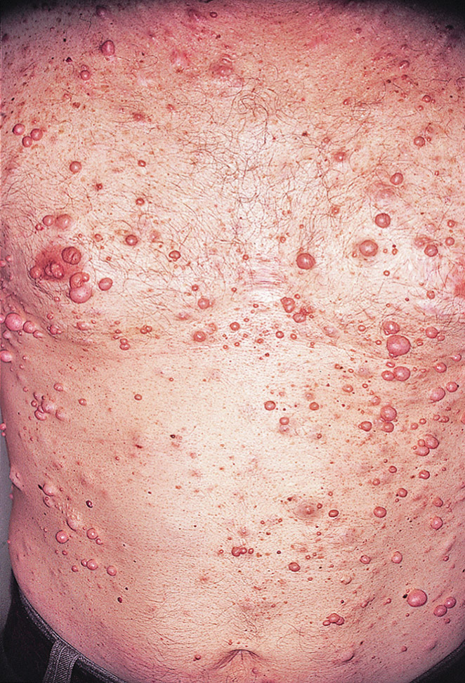
- 3.Multiple neurofibromas (Fig. E2) can be soft or firm; three subtypes:
- 4.Lisch nodule (small hamartoma of the iris) found in >90% of adult cases
- 5.Visual defects possibly related to optic gliomas (2% to 5%)
- 6.Neurodevelopment problems, such as learning disability and mental retardation (30%-40%)
- 7.Skeletal disorders including long bone dysplasia, pseudoarthrosis, scoliosis, short stature, and decreased bone mineral density3
- 1.Café-au-lait macules (100% of children by age 2 yr)
- •Common features of NF2-related SWN include:
- 1.Hearing loss and tinnitus related to bilateral acoustic neuromas (>90% of adults)
- 2.Cataracts (81%), retinal hamartoma, and epiretinal membranes
- 3.Headache, which may be due to intracranial meningiomas, present in 80% of patients
- 4.Unsteady gait
- 5.Cutaneous and subcutaneous neurofibromas but fewer than in NF1
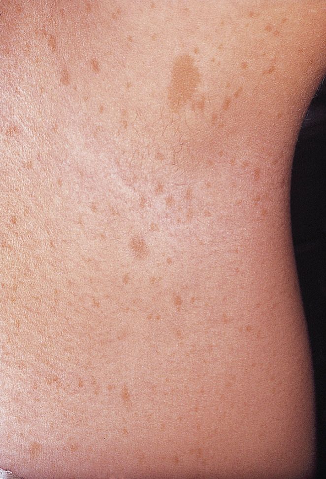
- 6.Café-au-lait macules (1%) (Fig. E3)3,4
- •Common features of SMARCB1-, LZTR1-, and 22q-related SWN include5:
Figure E1 Systemic features of neurofibromatosis type 1.
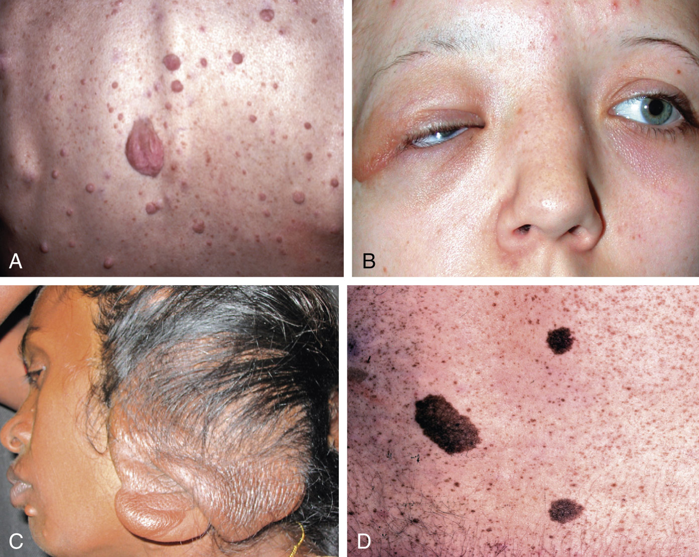
A, Discrete neurofibromas. B, Nodular plexiform neurofibroma of the eyelid. C, Elephantiasis nervosa. D, Café-au-lait spots.
(Courtesy S. Kumar Puri. Kanski JJ, Bowling B: Clinical ophthalmology: a systemic approach, ed 7, Philadelphia, 2010, Elsevier.)
- •NF1 is caused by DNA mutations located at 17q11.2 on chromosome 17, which is responsible for encoding the protein neurofibromin, a guanosine triphosphatase protein modulating signaling through the Ras/MAPK and PI3K/mTOR pathways.
- •NF2-related SWN is caused by DNA mutations located at 22q12.2 on chromosome 22 and is responsible for encoding the protein merlin, a potent inhibitor of glioma growth that modulates signaling through the PI3K/AKT, Raf/MEK/ERK, and mTOR pathways.
- •Both proteins are speculated to act as tumor suppressors.1
- •Over 50% de novo NF2-related SWN have mosaicism.4
- •Germline mutations in tumor suppressor genes SMARCB1 and LZTR1 on chromosome 22 account for 70% to 80% of non-NF2-related familial SWN. However, additional somatic mutations are required to trigger this condition. A four-hit hypothesis has been proposed for tumorigenesis in SMARCB1- and LZTR1-related SWN.
a If only café-au-lait macules and freckling are present, the diagnosis is most likely NF1 but, exceptionally, the person might have another diagnosis such as Legius syndrome. At least one of the two pigmentary findings (café-au-lait macules or freckling) should be bilateral.
b Sphenoid wing dysplasia is not a separate criterion in the case of an ipsilateral orbital plexiform neurofibroma.


