Author: Shuang Chen, MD and Shyam Joshi, MD

Hypereosinophilic syndrome (HES) refers to overproduction of eosinophils with sustained eosinophil counts over 1.5 × 10⁹/L on CBC on two separate examinations >1 mo apart in which eosinophilic infiltration of tissues (skin, lungs, heart, liver, spleen, bone marrow) and mediator release cause organ damage or dysfunction.1
- Primary (or neoplastic) hypereosinophilic syndrome (HESN)
- Secondary (or reactive) hypereosinophilic syndrome (HESR)
- Idiopathic hypereosinophilic syndrome (IHES)
| ICD-10CM CODE | |||
| D72.1 | Eosinophilia | ||
| D72.11 | Hypereosinophilic syndrome | ||
| D72.110 | Idiopathic HES | ||
| D72.111 | Lymphocytic variant HES | ||
| D72.118 | Other HES | ||
| D72.119 | HES, unspecified | ||
HES is rare, and the true prevalence is unknown.
Certain variants of HES, such as those linked to mutations in the genes for tyrosine kinase receptors PDGFRA and PDGFRB, predominantly occur in males.1 In contrast, other variants, including lymphocytic variant HES (L-HES) and HES of unknown origin, are equally distributed between males and females.
Usually occurs between the ages of 20 and 50 yr, although can develop in children. Patients with M-HES tend to present at a slightly younger age compared to other forms of HES.2 Children with HES are more likely to present with gastrointestinal complaints and have higher peak eosinophil counts than adults.
- •Clinical presentation of HES may vary from an incidental finding of eosinophilia to sudden onset of cardiac or neurologic symptoms.
- •The frequency of specific symptoms at presentation includes dermatologic (e.g., rash) in 37% of cases; pulmonary (cough and breathlessness) in 25%; gastrointestinal in 14%; cardiac in 5%; and neurologic in 4%. Additionally, 6% of patients presented with incidentally detected and clinically asymptomatic hypereosinophilia.1
- •Cutaneous manifestations (37%): Commonly include eczema (involving hands, flexural areas, or dispersed plaques), erythroderma, lichenification, dermographism, recurrent urticaria, and angioedema. Adult patients without a history of eczema who develop new-onset cutaneous symptoms should be investigated for cutaneous T-cell lymphoma. Patients who experience angioedema and urticaria, with or without other symptoms, are likely to have a benign course without cardiac or neurologic complications. Patients with mucosal ulcerations are more likely to have FIP1L1-PDGFRA-positive HES.
- •Pulmonary manifestations (25%): The most common presenting symptoms are dyspnea (45%), cough (39%), and wheezing (24%). Abnormal chest radiography or computed tomography (CT) findings are seen in 43% of patients, including parenchymal infiltrates (37%), pleural effusion (14%), intrathoracic lymphadenopathy (12%), and pulmonary emboli (4%).
- •GI manifestations (14%): Eosinophilic gastritis, enteritis, and/or colitis may occur and cause weight loss, abdominal pain, vomiting, and/or severe diarrhea. Hepatic involvement may manifest as chronic active hepatitis, focal hepatic lesions, eosinophilic cholangitis, or Budd-Chiari syndrome.
- •Cardiac manifestations (5%) include dyspnea, orthopnea, and signs and symptoms of congestive heart failure (CHF). Includes three stages: Acute necrotic stage secondary to endocardial infiltration of eosinophils, thrombus formation, and fibrotic stage. The latter two stages often result in a restrictive or dilated cardiomyopathy and/or valvular heart disease. The development of cardiac disease in HES is unpredictable. Some patients with sustained eosinophilia never develop cardiac involvement, and the severity of cardiac injury does not clearly correlate with the degree of peripheral eosinophilia. Platelet-derived growth factor receptor alpha (PDGFRA)-associated HES has an increased incidence of incapacitating and potentially lethal cardiac involvement in the absence of therapy.
- •Neurologic manifestations (4%) may present in three forms:
- •Ocular manifestations (23%) are thought to be the result of retinal microemboli.
- •Vascular manifestations include venous or arterial thrombosis of unknown mechanism, such as femoral artery occlusion, intracranial sinus thrombosis, and digital gangrene.
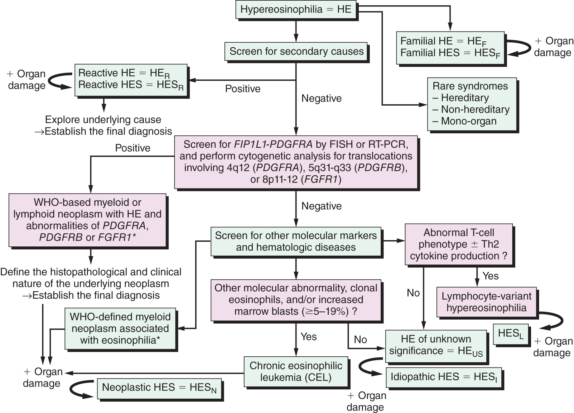
HES has been categorized into several clinically relevant variants: Primary/neoplastic HES, which includes myeloproliferative variants (M-HES) and T-lymphocytic variants (L-HES); secondary/reactive HES; familial HES; idiopathic HES (IHES); and organ-restricted hypereosinophilic syndromes.
- •Myeloproliferative variants of HES (M-HES) are classified into various subtypes. These include the FIP1L1-PDGFRA fusion gene (F/P-positive variant), which results from an interstitial chromosomal deletion on 4q12; PDGFRA fusion with other partner genes or point mutations; PDGFRB rearrangements such as t(5;12) (q31-35;p13); Janus kinase 2 (JAK2) point mutations, and translocations like t(8;9)(p22;p24); fibroblast growth factor receptor 1 (FGFR1) rearrangements such as (8p11-12); chronic eosinophilic leukemia (CEL); and HES with myeloid features without known mutations.
- •Lymphocytic variant HES (L-HES) is primarily characterized by skin and soft tissue involvement, with IL-5-producing T-cell subsets identified in the peripheral blood. The most commonly reported abnormal T-cell phenotype is CD3–CD4+. Besides IL-5, these abnormal T cells also produce other T helper type 2 (Th2) cytokines, such as IL-4 and IL-13, as well as GM-CSF. The molecular mechanisms of the disease remain largely unknown, and this disorder may progress to lymphoma.
- •Secondary (reactive) HES occurs in response to an identified stimulus such as an infectious or inflammatory process. Examples include helminth infections, episodic angioedema, and eosinophilic granulomatosis with polyangiitis (EGPA).
- •Familial HES has been reported when there is a family history of documented persistent hypereosinophilia of unknown cause. Autosomal dominant transmission of marked eosinophilia has been reported.
- •Idiopathic HES: Despite careful evaluation of HE, the etiology in as many as 75% of cases of presumed HES remains undefined.
- •Organ-restricted hypereosinophilic: When tissue hypereosinophilia is present, it causes end-organ damage, but HES blood criteria are unmet.