Author: Alison DeDent, MD and Erica Farrand, MD

DefinitionEosinophilic granulomatosis with polyangiitis (EGPA), formerly known as Churg-Strauss syndrome or as allergic granulomatosis and angiitis, refers to a multisystem, necrotizing, antineutrophil cytoplasmic antibody (ANCA)-associated vasculitis affecting small- and medium-sized vessels characterized by asthma and eosinophilia. ANCA reactivity may be positive or negative.1 Universal consensus for diagnostic criteria is lacking. Classification criteria for EGPA are described in Table E1.2
TABLE E1 *Classification Criteria for EGPA22
| CLINICAL CRITERIA | |
- •Obstructive airway disease
| +3 |
| +3 |
| +1 |
| LABORATORY AND BIOPSY CRITERIA |
- •Blood eosinophil count ≥1 × 10⁹/liter
| +5 |
- •Extravascular eosinophilic-predominant inflammation on biopsy
| +2 |
- •Positive c-ANCA or anti-PR3 antibodies
| –3 |
| –1 |
| Score ≥6 is 84.9% sensitive and 99.1% specific for EGPA | |
EGPA, Eosinophilic granulomatosis with polyangiitis; c-ANCA, c-antineutrophil cytoplasmic antibody; PR3, proteinase 3
Synonyms
- Allergic angiitis
- Allergic granulomatosis
- Allergic granulomatosis and angiitis
- Churg-Strauss syndrome
- EGPA
- CSS
| ICD-10CM CODE |
| M30.1 | Polyarteritis with lung involvement (Churg-Strauss) |
Epidemiology & Demographics
- •Epidemiology varies based on geographic location and applied classification criteria. Incidence and prevalence range from 0.18 to 4.0 cases per million person-yr and 2 to 30.4 per million person-yr, respectively.
- •Rarest of the ANCA-associated vasculitides.
- •Median age of onset is 49 to 59 yr.
- •No sex predominance.
Physical Findings & Clinical Presentation
- •The clinical picture of EGPA typically consists of three partially overlapping phases, which may or may not be sequential. See Table E2 for organ-specific manifestations.
- 1.Prodromal/allergic phase:6
- a.Main manifestation is asthma, present in 95% to 100% of patients
- b.Upper respiratory manifestations are common
- c.May precede development of systemic vasculitis by several years
- 2.Eosinophilic/tissue infiltration phase6:
- a.Characterized by peripheral eosinophilia and eosinophilic tissue infiltration leading to organ-specific signs and symptoms, most commonly with pulmonary, cardiac, and/or GI involvement
- 3.Systemic vasculitic phase6:
- a.Onset of necrotizing vasculitis leading to constitutional and organ-specific signs and symptoms, typically with cutaneous, renal, and nervous system manifestations
- •The presence of ANCA reactivity leads to distinct phenotypes that influence the frequency of organ-specific involvement6 (Table E2).
TABLE E2 Organ-Specific Manifestations and Phenotypes of EGPA4-7,9,13-16
| Organ Involvement | Disease Manifestations* | Phase | ANCA+ Phenotype | ANCA - Phenotype | Pathology |
| Ear, nose, and throat | - •Allergic rhinitis
- •Sinusitis
- •Nasal polyposis
| Prodromal | | | - •Eosinophilic infiltrates
|
| Pulmonary | - •Asthma
- •Pulmonary infiltrates (transient) or nodules
- •Pleural effusions
- •Alveolar hemorrhage (rare)
| Prodromal, eosinophilic, vasculitis | | x | - •Eosinophilic infiltration
- •Extravascular granulomas
- •Necrotizing vasculitis
|
| Cardiovascular | - •Cardiomyopathy
- •Pericarditis
- •Myocarditis
- •Myocardial infarction
- •Arrhythmia
- •Valvular defects
- •Endomyocardial fibrosis
- •Intracavitary thrombus
- •Arterial and venous thromboembolism
| Eosinophilic, vasculitis | | x | - •Endomyocardial and pericardial eosinophilic infiltration
- •Coronary vasculitis (rare)
|
| Gastrointestinal | - •Abdominal pain
- •Bleeding
- •Mesenteric vasculitis
| Eosinophilic, vasculitis | | | - •Eosinophilic infiltration
- •Vasculitis
|
| Renal | - •Microscopic hematuria
- •Proteinuria
- •Elevated creatinine
- •Rapidly progressive glomerulonephritis (less common)
| Vasculitis | x | | - •Crescentic and pauciimmune focal segmental necrotizing glomerulonephritis
- •Eosinophils and granulomas uncommon
|
| Nervous system | Peripheral nervous system:- •Mononeuritis multiplex
- •Polyneuropathy
Central nervous system (rare):- •Cerebral hemorrhage or infarct
| Vasculitis | x | | - •Necrotizing vasculitis
- •Eosinophilic infiltration less common
|
| Skin | - •Purpura
- •Dermal and subcutaneous nodules and papules
- •Ecchymosis
- •Livedo racemosa and/or hemorrhagic bullae
- •Urticaria
- •Ulcers
| Vasculitis | x | | - •Red granulomas
- •Dermal and subcutaneous vessel vasculitis
- •Eosinophilic infiltration
|
ANCA, Antineutrophil cytoplasmic antibodies.
EtiologyEtiology unknown; however, pathophysiology involves both T- and B-cell responses and eosinophil activation, with influence from genetic and environmental factors.5,6

The American College of Rheumatology (ACR) and European Alliance of Associations for Rheumatology (EULAR) have established validated classification criteria for EGPA (see Table E1). These criteria should be applied after a diagnosis of small- or medium-sized vasculitis has been established and alternative diagnoses have been excluded. A total of 6 or more points yields a sensitivity of 84.9% and a specificity of 99.1% for EGPA.2
Differential Diagnosis
- •Granulomatosis with polyangiitis (GPA) and microscopic polyangiitis (MPA)
- •Allergic bronchopulmonary aspergillosis (ABPA)
- •Chronic eosinophilic pneumonia (CEP)
- •Hypereosinophilic syndrome (HES)
- •IgG4-related disease
EGPA can be distinguished from the above diagnoses by the following:
- •In contrast to the other ANCA-associated vasculitides, including MPA and GPA, EGPA is associated with asthma and eosinophilia.
- •Kidney involvement, nasal/paranasal sinus bone erosions, and pulmonary nodule cavitation are much less common in EGPA compared to GPA.
- •CEP and ABPA are limited to lung involvement.
- •ANCAs, asthma, nasal polyps, and vasculitis are not features of HES, in contrast to EGPA.
- •The clinical characteristics of IgG4-related disease may mimic EGPA; however, vasculitis and eosinophilic granulomas are not seen on pathology.
Workup & Laboratory TestsInitial testing focuses on diagnosing EGPA and excluding alternative diseases:
- •CBC with differential: Eosinophilia ≥1 x 10⁹/liter is an ACR/EULAR classification criterion.
- •C-reactive protein and erythrocyte sedimentation rate are often elevated during the active phase.
- •Immunoglobulin E (IgE) is commonly elevated.
- •ANCA positivity is found in 30% to 47% of patients, and myeloperoxidase (MPO) antibody positivity with a perinuclear staining pattern (p-ANCA) is most common. Negative ANCA does not rule out EGPA.
- •Hypereosinophilia (eosinophils >1.5 × 10⁹) necessitates a comprehensive evaluation for causes of eosinophilia.
- •Presence of asthma and an elevated IgE suggest possible ABPA, and serum Aspergillus antibodies should be obtained.
Once EGPA is diagnosed, subsequent testing should screen for organ-specific involvement:
- •Cardiac: Brain natriuretic peptide (BNP) and troponin may be elevated, suggesting cardiac involvement. ECG may be abnormal, and echocardiogram may show wall motion abnormalities, left ventricular dysfunction, and effusions.
- •Renal: Creatinine may be mildly elevated, and urinalysis may show mild hematuria or proteinuria, suggesting renal involvement.
Imaging Studies
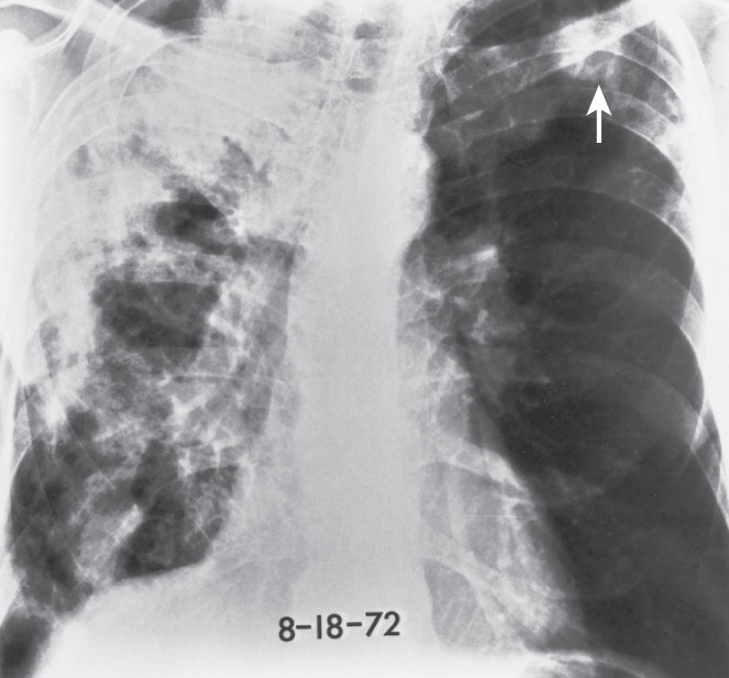
- •Chest radiograph may show bilateral, patchy, and migratory opacities.
- •Computed tomography (CT) scan is more sensitive and is the preferred imaging modality, though findings are nonspecific and may show ground-glass opacities or consolidation, bronchial wall thickening, centrilobular nodules, and pleural effusions (Fig. E1).
- •Paranasal sinus films may reveal sinusitis
- •Organ-specific symptoms warrant additional testing and may include angiography, pulmonary function tests, abdominal imaging, cardiac MRI, gastrointestinal endoscopy, and/or electromyogram and nerve conduction studies (EMG/NCS).
Figure E1 EGPA.
Thin-section CT scan at carina shows multifocal patchy ground-glass opacity around the patchy consolidation showing halo sign (arrow). Bronchial wall thickening is evident. CT, Computed tomography.
(From Choi YH et al: Thoracic manifestation of Churg-Strauss syndrome: radiologic and clinical findings, Chest 117[1]:117-124, 2000.)

