section name header

Basic Information ⬇

Author: Hannah Cain, MD

Definition

Osteoporosis is a skeletal disorder characterized by a progressive loss of bone mass and a decline in bone density and quality that results in increased bone fragility and a higher fracture risk. Poor bone mass acquisition during adolescence and bone loss during the sixth decade of life are the main processes responsible for osteoporosis. The various types are as follows:

Primary Osteoporosis:

Primary osteoporosis is the loss of bone mass due to aging and decreased gonadal function, not to any other chronic illness.

  • •Idiopathic osteoporosis: Unknown pathogenesis; may occur in children and young adults.
  • •Type I osteoporosis (postmenopausal women): Occurs after menopause due to an abrupt decline in estrogen production. It is characterized by accelerated and disproportionate trabecular bone loss and is associated with fractures of the spine, hip, and wrist. This is the most frequent kind of osteoporosis.
  • •Type II osteoporosis (involutional): Occurs in both men and women typically over the age of 75 yr due to the progressively negative balance between bone formation and resorption. It is characterized by both trabecular and cortical bone loss and is associated with fractures of the spine, long bones, and hip.
Secondary Osteoporosis:

Secondary osteoporosis is bone loss due to another chronic condition such as thyroxine excess, hyperparathyroidism, malignancies, gastrointestinal disease, medications, renal failure, and connective tissue diseases (see "Differential Diagnosis").

ICD-10CM CODES
M81.0Age-related osteoporosis without current pathological fracture
M81.4Drug-induced osteoporosis
M81.5Idiopathic osteoporosis
M81.6Localized osteoporosis
Epidemiology & Demographics
Prevalence (In U.S.):

  • •Affects more than 10 million people in the U.S.
  • •Annual incidence of osteoporotic fractures exceeds 1.5 million in the U.S. (70% women)
  • •Twice as common in women than in men
  • •Health care costs exceed $17 billion annually
  • •Health inequities in screening and treatment: Black women are less likely to be screened for osteoporosis and receive fewer prescriptions for osteoporosis treatment after diagnosis when compared with White women
Risk Factors:

  • •Female sex
  • •Postmenopausal state
  • •Advanced age
  • •Small body frame, low body weight (<58 kg)
  • •White or Hispanic ancestry
  • •Sedentary lifestyle
  • •Nulliparity
  • •Calcium deficiency
  • •Previous low-trauma fracture
  • •Parental history of hip fracture
  • •Tobacco use
  • •Excess alcohol use (> three drinks per day)
  • •Long-term glucocorticoid use
  • •Chronic disease states, e.g., primary ovarian insufficiency, diabetes mellitus, androgen deficiency, inflammatory bowel disease, hyperthyroidism, hypercortisolism

Conditions and drugs associated with osteoporosis are summarized in Table 1.

TABLE 1 Conditions and Drugs Associated With Osteoporosis

Inflammatory Disorders
  • •Rheumatoid arthritis
  • •Inflammatory bowel disease
  • •Cystic fibrosis

Bone Marrow Disorders
  • •Multiple myeloma
  • •Mastocytosis
  • •Leukemia

Hypogonadism
  • •Athletic amenorrhoea
  • •Hemochromatosis
  • •Turner syndrome
  • •Klinefelter syndrome
  • •Postchemotherapy
  • •Hypopituitarism

Malabsorption
  • •Celiac disease
  • •Gastrectomy
  • •Bariatric surgery
  • •Liver disease
  • •Total parenteral nutrition

Endocrine Disorders
  • •Cushing syndrome
  • •Diabetes
  • •Thyrotoxicosis
  • •Hyperparathyroidism
Low Body Weight
  • •Anorexia nervosa
  • •Human immunodeficiency virus infection

Immobilization
  • •Parkinson disease
  • •Poliomyelitis
  • •Cerebral palsy
  • •Paraplegia

Defective Synthesis of Connective Tissue
  • •Osteogenesis imperfecta
  • •Marfan syndrome
  • •Homocystinuria

Miscellaneous
  • •Chronic obstructive lung disease
  • •Congestive heart failure
  • •Pregnancy/lactation
  • •Ankylosing spondylitis
  • •Hypercalciuric nephrolithiasis
  • •Depression

Drugs
  • •Glucocorticoids, alcohol
  • •Medroxyprogesterone acetate
  • •Anticonvulsants, methotrexate
  • •Heparin, cyclosporin, omeprazole
  • •Aromatase inhibitors, glitazones
  • •Androgen deprivation therapy

From Robertson RP et al: DeGroot’s endocrinology, basic science and clinical practice, ed 8, Philadelphia, 2023, Elsevier.

Physical Findings & Clinical Presentation

  • •Most commonly asymptomatic.
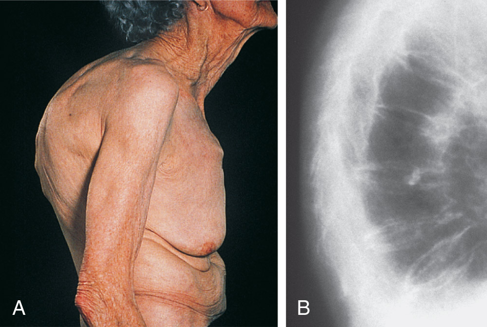
  • •Insidious and progressive development of dorsal kyphosis (dowager’s hump) (Fig. E1), loss of height, and skeletal pain typically associated with fracture; reduced gait speed or grip strength; other physical findings related to other conditions with associated increased risk for osteoporosis such as nodular thyroid, hepatic enlargement, jaundice, cushingoid features (see "Risk Factors" and Boxes 1 and 2).

BOX 2 Causes of Secondary Osteoporosis

Endocrine

  • •Hypogonadism in either sex, including untreated, premature menopause and treatment with aromatase inhibitors or androgen deprivation therapy
  • •Hyperthyroidism
  • •Hyperprolactinemia
  • •Cushing disease
  • •Diabetes
Gastrointestinal

  • •Celiac disease
  • •Inflammatory bowel disease
  • •Chronic liver disease
  • •Chronic pancreatitis
  • •Other causes of malabsorption
Rheumatologic

  • •Rheumatoid arthritis
  • •Other inflammatory arthropathies
Hematologic

  • •Multiple myeloma
  • •Hemoglobinopathies
  • •Systemic mastocytosis
  • •Chronic heparin treatment
Respiratory

  • •Cystic fibrosis
  • •Chronic obstructive pulmonary disease
Metabolic

  • •Homocystinuria
Chronic Renal Disease

Immobility

From Hochberg MC: Rheumatology, ed 7, Philadelphia, 2019, Elsevier.

BOX 1 Major Clinical Risk Factors for Osteoporotic Fracture

  • •Age
  • •Gender
  • •Previous fragility fracture
  • •Glucocorticoid therapy
  • •History of falls
  • •Family history of hip fracture
  • •Other causes of secondary osteoporosis
  • •Low body mass index
  • •Smoking
  • •High alcohol intake

From Hochberg MC: Rheumatology, ed 7, Philadelphia, 2019, Elsevier.

Figure E1 Dowager’s Hump

(A) Marked Thoracic Kyphosis Due to Multiple Osteoporotic Fractures in an Elderly Woman with (B) Corresponding x-Ray.

(From Geusens P: Osteoporosis: clinical features. In Hochberg MC et al [eds]: Rheumatology, ed 3, St. Louis, 2003, Mosby.)

Etiology

Normal bone turnover involves balance between process of bone resorption and bone formation. Osteoclasts resorb bone, and osteoblasts secrete bone matrix for building bone. In postmenopausal women, rate of bone turnover increases after loss of ovarian function, leading to progressive bone loss. Causes of osteoporosis in men are summarized in Box 3.

BOX 3 Causes of Osteoporosis in Men

  • Endocrine diseases
  • Hypogonadism
  • Cushing syndrome
  • Hyperthyroidism
  • Primary hyperparathyroidism
  • Hyperprolactinemia
  • Idiopathic renal hypercalciuriaa
  • Accompanying osteomalacia
  • Neoplastic disease
  • Multiple myeloma
  • Myeloproliferative and lymphoproliferative disease
  • Systemic mastocytosis
  • Diffuse bony metastasesa
  • Drugs/toxins
  • Glucocorticoidsa
  • Alcohol abusea
  • Excessive thyroid hormone replacement
  • Heparin
  • Anticonvulsantsa
  • Genetic collagen disorders
  • Osteogenesis imperfecta
  • Ehlers-Danlos syndrome
  • Marfan syndrome
  • Homocystinuria
  • Hemochromatosis
  • Other disorders
  • Skeletal sarcoidosis
  • Gaucher disease
  • Adult hypophosphatasia
  • Hemoglobulinopathies
  • Other factors
  • Chronic inflammatory diseases (rheumatoid arthritis, ankylosing spondylitis)
  • Prolonged immobilization
  • Malnutrition (including calcium deficiency and scurvy)
  • Gastrectomya
  • Chronic liver or kidney diseasea

From Hochberg MC et al: Rheumatology, ed 8, Philadelphia, 2023, Elsevier.

Several fracture risk calculation tools have been developed. Clinical risk factors used in the World Health Organization Fracture Risk Assessment Tool (WHO FRAX) 10-yr fracture risk calculator are summarized in Box 4.

BOX 4 Clinical Risk Factors Included in the FRAX Case-Finding Algorithm

  • •Age (50-90 yr)
  • •Sex
  • •Weight (in kilograms) and height (in centimeters). Body mass index is automatically computed from height and weight
  • •Previous fragility fracture (yes/no)
  • •Parental history of hip fracture (yes/no)
  • •Current tobacco smoking (yes/no)
  • •Long-term use of oral glucocorticoids (over 3 mo) (yes/no)
  • •Rheumatoid arthritis (yes/no)
  • •Alcohol consumption of 3 or more units daily (yes/no)
  • •Other causes of secondary osteoporosis (yes/no): Includes type 1 diabetes, osteogenesis imperfecta, longstanding untreated hyperthyroidism, hypogonadism or premature menopause (<45 yr), chronic malnutrition, malabsorption, chronic liver disease

From World Health Organization: WHO risk fracture assessment tool. www.sheffield.ac.uk/FRAX/. From Hochberg MC: Rheumatology, ed 7, Philadelphia, 2019, Elsevier.

a Most frequent causes.

Diagnosis ⬆ ⬇

Differential Diagnosis

  • •Malignancy (multiple myeloma, lymphoma, leukemia, metastatic carcinoma)
  • •Primary hyperparathyroidism
  • •Osteomalacia
  • •Paget disease
  • •Osteogenesis imperfecta: Types I, III, and IV
Screening

  • •The U.S. Preventive Services Task Force (USPSTF)1 recommends screening for older postmenopausal women (age ≥65) and in younger postmenopausal women (age 40-65) with at least one osteoporosis risk factor (i.e., cigarette smoking, excessive alcohol use, low body weight, parenteral history of hip fracture) with DXA scan. Evidence is insufficient to make a recommendation for or against screening men for osteoporosis to prevent fractures. Validated fracture and osteoporosis risk assessment tools include the following: Fracture Risk Calculator, Garvan Fracture Risk Calculator, Fracture Risk Assessment Tool (without inputting bone mineral density) Osteoporosis Risk Assessment Instrument, and Osteoporosis Self Assessment Tool.
  • •History and physical examination with appropriate evaluation for risk factors and secondary causes. Medications associated with osteoporosis are summarized in Table 2. Investigations for secondary osteoporosis are summarized in Box 5.
  • •WHO guidelines for the diagnosis of osteoporosis are based on bone mineral density (BMD) measurements of the hip or spine in g/cm2 and are reported as a T score.
  • •Dual-energy x-ray absorptiometry (DEXA) is the gold standard for screening and monitoring changes in BMD due to excellent precision, widespread availability, low cost, and minimal radiation exposure.
  • •DEXA (Fig. E2) is indicated in all women 65 yr and older and in postmenopausal women younger than 65 yr of age who are at risk for fracture (e.g., weight <127 lb, parental history of hip fracture, use of medications that cause bone loss, current smoking, excessive alcohol use, rheumatoid arthritis, or presence of diseases that cause bone loss). Clinical indications for bone densitometry are summarized in Table 3. Causes of erroneous bone mineral density measures by DEXA in the lumbar spine are summarized in Table 4.
  • •Fracture risk calculators: The relationships among clinical risk factors, BMD, and fracture incidence are complex. As such, several fracture risk calculators have been developed to facilitate the assessment of fracture risk. There are major differences in the variables that have been incorporated into the commonly used calculators, partly because different variables were available in the cohorts on which these calculators were based (Table 5). Both the FRAX and Garvan calculators can be used with or without entering BMD. For FRAX, inclusion of BMD makes little difference to its predictive efficacy.2
  • •FRAX questionnaire (Fig. E3): Use of FRAX calculator (www.sheffield.ac.uk/FRAX/) is proposed by the USPSTF to determine the need for screening in women between the ages of 50 and 64. If the FRAX 10-yr major osteoporotic risk is greater than or equal to 9.3%, the USPSTF recommends screening with DEXA scan.
  • •Currently, routine testing in men for osteoporosis is not recommended unless there are clinical manifestations of low bone mass.
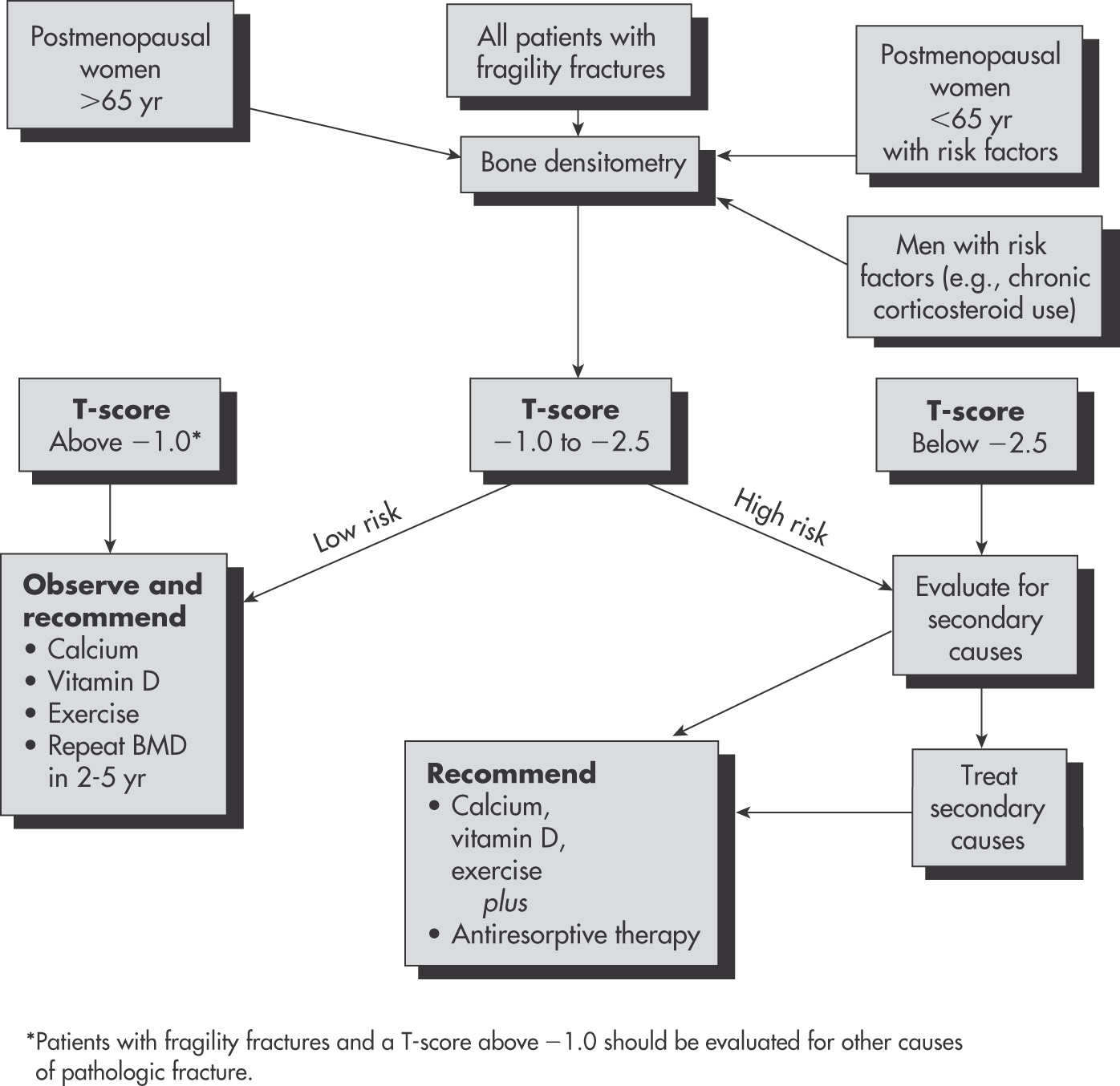
  • •Recommendations as to when to repeat bone density testing should be based on initial T scores (Fig. 4). Data from the Study of Osteoporotic Fractures indicates that in women with normal bone density or mild low bone mass, repeat testing might not be necessary for another 10 to 15 yr. For women with moderate low bone mass, a screening interval of 3 to 5 yr may be appropriate. For women with advanced bone loss/osteoporosis, testing every 1 to 2 yr is recommended.

BOX 5 Investigations for Secondary Osteoporosis in Older People with Low-Trauma Fractures or Low Bone Mineral Density

  • •CBC
  • •ESR or CRP
  • •Biochemical profile: Including renal function, adjusted serum calcium, and alkaline phosphatase
  • •Thyroid function tests
  • •Serum testosterone, sex hormone-binding globulin, LH, FSH (men)
  • •Serum and urine electrophoresis (vertebral fractures)
  • •Serum 25OHD and PTH

From Fillit HM: Brocklehurst’s textbook of geriatric medicine and gerontology, ed 8, Philadelphia, 2017, Elsevier.

TABLE 2 Medications Associated With Osteoporosis

SystemMedication
Endocrine
  • Aromatase inhibitors (e.g., anastrozole)
  • Excess thyroxine replacement
  • Glucocorticoids
  • Gonadotropin-releasing hormone agonists
  • Ovarian-suppressing drugs (e.g., medroxyprogesterone acetate)
  • Thiazolidinediones
  • SGLT2 inhibitors
GastrointestinalProton pump inhibitors
Hematologic
  • Heparin
  • Warfarin
Infectious diseaseAntiretroviral therapy
Immunosuppressant
  • Cyclosporine
  • Cytotoxic drugs
  • Tacrolimus
NeurologicAnticonvulsants-phenytoin, phenobarbital, carbamazepine
PsychiatricSelective serotonin reuptake inhibitors
RenalLoop diuretics (e.g., furosemide)

From Hochberg MC et al: Rheumatology, ed 8, Philadelphia, 2023, Elsevier.

TABLE 3 Clinical Indications for Bone Densitometry

  • All postmenopausal women <65 yr who have one or more additional risk factors for osteoporosis (besides menopause)
  • All women >65 yr regardless of additional risk factors
  • To document reduced bone density in patients with vertebral abnormalities or low bone mass on radiographs
  • Estrogen-deficient women at risk for low bone density who are considering use of estrogen or an alternative therapy, if bone density would influence the decision
  • Women who have been receiving estrogen replacement therapy for prolonged periods or to monitor the efficacy of a therapeutic intervention or interventions for osteoporosis
  • To diagnose low bone mass in people treated with glucocorticoids
  • To document low bone density in people with asymptomatic primary or secondary hyperparathyroidism

From Firestein GS et al: Firestein & Kelley’s textbook of rheumatology, ed 12, Philadelphia, 2024, Elsevier.

TABLE 4 Causes of Erroneous Bone Mineral Density Measures by DEXA in the Lumbar Spine

Overestimation of Bone Mineral Density
Extraneous calcification (lymph nodes, aorta)
Degenerative disk and spine disease (osteophytes)
Ankylosing spondylitis
Vertebral fracture
Sclerotic metastases
Vertebral hemangioma
Overlying metal artifacts (navel rings)
Surgical interventions (metallic rods, spinal fusion)
Vertebroplasty
Paget disease
Treatment with strontium ranelate
Underestimation of Bone Mineral Density
Laminectomy

DEXA, Dual-energy x-ray absorptiometry.

From Pope TL et al: Musculoskeletal imaging, ed 2, Philadelphia, 2015, Saunders.

Figure E2 (A) Posteroanterior Lumbar Spine Study Performed with a Ge Lunar Prodigy Dual-Energy x-Ray Absorptiometry (DEXA) Device on a 69-Year-Old Woman

Bone Mineral Density (BMD) and Standard Scores are Given for Each Vertebra, as Well as for Every Possible Combination of Contiguous Vertebrae. L1 to L4 BMD is Plotted on the Age-Regression Graph. (B) Left Proximal Femoral Study Performed with a Ge Lunar Prodigy Device on the Same Patient.
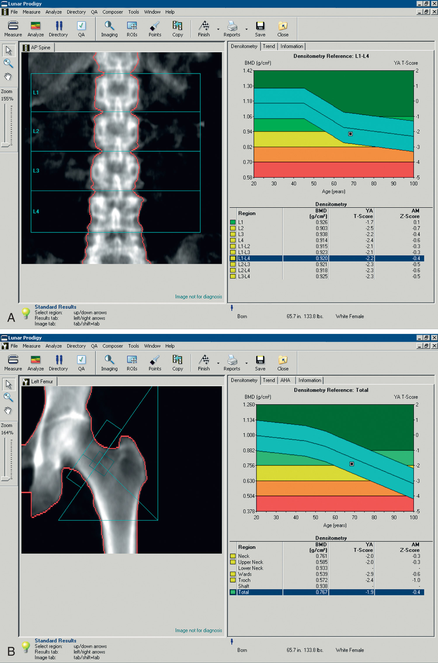
(From Hochberg MC et al: Rheumatology, ed 8, Philadelphia, 2023, Elsevier.)

TABLE 5 Characteristics of Two Widely Used Fracture Risk Calculators

FRAXGarvan
Clinical risk factors
  • Age (40-90 yr)
  • Sex
  • Weight
  • Height
  • Previous fracture in adulthood (yes or no)
  • Parent fractured hip
  • Current smoking
  • Glucocorticoid use
  • Rheumatoid arthritis
  • Secondary osteoporosis
  • Alcohol
  • Bone mineral density (optional)
  • Country ± ethnicity
  • Age (50-96 yr)
  • Sex
  • Fractures since age of 50 yr (up to three)
  • Falls over last 12 mo (up to three)
  • Bone mineral density or weight
Competing risk of deathAllowed forNot allowed for
Fracture risk outputs10-yr risk of hip fracture or of a major osteoporotic fracture (i.e., hip, clinical spine, humerus, or forearm)5- or 10-yr risk of hip or any fragility fracture

From Robertson RP et al: DeGroot’s endocrinology, basic science and clinical practice, ed 8, Philadelphia, 2023, Elsevier.

Figure E3 FRAX questionnaire.
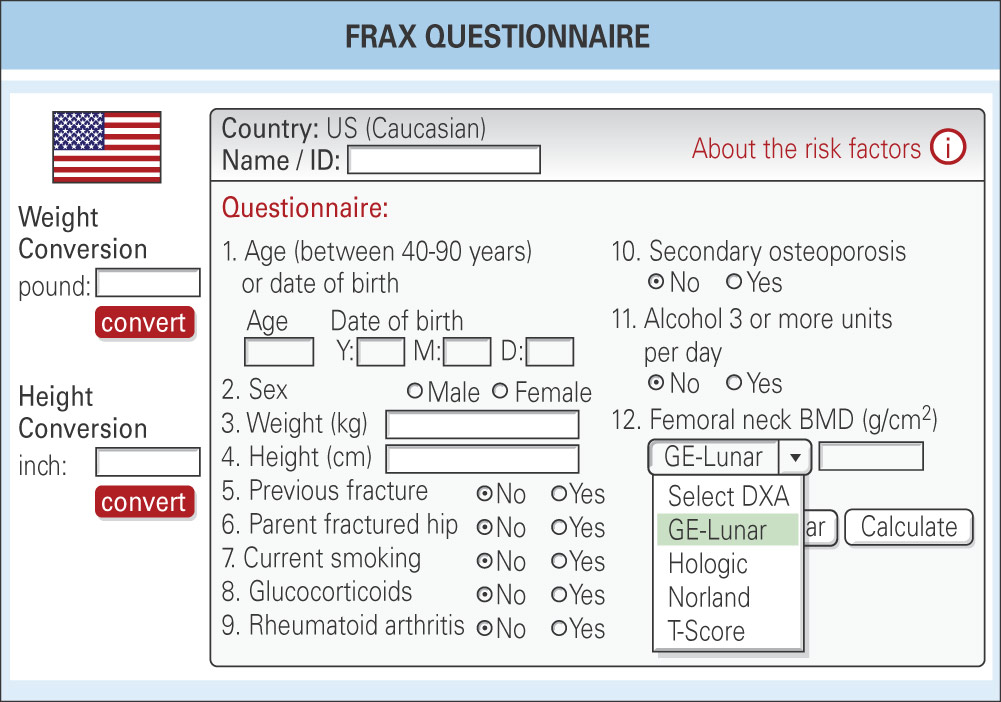
(From Hochberg MC et al: Rheumatology, ed 8, Philadelphia, 2023, Elsevier.)

Figure 4 Diagnosis and management of osteoporosis.

!!flowchart!!

Laboratory Tests (Box 6

  • •Comprehensive metabolic panel
  • •Complete blood count
  • •Thyroid-stimulating hormone
  • •24-h urinary calcium levels and 26-hydroxy-vitamin D level may be helpful in evaluating for secondary causes of osteoporosis
  • •Consider celiac panel and serum protein electrophoresis
  • •Biochemical markers of bone remodeling may be useful to predict rate of bone loss and/or follow therapy response. Specific biochemical markers (listed below) are followed to document response to therapy
    1. 1.High-turnover osteoporosis: High levels of resorption markers (lysyl pyridinoline, deoxy lysyl pyridinoline, n-telopeptide of collagen cross-links, C-telopeptide of collagen cross-links) and formation markers (osteocalcin and bone-specific alkaline phosphatase); indicates accelerated bone loss responding best to antiresorptive therapy
    2. 2.Low or normal-turnover osteoporosis: Normal or low levels of the markers of resorption and formation (see "high-turnover osteoporosis" listed previously); no accelerated bone loss; responds best to drugs that enhance bone formation

BOX 6 Recommended Laboratory Investigations for Individuals With Osteoporosis

Recommended Screening in All Patients

  • •Serum calcium, albumin, phosphorus*,†,‡,§
  • •Serum creatinine*,†,‡,§
  • •Liver function tests*
  • •Bicarbonate*
  • •Complete blood count*
  • •24-h urinary calcium level*,†,‡,§
  • •25-hydroxyvitamin D level*,†,‡,§
  • •Thyroid-stimulating hormone level*,†,‡,§
Other Testing, if Appropriate

  • •Biochemical markers of bone turnover*,†,‡,§
  • •Cortisol levels*,‡
  • •Protein electrophoresis*,†,§
  • •Parathyroid hormone level*,§

From Hochberg MC: Rheumatology, ed 7, Philadelphia, 2019, Elsevier.

Imaging Studies

  • •Bone mineral density (BMD) determination (see "Workup") should be performed on all women with determined risk factors and/or associated secondary causes. Criteria for the diagnosis of osteoporosis based on measurement of bone density and T score are summarized in Table 6.
    1. 1.Normal: BMD <1 standard deviation (SD) below the young adult reference mean
    2. 2.Low bone mass: BMD 1 to 2.5 SD below the young adult reference mean
    3. 3.Osteoporosis: BMD >2.5 SD below the young adult reference mean
  • •For patients undergoing treatment: The frequency of BMD monitoring is controversial, and many experts recommend that clinicians should not monitor BMD during the initial 5-yr drug treatment period because no studies have proven that such monitoring improves fracture outcomes.
    1. 1.X-ray exam of appropriate part of skeleton (Figs. E5 and E6) is indicated to evaluate clinical osteoporotic fracture only.

TABLE 6 Diagnostic Categories for Osteoporosis Based on World Health Organization Criteria

CategoryDefinition
NormalBMD not more than 1 SD below the young adult mean value
Low bone mass (osteopenia)BMD lying between 1 and 2.5 SD below the young adult mean value
OsteoporosisBMD more than 2.5 SD below the young adult mean value

BMD, Bone mineral density; SD, standard deviation.

From World Health Organization data, 1994. Hochberg MC et al: Rheumatology, ed 8, Philadelphia, 2023, Elsevier.

Figure E5 Regional osteoporosis.
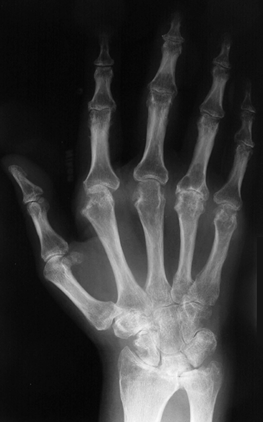
Hand radiograph in early rheumatoid arthritis (RA) shows periarticular low bone mass at the metacarpophalangeal and interphalangeal joints, with joint space narrowing and juxtaarticular erosions. The periarticular low bone mass is the earliest radiographic feature of RA and is related to hyperemia, synovial inflammation, and local cytokines that stimulate osteoclastic bone resorption.

(From Pope TL et al: Musculoskeletal imaging, ed 2, Philadelphia, 2015, Saunders.)

Figure E6 General osteoporosis.
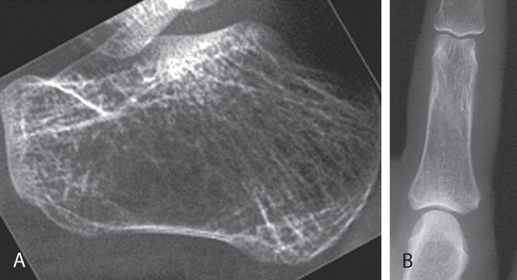
Radiographic features include reduced radiographic density (low bone mass) with reduction in the number of trabeculae, which may be destroyed completely, and the bone cortex becomes thinned as evident in the lateral radiograph of the calcaneus (A) and radiograph of the phalanx (B). When these features are present, bone densitometry using dual-energy x-ray absorptiometry (DEXA) should be suggested.

(From Pope TL et al: Musculoskeletal imaging, ed 2, Philadelphia, 2015, Saunders.)

* American Association of Clinical Endocrinologists guidelines.

† National Osteoporosis Foundation Physician’s Guide.

‡ U.S. Surgeon General’s report.

§ American College of Obstetricians and Gynecologists.

Treatment ⬆ ⬇

When to treat:

Nonpharmacologic Therapy

Prevention:

  • •Identification and minimization of risk factors
  • •Appropriate diagnosis and treatment of secondary causes
  • •Behavioral modification: Proper nutrition, physical activity (specifically weight-bearing activity), fracture prevention strategies, fall risk reduction
Acute General Rx

  • •Vitamin D supplement: 600 IU/day for persons 19 to 70 yr of age and 800 IU/day for persons 71 yr and older. Recommended supplementation of calcium and vitamin D is summarized in Table 6.
  • •Calcium supplement: The recommended dietary intake of calcium for women 19 to 50 yr of age and men 19 to 70 yr of age is 1000 mg/day; women older than age 50 and men older than age 70 require 1200 mg/day. Calcium intake above 2500 mg/day (2000 mg/day in persons >50 yr of age) should be avoided. Consumption of calcium-rich foods and beverages is the preferred approach to ensuring adequate calcium intake.3
  • •Bisphosphonates have been used in osteoporosis and remain the most widely used agents, being joined more recently by denosumab (Table 7). Oral bisphosphonates (alendronate, risedronate): Decrease bone resorption by attenuating osteoclast activity. They are first-line therapy for the treatment of most patients with osteoporosis, with proven efficacy to reduce fracture risk. The bisphosphonates differ in binding affinity, dose frequency, and route of administration. To facilitate absorption, most oral bisphosphonates are taken on an empty stomach with a full glass of water. Patients are instructed to remain in a sitting or standing position for 30 to 60 min. Contraindications include esophageal disorders, inability to remain standing for 30 to 60 min after taking the medication, chronic kidney disease, and Roux-en-Y gastric bypass procedures. Adverse side effects include gastroesophageal reflux disease, esophagitis, transient hypocalcemia, musculoskeletal pain, renal impairment, ocular side effects, jaw necrosis, and atypical femur fracture.
  • •Biologic agents: Denosumab is a human monoclonal antibody that inhibits osteoclast formation and prevents resorption for treatment of postmenopausal osteoporosis. It is used as a second-line pharmacologic treatment to reduce the risk of fractures in post-menopausal females diagnosed with primary osteoporosis who have contraindications to or experience adverse effects of biphosphonates.3 Dosage is 60 mg subcutaneously every 6 mo. Romosozumab is a monoclonal antibody that increases bone formation and decreases bone resorption by binding to sclerostin. It may be considered for patients who are at high risk for fracture and cannot tolerate any other osteoporotic therapies. Dosage is two consecutive subcutaneous injections (105 mg each) for a total dose of 210 mg once monthly. It should not be used in patients who have had a myocardial infarction or stroke in the past year.
  • •Zoledronic acid: A bisphosphonate given by IV infusion over at least 15 min, 5 mg once per year may be used in patients who are unable to tolerate oral bisphosphonates. It is contraindicated in patients with acute renal failure.
  • •Teriparatide is a recombinant human parathyroid hormone used for postmenopausal women with osteoporosis who are at high risk for fracture, especially vertebral fractures. It is also used in men with primary or hypogonadal osteoporosis who are at high risk of fracture. It is administered by injection 20 mcg daily, subcutaneously into the thigh or abdominal wall. Use for more than 2 yr is not recommended. It stimulates bone formation and reduces the risk of fracture but may increase the risk of stroke in older women with osteoporosis. Common side effects include headaches, myalgia, hypercalcemia, and hypercalciuria. Trials involving abaloparatide, a selective activator of the parathyroid hormone type 1 receptor, have also shown reduced risk of new vertebral and nonvertebral fractures in postmenopausal women with osteoporosis. Use of teriparatide is followed by a bisphosphonate upon its discontinuation in females with primary osteoporosis and a very high risk of fractures.4
  • •Estrogen prescription drugs or raloxifene should not be prescribed to treat women with osteoporosis.

TABLE 7 Principal Medications for Management of Osteoporosis

MedicationDoseIndicationsDosing InstructionsComments
Alendronate
  • 70 mg/wk PO
  • 10 mg/d PO
Treatment of postmenopausal, male and glucocorticoid osteoporosisTake fasting with a glass of water while sitting or standing. Remain upright and fasting for 30 min
  • 35 mg/wk PO
  • 5 mg/d PO
Prevention of postmenopausal osteoporosis
Risedronate5 mg/d, 35 mg/wk, 75 mg ×2 per mo, 150 mg/mo, all POTreatment or prevention of postmenopausal osteoporosis; treatment of male and glucocorticoid osteoporosis
Ibandronate150 mg/mo POTreatment or prevention of postmenopausal osteoporosisAs above, but remain upright and fasting for 60 min
3 mg/3 mo IVTreatment of postmenopausal osteoporosisInject over 15-30 sec
Zoledronate or zoledronic acid5 mg/yr IVTreatment of postmenopausal, male, and glucocorticoid osteoporosis, fracture prevention after hip fractureIV infusion in 100 ml over at least 15 min
5 mg every 2 yrPrevention of postmenopausal osteoporosis
Raloxifene60 mg/d POPrevention and treatment of postmenopausal osteoporosis
Denosumab60 mg every 6 mo SCTreatment of postmenopausal, male, and glucocorticoid osteoporosis patients at high fracture risk
Teriparatide20 μg/d SC for 18-24 moTreatment of postmenopausal, male, and glucocorticoid osteoporosis patients at high fracture riskFollow with antiresorptive agentFDA recommends considering >2 yr of cumulative use during a patient’s lifetime only if fracture risk remains high
Abaloparatide80 μg/d SC for 18-24 moTreatment of postmenopausal osteoporosis patients at high fracture risk
  • Inject into the periumbilical region. Administer initial doses where the patient can lie down if orthostatic hypotension occurs
  • Follow with antiresorptive agent
  • See above
  • Declined registration in Europe
Romosozumab210 mg/mo SC for 12 moTreatment of postmenopausal osteoporosis at high fracture risk or patients who have failed or are intolerant to other available osteoporosis therapyFollow with antiresorptive agent

FDA, U.S. Food and Drug Administration; IV, intravenously; PO, by mouth; SC, subcutaneously.

From Robertson RP et al: DeGroot’s endocrinology, basic science and clinical practice, ed 8, Philadelphia, 2023, Elsevier.

Chronic Rx

  • •Lifelong attention to behavior modification (nutrition, physical activity, fracture prevention strategies) and compliance with pharmacologic intervention. Recommendations include weight-bearing and muscle-strengthening exercises, smoking cessation, reduced alcohol intake, and adoption of fall prevention strategies.
  • •There is little evidence to guide physicians about long-term bisphosphonate therapy. The decision to continue drug therapy beyond 5 yr should reflect reassessment of risk and benefit. The risks of atypical fracture of the femur and osteonecrosis of the jaw (ONJ)4 increase after 5 yr of bisphosphonate use. It is reasonable to consider a drug holiday in postmenopausal women who are not at high fracture risk after 3 yr (IV) to 5 yr (oral) of bisphosphonate therapy. Continued treatment may be advisable in those at highest risk.
  • •Continuing need to eliminate high-risk factors when possible and to optimally manage secondary causes of osteoporosis.
Disposition

Goals for diagnosis and treatment include identification of women at risk; initiation of lifelong preventive measures for all women; institution of treatment modalities that will result in a decrease in fracture risk; and reduction of morbidity, mortality, and unnecessary institutionalization, thereby improving quality of independent life and productivity. Table 8 summarizes the effect of major treatment options on the risk of vertebral, nonvertebral, and hip fractures.

TABLE 8 Effect of Major Treatment Options on the Risk of Vertebral, Nonvertebral, and Hip Fractures

Vertebral FracturesNonvertebral FracturesHip Fractures
AlendronateAAA
EtidronateANDND
RisedronateAAA
RaloxifeneANDND
Strontium ranelateAA(A)
TeriparatideAAND
DenosumabAAA
Zoledronate*AAA
Ibandronate*A(A)ND
Calcium and vitamin D*NDAA

* A indicates evidence from randomized, controlled trials and/or meta-analysis; (A) reflects that a beneficial effect on fracture risk was found only in post hoc subgroup analysis; ND indicates that fracture reduction has not been demonstrated.

From Fillit HM: Brocklehurst’s textbook of geriatric medicine and gerontology, ed 8, Philadelphia, 2017, Elsevier.

Reference(s) ⬆

Available at Ebooks.health.elsevier.com.

  1. Screening for osteoporosis to prevent fractures: U.S. Preventive Services Task Force recommendation statementJAMA. 333(6):498-508, 2025.
  2. Robertson RP : DeGroot’s endocrinology, basic science and clinical practice. ed 8Elsevier-Philadelphia, 2023.
  3. Bauer DC : Calcium supplements and fracture preventionN Engl J Med. 369:1537-1543, 2013.
  4. Ayers C : Effectiveness and safety of treatments to prevent fractures in people with low bone mass or primary osteoporosis: a living systematic review and network meta-analysis for the American College of PhysiciansAnn Intern Med. 176(2):182-195, 2023.