section name header

Basic Information ⬇

Author: Tara C. Bouton, MD, MPH, TM and Glenn G. Fort, MD, MPH

Definition

Pulmonary tuberculosis (TB) is an infection of the lung and occasionally surrounding structures, caused by the bacterium Mycobacterium tuberculosis (Mtb). Two states of M. tuberculosis infection are recognized: Latent tuberculosis infection (LTBI) and acute tuberculosis disease, although infection and immunologic control exist across a spectrum. LTBI is a state of persistent immune response to stimulation by M. tuberculosis antigens without evidence of clinically manifested active TB and with bacillary replication absent or below some undefined threshold as a result of immunologic control. Most persons with LTBI never become sick with TB; however, 5% to 15% have progression to tuberculosis disease.1 Multidrug-resistant (MDR) TB is defined as disease caused by strains of Mtb that are at least resistant to treatment with isoniazid (INH) and rifampin (RIF) (two of the most effective first-line drugs); extensively drug-resistant (XDR) TB refers to disease caused by MDR strains that are also resistant to treatment with any fluoroquinolone and bedaquiline or linezolid.

Synonym

  • TB
ICD-10CM CODES
A15.0Tuberculosis of lung
A15.7Primary respiratory tuberculosis
Epidemiology & Demographics
Incidence (Worldwide):

  • •One fourth of the world’s population is infected with TB, and it is one of the world’s leading infectious disease killers.
  • •In 2017, there were an estimated 10 million new cases of active TB; 9% involved coinfection with HIV; 1.3 million deaths from TB, including 300,000 among HIV-infected patients; 457,560 incident cases of multidrug-resistant TB worldwide.
Incidence (In U.S.):

  • •In 2021, a total of 7882 new TB cases were reported in the U.S. The U.S. TB incidence in 2021 was 2.4/100,000 persons.
  • •The rate among non-U.S.-born persons was >14 times that in the U.S.-born persons.
  • •Since 1993, TB case counts and rates have declined in the U.S. As the number of cases decreases overall, an increasing percentage of cases occurs among non-U.S.-born persons.
  • •More than 90% of new cases each yr are from reactivated prior infections, and 9% represent new infections.
  • •Only 10% of patients with purified protein derivative (PPD) conversions will develop TB, most within 1 to 2 yr, though this is higher in HIV-positive patients (8%/yr).
  • •In 2021 in the U.S., 1.3% of cases were MDR TB, which is a decline from 8.2% in 2008.
Prevalence (In U.S.):

  • •Between 3.1% and 5.0% of the U.S. population is estimated to be infected with latent TB infection (LTBI)
  • •Varies widely among population groups
Predominant Sex:

Globally, in 2017, 6 million men contracted TB compared to 3.2 million women

Predominant Age:

Ages 24 to 45

Peak Incidence:

  • •Infancy
  • •Teenage years
  • •Pregnancy
  • •Elderly
  • •HIV-positive patients at highest risk regardless of age. Table E1 compares clinical manifestations of active TB in early versus late HIV infection

TABLE E1 Clinical Manifestations of Active Tuberculosis in Early Versus Late HIV Infectiona

Early HIV InfectionLate HIV Infection
Tuberculin test resultUsually positiveUsually negative
AdenopathyCommonUnusual
Pulmonary distributionUpper lobeLower and middle lobe
CavitationOften presentTypically absent
Extrapulmonary disease10%-15% of cases50% of cases

HIV, Human immunodeficiency virus.

a For practical purposes, "early" and "late" may be defined as CD4+ cell counts greater than 300 cells/mm3 and less than 200 cells/mm3, respectively.

From Bennett JE et al: Mandell, Douglas, and Bennett’s principles and practice of infectious diseases, ed 9, Philadelphia, 2020, Elsevier.

Physical Findings & Clinical Presentation

  • •See "Etiology."
  • •Primary pulmonary TB infection generally asymptomatic
  • •Reactivation pulmonary TB:
    1. 1.Fever
    2. 2.Night sweats
    3. 3.Cough
    4. 4.Hemoptysis
    5. 5.Scanty nonpurulent sputum
    6. 6.Weight loss
    7. 7.Chest pain
  • •Progressive primary pulmonary TB disease: Same as reactivation pulmonary TB
  • •TB pleurisy:
    1. 1.Pleuritic chest pain
    2. 2.Fever
    3. 3.Shortness of breath
  • •Rare massive, suffocating, fatal hemoptysis secondary to erosion of pulmonary artery within a cavity (Rasmussen aneurysm)
  • •Chest examination:
    1. 1.Not specific
    2. 2.Usually underestimates extent of disease
    3. 3.Rales accentuated after a cough (posttussive rales)
Etiology

  • •Mtb, a slow-growing, aerobic, non-spore-forming, nonmotile bacillus, with a lipid-rich cell wall:
    1. 1.Lacks pigment
    2. 2.Produces niacin
    3. 3.Reduces nitrate
    4. 4.Produces heat-labile catalase
  • •Mtb staining: Acid-fast and acid-alcohol fast by Ziehl-Neelsen method, appearing as red, slightly bent, beaded rods 2 to 4 microns long (acid-fast bacilli [AFB]), against a blue background
  • •Polymerase chain reaction (PCR) to detect <10 organisms/ml in sputum (compared with the requisite 10,000 organisms/ml for AFB smear detection)
  • •Culture:
    1. 1.Growth on solid media (Löwenstein-Jensen; Middlebrook 7H11) in 2 to 6 wk
    2. 2.Growth in liquid media (BACTEC, using a radioactive carbon source for early growth detection) often in 9 to 16 days
    3. 3.Enhanced in a 5% to 10% carbon dioxide atmosphere
  • •Genome sequencing:
    1. 1.Facilitates immediate identification of Mtb strains and resistance in early growing cultures
    2. 2.False negatives possible if growth suboptimal
  • •Humans are the only reservoir for Mtb
  • •Transmission:
    1. 1.Aerosolized droplets containing AFB generated through cough, speaking, singing, bronchoscopy, or autopsy are inhaled directly into alveoli
    2. 2.Facilitated by close exposure to high-velocity cough (unprotected by proper mask or respirators) from patients with AFB-positive sputum and cavitary lesions
    3. 3.Locations of increased TB transmission risk include hospitals, homeless shelters, correctional facilities, nursing homes, and residential homes for those with HIV
  • •Pathogenesis:
    1. 1.Mtb are ingested by macrophages in alveoli, then transported to regional lymph nodes, where spread is contained
    2. 2.Some Mtb may reach bloodstream and disseminate widely
    3. 3.Primary TB (often asymptomatic, minimal pneumonitis in lower or midlung fields, with hilar lymphadenopathy) is essentially an intracellular infection, with multiplication of organisms continuing 2 to 12 wk after primary exposure, until cell-mediated hypersensitivity (detected by positive skin test reaction to tuberculin PPD) matures, and results in subsequent containment of infection
    4. 4.Local and disseminated Mtb are thus contained by the following T-cell-mediated immune responses:
      1. a.Recruitment of monocytes
      2. b.Transformation of lymphocytes with secretion of lymphokines
      3. c.Activation of macrophages and histiocytes
      4. d.Organization into granulomas, where Mtb may survive within macrophages (Langhans giant cells), but within which multiplication essentially ceases (95%) and from which spread is prohibited
    5. 5.Progressive primary pulmonary disease:
      1. a.May immediately follow the asymptomatic phase
      2. b.Necrotizing pulmonary infiltrates
      3. c.Tuberculous bronchopneumonia
      4. d.Endobronchial TB
      5. e.Interstitial TB
      6. f.Widespread miliary lung lesions
    6. 6.Postprimary TB pleurisy with pleural effusion:
      1. a.Develops after early primary infection, although often before conversion to positive PPD
      2. b.Results from pleural seeding from a peripheral lung lesion or rupture of lymph node into pleural space
      3. c.May produce a large (sometimes hemorrhagic) exudative effusion (with polymorphonuclear cells early, rapidly replaced by lymphocytes), frequently without pulmonary infiltrates
      4. d.Generally resolves without treatment
      5. e.Portends a high risk of subsequent clinical disease and therefore must be diagnosed and treated early (pleural biopsy and culture) to prevent future catastrophic TB illness
      6. f.May result in disseminated extrapulmonary infection
    7. 7.Reactivation pulmonary TB:
      1. a.Occurs months to decades after primary TB
      2. b.Preferentially involves the apical posterior segments of the upper lobes and superior segments of the lower lobes
      3. c.Associated with necrosis and cavitation of involved lung, hemoptysis, chronic fever, night sweats, weight loss
      4. d.Spread within lung occurs via cough and inhalation
    8. 8.Reinfection TB:
      1. a.May mimic reactivation TB
      2. b.Ruptured caseous foci and cavities, which may produce endobronchial spread
    9. 9.Mtb in both progressive primary and reactivation pulmonary TB:
      1. a.Intracellular (macrophage) lesions (undergoing slow multiplication)
      2. b.Closed caseous lesions (undergoing slow multiplication)
      3. c.Extracellular, open cavities (undergoing rapid multiplication)
      4. d.INH and RIF are bactericidal in all three sites
      5. e.Pyrazinamide (PZA) especially active within acidic macrophage environment
      6. f.Extrapulmonary reactivation disease also possible
    10. 10.Rapid local progression and dissemination in infants with devastating illness before PPD conversion occurs
    11. 11.Most symptoms (fever, weight loss, anorexia) and tissue destruction (caseous necrosis) from cytokines and cell-mediated immune responses
    12. 12.Mtb has no important endotoxins or exotoxins
    13. 13.Granuloma formation related to tumor necrosis factor (TNF) secreted by activated macrophages

Diagnosis ⬆ ⬇

Differential Diagnosis

  • •Necrotizing pneumonia (anaerobic, gram-negative)
  • •Histoplasmosis
  • •Coccidioidomycosis
  • •Melioidosis
  • •Interstitial lung diseases (rarely)
  • •Cancer
  • •Sarcoidosis
  • •Silicosis
  • •Rare pneumonias:
    1. 1.Rhodococcus equi (cavitation)
    2. 2.Bacillus cereus (50% hemoptysis)
    3. 3.Eikenella corrodens (cavitation)
Workup

  • •Sputum for AFB stains and Mtb culture.
  • •The Xpert MTB/RIF: An automated molecular test for Mtb and resistance to RIF that provides sensitive detection of TB and RIF resistance directly from untreated sputum in less than 2 h with minimal hands-on time.
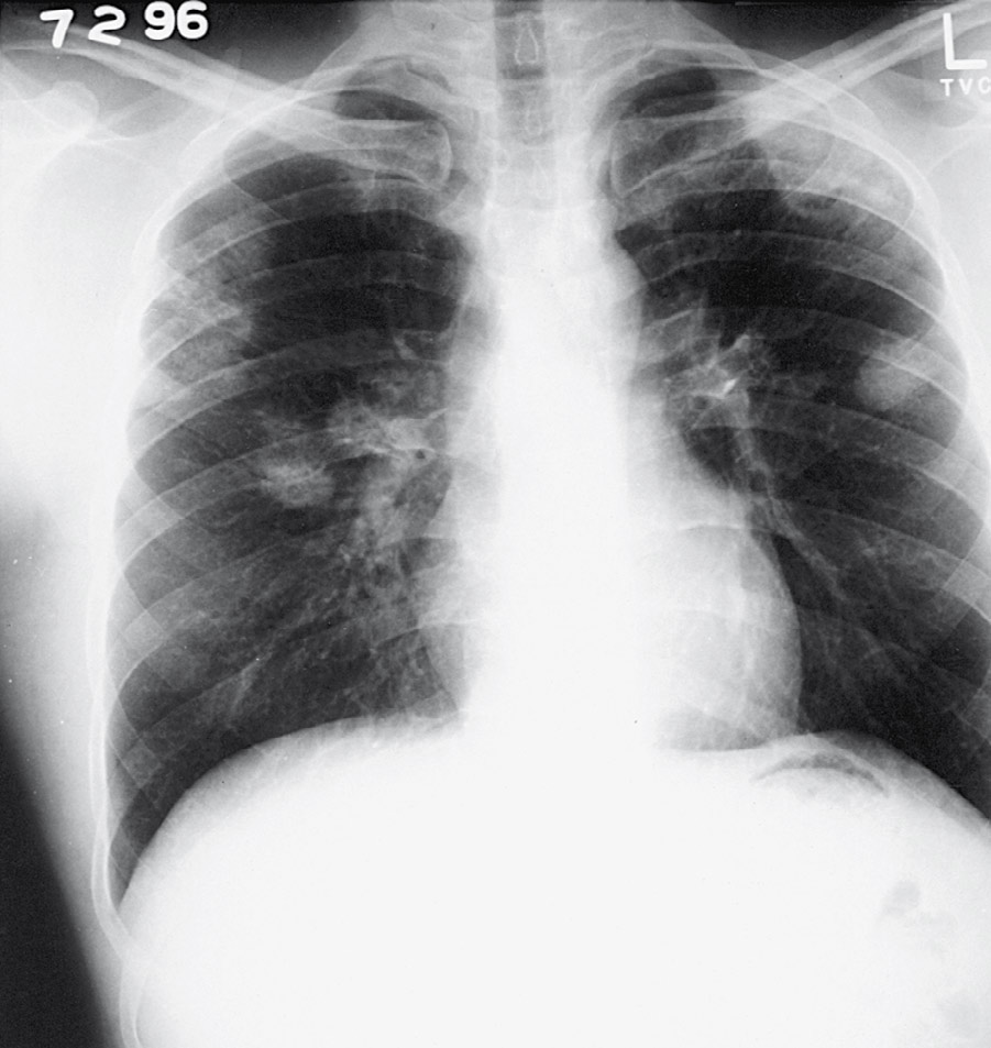
  • •Chest x-ray (Fig. E1).
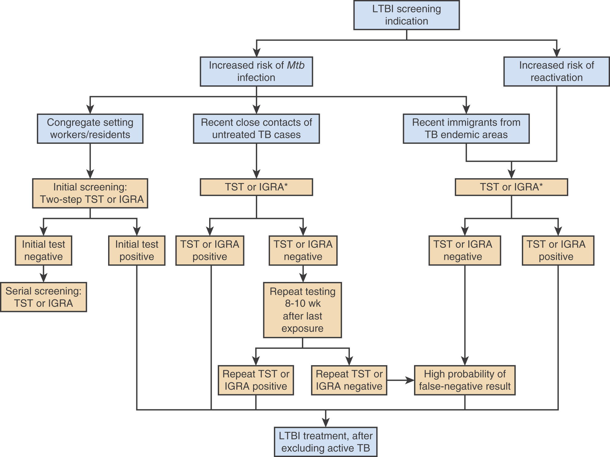
  • •Target groups for LTBI screening are summarized in Table E2. PPD (tuberculin skin test), or interferon gamma release assays (IGRA) can be used for screening of LTBI (Fig. E2):
    1. 1.Recent conversion from negative to positive within 3 mo of exposure is highly suggestive of recent infection.
    2. 2.Negative PPD never rules out acute TB.
    3. 3.Prior positive PPD may become negative after several years and return to positive only after second repeated PPD; repeat second PPD within 1 wk, which thus may mimic skin test conversion.
    4. 4.Positive PPD reaction is determined as follows:
      1. a.Induration 48-72 h after intradermal injection of 0.1 ml of 5 tuberculin PPD.
      2. b.5-mm induration in persons living with HIV (or other severe immunosuppressed state affecting cellular immune function), close contact of active TB, fibrotic chest lesions.
      3. c.10-mm induration if in high-medical risk groups (renal failure, gastrectomy, silicosis, diabetes, <90% ideal body weight), foreign-born where TB rates are high (Southeast Asia, Latin America, Africa, India), living in a congregate setting, people who abuse drugs, TB lab workers, children <5 yrs, children exposed to adults of high-risk.
      4. d.15-mm induration if low risk.
    5. 5.PPD cannot distinguish between TB disease and TB infection.
  • •IGRAs: Diagnostic test for LTBI, known as the QuantiFERON TB Gold test (QFT-G) and T-SPOT.TB Assay. These blood tests measure interferon response to specific Mtb antigens. The test may assist in distinguishing true positive reactions from individuals with latent TB from PPD reactions related to nontuberculous mycobacteria, prior bacillus Calmette-Guérin vaccination, or difficult-to-interpret skin-test results from people with dermatologic conditions or immediate allergic reactions to PPD. IGRAs have a specificity >95% for diagnosis of latent TB. The sensitivity of the T-SPOT assay (90%) appears to be higher than the QFT-G test (80%). It should be noted that IGRA sensitivity is diminished by HIV infection. As with PPD, negative tests do not rule out active TB disease, and positive tests cannot distinguish between TB infection and disease.

Figure E1 Chest Radiograph Demonstrating Multiple Bilateral Pulmonary Tuberculomas in an Asymptomatic 35-Yr-Old Man from Poland

(From Bennett JE et al: Mandell, Douglas, and Bennett’s principles and practice of infectious diseases, ed 9, Philadelphia, 2020, Elsevier.)

TABLE E2 Target Groups for Latent Tuberculosis Infection Screening

Individuals With Increased Risk of Infection
Contacts of individuals with untreated infectious active tuberculosis
Individuals who have immigrated to the United States within the past 5 yr from tuberculosis endemic areas
Individuals who work and/or reside in high-risk congregate settings (e.g., hospitals, homeless shelters, prisons, nursing homes)
Individuals With Conditions Associated With Increased Risk for Reactivation
High Risk for Reactivation (Risk of Reactivation Is at Least Six Times Higher Than for Healthy Individuals)
Human immunodeficiency virus infection
Severe immunosuppression (e.g., individuals receiving medication for solid-organ transplantation, chemotherapy, tumor necrosis factor-α inhibitors)
Certain malignancies (e.g., hematologic malignancies, head and neck cancers)
Silicosis
End-stage renal disease
Radiographic evidence of prior granulomatous disease (e.g., fibrotic lesions on chest imaging)
Children <5 yr with a positive tuberculin skin test reaction
Moderate Risk for Reactivation (Risk of Reactivation Is Less Than Six Times Higher Than for Healthy Individuals)
Corticosteroid use (≥15 mg daily for ≥1 mo)
Diabetes mellitus
Underweight or malnourished individuals (includes malabsorptive conditions, such as gastrectomy, jejunoileal bypass surgeries)
Substance abuse (e.g., smoking, alcohol abuse, injection drug use)
Radiographic evidence of solitary or small granulomas

From Broaddus VC et al: Murray & Nadel’s textbook of respiratory medicine, ed 7, Philadelphia, 2022, Elsevier.

Figure E2 Latent tuberculosis infection screening algorithm.

!!flowchart!!

Individuals at high likelihood of having LTBI, due to demographic or specific exposure history or an untreated previously positive TST or IGRA, and a high probability of a presently false-negative result, due to immunodeficiency or immunosuppression, should be considered for treatment. *IGRA preferred for individuals with prior bacillus Calmette-Guérin and individuals at high risk for becoming lost to follow-up. TST preferred for children younger than 5 yr. Dual testing (not shown) may be considered for certain situations. IGRA, Interferon gamma release assay; LTBI, latent tuberculosis infection; Mtb, Mycobacterium tuberculosis; TB, tuberculosis; TST, tuberculin skin test.

(From Broaddus VC et al: Murray & Nadel’s textbook of respiratory medicine, ed 7, Philadelphia, 2022, Elsevier.)

Laboratory Tests (Table E3

  • •Sputum for TB PCR, AFB stains, and mycobacterial culture:
    1. 1.Induced sputum if patient not coughing productively
  • •Sputum from bronchoscopy if high suspicion of TB in patients unable to produce expectorated induced sputum for AFB:
    1. 1.Postbronchoscopy cough specimen may improve yield
    2. 2.Consider lung biopsy if sputum negative, especially if infiltrates are predominantly interstitial
  • •AFB stain-negative sputum may grow Mtb subsequently (especially in HIV).
  • •Gastric aspirates may be helpful, especially in children.
  • •CBC:
    1. 1.Variable values:
      1. a.White blood cells (WBCs): Low, normal, or elevated (including leukemoid reaction: >50,000)
      2. b.Normocytic, normochromic anemia often
  • •Rarely helpful diagnostically.
  • •Erythrocyte sedimentation rate usually elevated.
  • •Thoracentesis:
    1. 1.Exudative effusion:
      1. a.Elevated protein and adenosine deaminase
      2. b.Decreased glucose
      3. c.Elevated WBCs (polymorphonuclear leukocytes early, replaced later by lymphocytes)
      4. d.May be hemorrhagic
    2. 2.Pleural fluid usually AFB-negative
    3. 3.Pleural biopsy often diagnostic-may need to be repeated for diagnosis
    4. 4.Culture pleural biopsy tissue for AFB
  • •Bone marrow biopsy is often diagnostic in difficult-to-diagnose cases, especially miliary TB.

TABLE E3 Comparison of Assays Used in the Diagnosis of Active Tuberculosis

Clinical Laboratory QuestionDiagnostic AssayAdvantagesLimitations
Are mycobacteria present in a clinical specimen?Culture on solid media (Löwenstein-Jensen egg-based or Middlebrook agar-based media)Gold standard for isolating Mycobacterium tuberculosis; detects 10-100 organisms/ml; shows colony morphology, detects mixed infection, allows quantification of growth; provides organisms for speciation, strain identification, susceptibility testingVisible growth takes 3-8 wk
Culture in liquid brothSensitivity and specificity similar to solid media; automated systems decrease workload; provides organisms for speciation, strain identification, and susceptibility testing; turn positive in a mean of 10 days for smear-positive and 20 days for smear-negative specimensDoes not show colony morphology, detect mixed cultures, or quantify growth
Acid-fast stain (Ziehl-Neelsen, Kinyoun, auramine rhodamine)Same-day results; simple technology; inexpensive light microscope; fluorescence microscope required for auramine-rhodamine stainLess sensitive than culture, requiring 10,000 organisms/ml; cannot distinguish M. tuberculosis from other mycobacteria
Nucleic acid amplification (e.g., PCR)Same-day results; sensitivity intermediate between acid-fast stain and culture; identifies organisms as members of M. tuberculosis complexRequires advanced laboratory techniques; cannot distinguish dead from viable organisms; culture still needed for speciation, strain identification, and susceptibility testing
Nucleic acid amplification with GeneXpert MTB/RIF and Xpert MTB/RIF Ultra
  • Sensitivity of Xpert MTB/RIF roughly 100 CFUs/ml, requires 100 min, detects rifampin resistance mutations with sensitivity of 95% and specificity of 98%
  • Xpert MTB/RIF Ultra is more sensitive than Xpert MTB/RIF but with slightly less specificity
Expensive equipment; detects dead bacilli; culture needed for other susceptibility testing
Urinary antigen detection (e.g., ELISA for LAM)Point-of-care testing; relatively high sensitivity in advanced untreated AIDS (<50 CD4 T cells/mm3)Sensitivity very low in situations other than advanced AIDS
Is a mycobacterium isolated from a clinical specimen a member of M. tuberculosis complex? (M. tuberculosis, Mycobacterium bovis, M. bovis-BCG, Mycobacterium africanum, Mycobacterium microti, or Mycobacterium canettii )Nucleic acid amplification(See above)(See above)
Nucleic acid probesResults available in 2 h; sensitivity and specificity approach 100%; does not require amplificationRequires at least 105 organisms; most useful for pure culture, not directly on clinical specimen; cannot distinguish among members of M. tuberculosis complex
BACTEC p-nitroacetyl-aminohydroxypropiophenone assay (NAP)Provides preliminary identification of M. tuberculosis complex (NAP susceptible)Need for paired cultures increases cost (growth with and without NAP)
High-performance liquid chromatography (HPLC)Same-day results; sensitivity and specificity approach 100%; can distinguish M. bovis BCG from other members of M. tuberculosis complexRequires HPLC technology; only useful with pure culture
To which species of M. tuberculosis complex does a clinical isolate belong?Colony morphology and biochemical assays (e.g., niacin test, heat-sensitive catalase, nitrate reduction, pyrazinamide monodrug resistance)Classic approach for speciation of M. tuberculosisTime-consuming and labor-intensive
PCR genomic analysisMay rapidly distinguish among M. tuberculosis complex speciesNot yet commercially available for this purpose
Do different M. tuberculosis isolates represent the same strain?Genotyping by restriction fragment length polymorphism analysis, spoligotyping, mycobacterial interspersed repetitive unit analysis, and whole-genome sequencingThe CDC offers free strain typing through the National Tuberculosis Genotyping and Surveillance NetworkSophisticated assay available only at specialized centers
Is an M. tuberculosis isolate drug resistant?Agar proportion methodQuantifies the proportion of organisms resistant to a drugRequires as long as 8 wk
Liquid BACTEC methodResults within 5-14 daysDoes not quantify proportion of resistance
Molecular line probe assays and whole-genome sequencing for chromosomal mutations associated with drug resistance (see above for GeneXpert MTB/RIF)Allow same-day determination of drug resistance; line probe assay can be performed directly on smear positive sputum samples or culture isolates; can detect resistance to isoniazid, rifampin, quinolones, and second line injectablesNeed to be validated in multiple clinical settings and not yet FDA approved

AIDS, Acquired immunodeficiency syndrome; BCG, bacillus Calmette-Guérin; CDC, Centers for Disease Control and Prevention; CFU, colony-forming unit; ELISA, enzyme-linked immunosorbent assay; FDA, US Food and Drug Administration; LAM, lipopolysaccharide lipoarabinomannan; PCR, polymerase chain reaction.

From Bennett JE et al: Mandell, Douglas, and Bennett’s principles and practice of infectious diseases, ed 9, Philadelphia, 2020, Elsevier.

Imaging Studies

  • •Chest x-ray examination:
    1. 1.Primary infection reflected by calcified peripheral lung nodule with calcified hilar lymph node
    2. 2.Reactivation pulmonary TB:
      1. a.Necrosis
      2. b.Cavitation (especially on apical lordotic views)
      3. c.Fibrosis and hilar retraction
      4. d.Bronchopneumonia
      5. e.Interstitial infiltrates
      6. f.Miliary pattern
      7. g.Many of the previous findings may also accompany progressive primary TB
    3. 3.TB pleurisy:
      1. a.Pleural effusion, often rapidly accumulating and massive
    4. 4.TB activity not established by single chest x-ray examination
    5. 5.Serial chest x-ray examinations are excellent indicators of progression or regression

Treatment ⬆ ⬇

Nonpharmacologic Therapy

  • •High-calorie, high-protein diet to reverse malnutrition and enhance immune response to TB
  • •Isolation in negative-pressure rooms with high-volume air replacement and circulation, with health care provider wearing proper protective 0.5- to 1-micron filter respirators, until three consecutive sputum AFB smears are negative during workup (taken in 8- to 24-h intervals, with at least one being an early morning specimen) or a diagnosed patient has been on standard multidrug treatment for at least 2 wk and is clinically improving
Acute General Rx

  • •Generally, treatment of active TB consists of two phases: An intensive phase, followed by a continuation phase
  • •Compliance (rigid adherence to treatment regimen) chief determinant of success:
    1. 1.Supervised directly observed therapy (DOT) recommended for all patients and mandatory for patients with high likelihood of loss to follow-up
  • •Adult initial regimen:
    1. 1.INH 5 mg/kg (INH; max 300 mg) PO, RIF 10 mg/kg (RIF; max 600 mg) PO, ethambutol (EMB) by weight increments (EMB: 40 to 55 kg, 800 mg; 56 to 75 kg,1200 mg; 76 to 90 kg, 1600 mg) PO, and PZA by weight increments (PZA: 40 to 55 kg, 1000 mg; 56 to 75 kg, 1500 mg; 76 to 90 kg, 2000 mg) PO dosed daily
    2. 2.Pyridoxine (vitamin B6, 25 mg/day) should be given with INH to prevent peripheral neuropathy
    3. 3.Usually decreased to INH and RIF after 8 wk depending on susceptibilities, with ultimate duration of therapy dependent on HIV treatment status, extent of disease, regimen used, and treatment response, but generally at least 6 mo
    4. 4.Alternative, more complicated DOT regimens
    5. 5.In patients with rifampin-susceptible TB, a strategy involving initial treatment with an 8-wk bedaquiline-linezolid regimen was shown to be noninferior to standard treatment for TB with respect to clinical outcomes. This strategy was associated with a shorter duration of treatment and with no evident safety concerns2
  • •Monitor for clinical toxicity (especially hepatitis). Pharmacology and adverse effects of antituberculosis medications are summarized in Table E4
    1. 1.Patient and physician awareness that anorexia, nausea, right upper quadrant pain, and unexplained malaise require immediate cessation of treatment
    2. 2.Evaluation of liver function testing:
      1. a.Minimal aspartate aminotransferase/alanine transaminase elevations without symptoms generally transient and not clinically significant
  • •Drug resistance (often multiple drug resistance TB [MDRTB]) increased by:
    1. 1.Prior treatment
    2. 2.Acquisition of TB in countries with high TB burden
    3. 3.Known contact with MDR TB
    4. 4.Other factors associated with increased risk: Homelessness, incarceration, AIDS, IV drug use
  • •Other medications used in MDR or XDR TB include levofloxacin, moxifloxacin, cycloserine, aminoglycosides such as amikacin or kanamycin, PAS, clofazimine, bedaquiline, delamanid, pretomanid, linezolid, and ethionamide. A recent trial with the combination of bedaquiline, pretomanid, and linezolid revealed a favorable outcome at 6 mo after the end of therapy in a high percentage of patients with highly drug-resistant forms of TB. Other trials3 have shown that a 6-mo bedaquiline pretomanid-moxifloxacin regimen that included linezolid at a dose of 600 mg daily may be used in place of longer regimens in persons older than 14 yr of age who have multidrug-resistant or rifampin-resistant TB, regardless of their HIV status
  • •Drug-resistant TB should be treated in consultation with a TB expert
  • •Preventive treatment for PPD conversion only (infection without disease):
    1. 1.Must be certain that chest x-ray examination is negative and patient has no symptoms of TB
    2. 2.Most important groups:
      1. a.HIV-positive and other severely immunocompromised patients
      2. b.Close contact with active TB
      3. c.Recent converter
      4. d.Old TB on chest x-ray examination
      5. e.IV drug addict
      6. f.Medical risk factor
      7. g.High-risk foreign country
      8. h.Homeless
      9. i.Box E1 summarizes persons in whom preventive therapy should be initiated to prevent progression to TB
    3. 3.Treatment of latent M. tuberculosis (Tables E5 and E6)
  • •Infants generally given prophylaxis immediately if recent contact with active TB (even if infant PPD negative), then retested with PPD in 3 mo (continuing INH if PPD becomes positive and stopping preventive treatment if PPD remains negative)

TABLE E4 Pharmacology and Adverse Effects of Antituberculosis Medications

DrugPharmacologyAdverse Effects
INH
  • Prodrug, activated by bacterial catalase/peroxidase, KatG
  • Peak plasma concentration: 1-2 after ingestion
  • Cmax: 3-5 μg/ml
  • Half-life: 2-5 h (slow acetylators), 0.5-2 h (fast acetylators)
  • Metabolism: Hepatic acetylation (NAT2 enzyme) and excreted in urine
  • Liver injury
  • Peripheral neuropathy due to pyridoxine deficiency; give pyridoxine (25-50 mg) with each INH dose if at risk (patients with diabetes, HIV, renal disease, alcohol abuse, malnutrition, pregnancy) or if neuropathy develops
  • CNS toxicity: Headache, poor concentration, depression, seizures, optic neuritis
  • Rash
  • Hematologic abnormalities
  • INH inhibition of cytochrome P450 causes increases in serum concentrations of certain medications, including phenytoin, carbamazepine, valproic acid, clopidogrel, warfarin, theophylline, ketoconazole
RIF
  • Peak plasma concentration: 1-4 h after ingestion
  • Cmax: 8-24 μg/ml
  • Half-life: 2-3 h; prolonged with liver disease
  • Metabolism: Hepatic deacetylation to enterohepatically recirculated active metabolite
  • Liver injury: Usually cholestatic pattern of injury
  • GI upset
  • Hypersensitivity (flulike syndrome); symptoms include fever, headache, malaise, myalgias that start 1-2 h postadministration; may resolve with change from intermittent to daily RIF therapy
  • Rash
  • Hematologic abnormalities
  • Discoloration of body fluids (e.g., urine, feces, tears)
  • Cytochrome P450 induction causes decrease in serum concentrations of many medications, including antiretrovirals, anticonvulsants, anticoagulation, immunosuppressants, chemotherapy, methadone, oral contraceptives, levothyroxine, and antihypertensives
PZA
  • Prodrug, activated by bacterial pyrazinamidase enzymes
  • Peak plasma concentration: 1-4 h after ingestion
  • Cmax: 20-40 μg/ml
  • Half-life: 10 h
  • Metabolism: Hydrolyzed by liver, excreted in urine
  • Liver injury
  • Arthralgias, gout: Consider avoiding in patients with gout
  • GI upset
  • Rash
  • Photosensitivity
EMB
  • Peak plasma concentration: 2-4 h after ingestion
  • Cmax: 2-6 μg/ml
  • Half-life: 3-4 h
  • Primarily excreted through kidneys in unchanged form
  • Reduce dose in patients with impaired renal function
  • Optic neuritis: Perform patient education, baseline and monthly assessment of visual acuity, and color discrimination while receiving EMB
  • Rash
  • GI upset
Levofloxacin
  • Peak plasma concentration: 1-2 h postingestion
  • Cmax: 8-12 μg/ml
  • Half-life: 6-8 h
  • Primarily excreted through kidneys in unchanged form; if CrCl <30 ml/min, administer tiw (not daily)
  • GI: Nausea, vomiting, diarrhea, abdominal pain
  • CNS toxicity: Headache, insomnia, dizziness, tremulousness
  • QT prolongation
  • Tendon effects: Tendonitis, tendon rupture (more common in elderly)
  • Arthralgias
  • Peripheral neuropathy
  • Rash
Moxifloxacin
  • Peak plasma concentration: 1-3 h postingestion
  • Cmax: 3-5 μg/ml
  • Half-life: 11-13 h
  • Hepatic metabolism via glucuronide and sulfate conjugation; approximately 45% excreted unchanged in urine and feces
  • GI: Nausea, vomiting, diarrhea, abdominal pain
  • CNS toxicity: Dizziness, headache, insomnia, tremulousness, confusion
  • QT prolongation
  • Tendon effects: Tendonitis, tendon rupture (more common in elderly)
  • Arthralgias
  • Peripheral neuropathy
  • Rash
  • Liver injury (rare)
Bedaquiline
  • Peak plasma concentration: 5 h after ingestion (should be taken with food, which increases bioavailability)
  • Cmax: 2.7 μg/ml (200-mg dose)
  • Half-life: 5.5 mo
  • Hepatic metabolism via CYP3A4, excreted in feces
  • Nausea
  • QT prolongation
  • Headache
  • Rash
  • Arthralgia
  • Transaminitis
Linezolid
  • Peak plasma concentration: 1-2 h after ingestion
  • Cmax: 12-24 μg/ml
  • Half-life: 5 h
  • Hepatic metabolism, urinary excretion
  • Peripheral neuropathy
  • Hematologic: Thrombocytopenia, leukopenia, anemia
  • GI: Diarrhea, nausea
  • Optic neuropathy
  • Serotonin syndrome: Risk increased in presence of other serotonergic medications
Clofazimine
  • Peak plasma concentration: 4-8 h if taken with food (take with food to improve absorption and tolerability)
  • Cmax: 0.5-2.0 μg/ml
  • Half-life: 70 days
  • Hepatic metabolism, excretion unknown
  • Skin discoloration (reversible) in 75%-100% of patients
  • Photosensitivity
  • GI: Abdominal pain, nausea, splenic infarction
  • Dry skin
  • QT prolongation
Cycloserine
  • Peak plasma concentration: 2-4 h after ingestion, Cmax: 20-35 μg/ml
  • Half-life: 12 h
  • Hepatic metabolism; approximately 65% excreted unchanged in urine
  • CNS toxicity: Lethargy, difficulty with concentration, depression, confusion, psychosis, seizures; pyridoxine may prevent/treat symptoms
  • Peripheral neuropathy
  • Rash
Delamanid
  • Prodrug, activated by bacterial nitroreductase
  • Peak plasma concentration: 4 h after ingestion (take with food, which increases bioavailability threefold)
  • Cmax: 0.37 μg/ml
  • Half-life: 30-38 h
  • Metabolized by plasma albumin, excreted in feces
  • GI: Nausea, vomiting, abdominal pain
  • Headache, insomnia, dizziness, tinnitus
  • QT prolongation
  • Palpitations
Pretomanid
  • Prodrug, activated by bacterial nitroreductase
  • Peak plasma concentration: 4-5 h after ingestion (take with food, which increases bioavailability 75%)
  • Cmax: 2 μg/ml
  • Half-life: 16-20 h
  • No single major metabolic pathway identified, although CYP3A4 contributes ∼20% to metabolism; excreted in urine and feces
  • Adverse effects reported based on combination treatment with bedaquiline and linezolid: Peripheral and optic neuropathy, QT prolongation
  • Myelosuppression: Hematologic:
  • Thrombocytopenia, leukopenia, anemia
  • Transaminitis, hepatic toxicity
  • Lactic acidosis
  • GI: Diarrhea, nausea
  • Rash
  • Headache

Cmax, Maximum concentration; CNS, central nervous system; CrCl, creatinine clearance; CYP3A4, cytochrome P450 enzyme; EMB, ethambutol; GI, gastrointestinal; HIV, human immunodeficiency virus; INH, isoniazid; NAT2, N-acetyltransferase 2; RIF, rifampin; PZA, pyrazinamide; tiw, three times weekly.

From Broaddus VC et al: Murray & Nadel’s textbook of respiratory medicine, ed 7, Philadelphia 2022, Elsevier.

BOX E1 Persons in Whom Treatment Should Be Initiated to Prevent Progression to Tuberculosis

  • Household members and other close associates of persons with potentially infectious tuberculosis
  • Contacts of any age with a Mantoux tuberculin skin test reading of 5 mm or greater and no documented history of reaction in the past, or with a positive interferon gamma release assay (IGRA), should be considered recently infected and receive therapy if they have not been treated previously
  • Newly infected people, regardless of age, who have had a tuberculin skin test or IGRA conversion within the past 2 yr
  • People with HIV infection who have a reaction of 5 mm or greater to a Mantoux test or positive IGRA
  • People of any age with past tuberculosis infection who received inadequate treatment
  • People of any age with a positive tuberculin skin test or positive IGRA and an abnormal but stable chest radiograph
  • People with significant tuberculin reactions or positive IGRAs who have special clinical situations, including silicosis, diabetes mellitus, prolonged corticosteroid therapy, immunosuppressive therapy, hematologic malignant disease, or end-stage renal disease
  • All children and adolescents with a positive tuberculin skin test reaction or interferon release assay result

Adapted from Cherry JD et al: Feigin and Cherry’s textbook of pediatric infectious diseases, ed 8, Philadelphia, 2019, Elsevier.

TABLE E5 Recommendations for Regimens to Treat Latent Tuberculosis Infection

Priority Rank*RegimenRecommendation (strong or conditional)Evidence (high, moderate, low, or very low)
Preferred3 mo INH plus rifapentine given once weeklyStrongModerate
Preferred4 mo RIF given dailyStrongModerate (HIV negative)†
Preferred3 mo INH plus RIF given dailyConditionalVery low (HIV negative)
ConditionalLow (HIV positive)
Alternative6 mo INH given dailyStrong§Moderate (HIV negative)
ConditionalModerate (HIV positive)
Alternative9 mo isoniazid given dailyConditionalModerate

HIV, Human immunodeficiency virus; INH, isoniazid; RIF, rifampin.

* Preferred: Excellent tolerability and efficacy, shorter treatment duration, higher completion rates than longer regimens and therefore higher effectiveness. Alternative: Excellent efficacy but concerns regarding longer treatment duration, lower completion rates, and therefore lower effectiveness.

† No evidence reported in HIV-positive persons.

§ Strong recommendation for those persons unable to take a preferred regimen (e.g., due to drug intolerability or drug-drug interactions).

From Sterling TR et al: Guidelines for the treatment of latent tuberculosis infection: recommendations from the National Tuberculosis Controllers Association and CDC, 2020, MMWR Morb Mortal Wkly Rep 69(1):1-11, 2020, Table 3.

TABLE E6 Dosages for Recommended Latent Tuberculosis Infection Treatment Regimens

DrugDurationDose and Age GroupFrequencyTotal Doses
INH* and rifapentine†3 moAdults and children aged ≥12 yrOnce weekly12
INH: 15 mg/kg rounded up to the nearest 50 or 100 mg; 900 mg maximum
Rifapentine:
10-14 kg, 300 mg
14.1-25 kg, 450 mg
25.1-32 kg, 600 mg
32.1-49.9 kg, 750 mg
≥50 kg, 900 mg maximum
Children aged 2-11 yr
INH*: 25 mg/kg; 900 mg maximum
Rifapentine†: See earlier
RIF4 moAdults: 10 mg/kgDaily120
Children: 15-20 mg/kg**
Maximum dose: 600 mg
INH* and RIF3 moAdultsDaily90
INH*: 5 mg/kg; 300 mg maximum
RIF: 10 mg/kg; 600 mg maximum
Children
INH*: 10-20 mg/kg††; 300 mg maximum
RIF: 15-20 mg/kg; 600 mg maximum
INH*6 moAdults: 5 mg/kgDaily180
Children: 10-20 mg/kg††
Maximum dose: 300 mg
Adults: 15 mg/kgTwice weekly§52
Children: 20-40 mg/kg††
Maximum dose: 900 mg
9 moAdults: 5 mg/kgDaily270
Children: 10-20 mg/kg††
Maximum dose: 300 mg
Adults: 15 mg/kgTwice weekly§76
Children: 20-40 mg/kg††
Maximum dose: 900 mg

INH, Isoniazid; RIF, rifampin.

* Isoniazid is formulated as 100- and 300-mg tablets.

† Rifapentine is formulated as 150-mg tablets in blister packs that should be kept sealed until use.

§ Intermittent regimens must be provided via directly observed therapy (i.e., a health care worker observes the ingestion of medication).

RIF (rifampicin) is formulated as 150- and 300-mg capsules.

** The American Academy of Pediatrics acknowledges that some experts use rifampin at 20-30 mg/kg for the daily regimen when prescribing for infants and toddlers. (From American Academy of Pediatrics: Tuberculosis. In Kimberlin DW et al [eds]: Red book: 2018 report of the Committee on Infectious Diseases, ed 31, Itasca, IL, 2018, American Academy of Pediatrics, pp. 829-853.)

†† The American Academy of Pediatrics recommends an isoniazid dosage of 10-15 mg/kg for the daily regimen and 20-30 mg/kg for the twice-weekly regimen.

From Sterling TR et al: Guidelines for the treatment of latent tuberculosis infection: recommendations from the National Tuberculosis Controllers Association and CDC, 2020, MMWR Morb Mortal Wkly Rep 69(1):1-11, 2020, Table 4.

Chronic Rx

Generally not indicated beyond treatment described previously

Disposition

  • •Monthly follow-up by physician experienced in TB treatment.
  • •Confirm sensitivity testing and alter treatment appropriately.
  • •Frequent sputum samples until culture is negative.
  • •Confirm regression on chest x-ray examination at 2 to 3 mo.
  • •Approximately 5% of patients with drug-susceptible TB have a relapse after 6 mo of first-line therapy. Higher minimum inhibitory concentration values of INH or RIF in predominant isolates of M. tuberculosis are associated with a greater risk of relapse.
Referral

  • •To infectious disease expert for:
    1. 1.HIV-positive patient
    2. 2.Patient with suspected drug-resistant TB
    3. 3.Patients previously treated for TB
    4. 4.Patients whose fever has not decreased and sputum has not converted to negative in 2 to 4 wk
    5. 5.Patients with overwhelming pulmonary or extrapulmonary TB
  • •To pulmonologist for bronchoscopy or pleural biopsy, if necessary for diagnosis

Pearls & Considerations ⬆ ⬇

Comments

  • •All contacts (especially close household contacts and infants) should be properly tested for PPD or IGRA conversions during 3 mo after exposure.
  • •Those with positive PPD or IGRA should be evaluated for active TB and properly treated or given prophylaxis.
  • •Previous treatment is a common risk factor for XDR and MDR TB.
  • •In treatment of LTBIs, evidence exists for the efficacy and safety of INH monotherapy, rifampicin monotherapy, and combination therapies.
  • •Left untreated, 5% to 10% of patients with latent TB develop active TB. Updated guidelines from the CDC and the National Tuberculosis Controllers Association for latent TB treatment include any of the following regimens:
    1. 1.Once-weekly INH plus rifapentine for 3 mo administered under direct observation. The guideline recommends this regimen for adults and children (age <2 yr), including patients who are HIV-positive.
    2. 2.Daily RIF for 4 mo is also strongly recommended, especially for HIV-negative people, and perhaps has the lowest toxicity.
    3. 3.Daily RIF plus INH for 3 mo is another alternative.
    4. 4.A recent 4 mo trial of rifapentine regimens with or without moxifloxacin for tuberculosis revealed that the efficacy of a 4-mo rifapentine-based regimen containing moxifloxacin was noninferior to the standard 6-mo regimen in the treatment of tuberculosis.1

Reference(s) ⬆

  1. Morris AM : Sigmoid diverticulitis: a systematic reviewJAMA. 311(3):287-297, 2014.
  2. Paton NI : Treatment strategy for rifampin-susceptible tuberculosisN Engl J Med. 388(10):873-887, 2023.
  3. Thwaites G, Nguyen NV : Linezolid for drug-resistant tuberculosisN Engl J Med. 387(9):842-843, 2022.