Author: Spyridoula Tsetsou, MD
TABLE E1 Overview of Classification of Traumatic Brain Injury
CN, Cranial nerve; CSF, cerebrospinal fluid; GCS, Glasgow Coma Scale; GSW, gunshot wound; MVC, motor vehicle collision.
Adapted from Jankovic J et al: Bradley and Daroffs neurology in clinical practice, ed 8, Philadelphia, 2022, Elsevier.
TABLE E2 Classification of Traumatic Brain Injury
| Mild | Moderate | Severe | |
| Structural imaging | Normal | Normal or abnormal | Normal or abnormal |
| Loss of consciousness | >30 mina | 30 min to 24 h | >24 h |
| Post-traumatic amnesia | 0-1 day | >1 day and >7 days | >7 days |
| Glasgow Coma Scale score | 13-15 | 9-12 | 3-8 |
| Abbreviated Injury Scale score: Head | 1-2 | 3 | 4-6 |
a Any alteration in mental state at the time of the accident.
From Stern TA et al: Massachusetts General Hospital comprehensive clinical psychiatry, ed 3, Philadelphia, 2025. Elsevier.
| ICD-10CM CODES | |||
| S06.9 X0A | Intracranial injury | ||
| S06.1X7A | Traumatic cerebral edema with loss of consciousness of any duration with death due to brain injury prior to regaining consciousness, initial encounter | ||
| S06.2X9A | Diffuse traumatic brain injury with loss of consciousness of unspecified duration, initial encounter | ||
| S06.300A | Unspecified focal traumatic brain injury without loss of consciousness, initial encounter | ||
| S06.305A | Unspecified focal traumatic brain injury with loss of consciousness greater than 24 h with return to preexisting conscious level, initial encounter | ||
| S06.309A | Unspecified focal traumatic brain injury with loss of consciousness of unspecified duration, initial encounter | ||
| S09.90 | Unspecified injury of the head | ||
Traumatic brain injury (TBI) is a leading cause of mortality in the young and elderly worldwide. In the 2022 Centers for Disease Control and Prevention (CDC) surveillance report, suicide, unintentional falls, and motor vehicle accidents (MVAs) caused the majority of TBI-related deaths.4
Globally, more than 69 million people suffer from TBI each yr, with North America and Europe showing highest incidence and Southeast Asian and Western Pacific countries experiencing the greatest burden of disease. According to one estimate, up to 4.6 million individuals suffer from TBI in the U.S. and Canada each yr.5 In 2014, about 2.87 million emergency department visits, including deaths and hospitalizations, were associated with TBI. The financial burden of TBI has been estimated to be greater than $80 billion/yr in the United States and is approaching $400 billion worldwide in direct and indirect costs.6
In 2016, the global prevalence of TBI was estimated at 55.5 million. From 1990 to 2016, the age-standardized prevalence of TBI increased by 8.4%.7
TBI occurs more commonly in males and hospitalizations/deaths are the highest in adults >75 yr of age.4
TBI and Apo E Ε4 synergistically are associated with a tenfold increased risk for Alzheimer disease. Apo E Ε4 is also associated with larger intracerebral hematomas and greater ischemia after TBI.8
TBI patients present with a spectrum of clinical symptoms (Table E3) including nausea, vomiting, headache, seizures, altered mental status, and/or coma. Stigmata of trauma, including bruises, scalp lacerations, and periorbital or mastoid ecchymosis suggesting skull base fractures, can be telltale signs of underlying traumatic brain injury. The spectrum of TBI is most commonly assessed using the GCS (Table E4), which ranges from 3 to 15 and utilizes eye, motor, and verbal exams (Table E5).
TABLE E3 Symptoms of Mild Traumatic Brain Injury
| Physical | Cognitive | Behavior |
From Stern TA et al: Massachusetts General Hospital comprehensive clinical psychiatry, ed 3, Philadelphia, 2025. Elsevier.
| Response | Points | ||
| Speech | |||
| Alert, oriented, and conversant | 5 | ||
| Confused, disoriented, but conversant | 4 | ||
| Intelligible words, not conversant | 3 | ||
| Unintelligible sounds | 2 | ||
| No verbalization, even with painful stimulus | 1 | ||
| Eye Opening | |||
| Spontaneous | 4 | ||
| To verbal stimuli | 3 | ||
| To painful stimuli | 2 | ||
| None, even with painful stimuli | 1 | ||
| Motor | |||
| Follows commands | 6 | ||
| Localizes painful stimulus | 5 | ||
| Withdraws from painful stimulus | 4 | ||
| Flexor posturing with central pain | 3 | ||
| Extensor posturing with central pain | 2 | ||
| No response to painful stimulus | 1 | ||
Data from Teasdale G, Jennett B: Assessment of coma and impaired consciousness: a practical scale, Lancet 2:81-84, 1974. In Vincent JL et al: Textbook of critical care, ed 8, Philadelphia, 2024, Elsevier.
TABLE E5 Useful Criteria to Assess the Severity of Head Injurya
| Quantifying the Degree of Head Injury | Glasgow Coma Scale (GCS) | Score |
| Eye Opening | ||
| Spontaneous | 4 | |
| To speech | 3 | |
| To pain | 2 | |
| None | 1 | |
| Verbal Response | ||
| Oriented | 5 | |
| Confused conversation | 4 | |
| Inappropriate words | 3 | |
| Incomprehensible sounds | 2 | |
| None | 1 | |
| Best Motor Response | ||
| Obeys commands | 6 | |
| Localizes pain | 5 | |
| Flexion withdrawal to pain | 4 | |
| Abnormal flexion (decorticate) | 3 | |
| Extension (decerebrate) | 2 | |
| None (flaccid) | 1 |
TBI, Traumatic brain injury.
a Consultation with other specialists (i.e., neurosurgeon) may be valuable for the orthopedic surgeon unfamiliar with the process of "clearing" the patients head injury for ischemic monomelic neuropathy (IMN) fixation.
From Browner B et al: Skeletal trauma: basic science, management, and reconstruction, ed 6, Philadelphia, 2019, Elsevier.
Figure E1 In This Drawing, a Hammer Blow to the Back of the Head Inflicts a Coup Injury to the Occipital Region and, as is Typical, a More Extensive Contrecoup Injury to the Inferior Surface of the Frontal and Anterior Tips of the Temporal Lobes
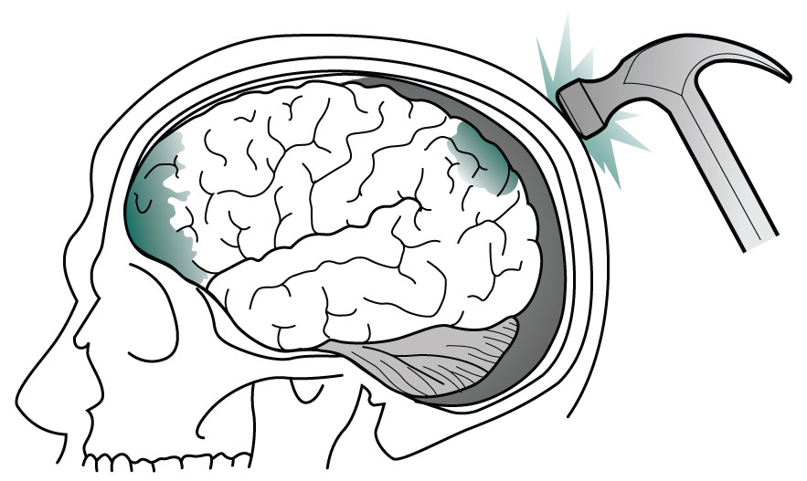
(From Kaufman DM et al: Kaufmans clinical neurology for psychiatrists, ed 9, Philadelphia, 2023, Elsevier.)
Figure E2 Meningeal Arterial Bleeding, Which Usually Results from a Blow Forceful Enough to Fracture the Skull, Causes an Epidural Hematoma
In Contrast, Venous Bleeding, Usually Slower and Under Less Pressure, Causes Subdural Hematoma. Ruptured Aneurysms and Head Trauma Often Cause Subarachnoid Hemorrhage (SAH). In SAH, Blood Spreads Within the Subarachnoid Space over the Convexities, Between the Gyri, into the Interhemispheric Fissure, and Down into the Spinal Canal.
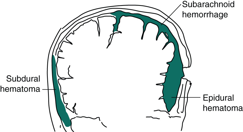
(From Kaufman DM et al: Kaufmans clinical neurology for psychiatrists, ed 9, Philadelphia, 2023, Elsevier.)
Figure E3 Image of a gunshot wound to the head-a penetrating injury.
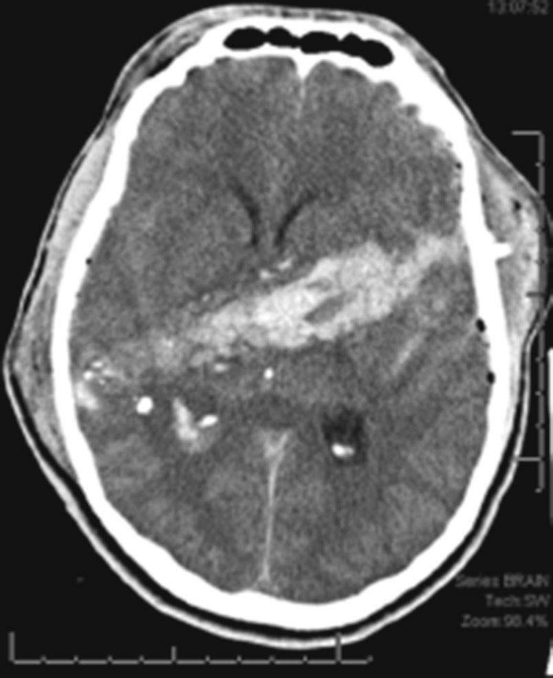
The bullet leaves a path of destructions marked by blood, bone, and bullet fragments. Brain tissue around the bullet path is also damaged by the propagating pressure wave. However, this is not visible on computed tomography (axial computed tomography).
(From Jankovic J et al: Bradley and Daroffs neurology in clinical practice, ed 8, Philadelphia, 2022, Elsevier.)
Figure E4 Another Form of Penetrating Injury is Shown on This Anteroposterior x-Ray of the Skull, with Two Nails from a Nail Gun Stuck in the Skull and Penetrating the Right Frontal Lobe
This Patient was Neurologically Intact on Presentation. The Difficulty in Such a Case is that Bleeding Can Occur Deep in the Brain When the Nails are Removed. This Patient was Also at High Risk for Developing Pseudoaneurysm Later on.
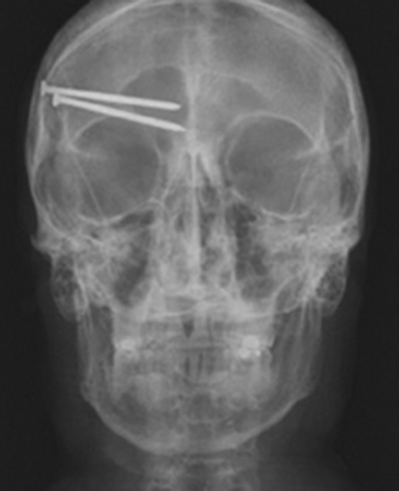
(From Jankovic J et al: Bradley and Daroffs neurology in clinical practice, ed 8, Philadelphia, 2022, Elsevier.)
Differential diagnosis of TBI is quite limited; however, there are several considerations and diagnoses that should be considered, especially regarding mechanism of fall or circumstances that led to an MVA. Prodromes, signs, and symptoms should be looked for, followed by detailed workup as needed.
TBI workup is always a part of the advanced trauma life support (ATLS) protocol. Primary and secondary survey followed by imaging studies constitutes the standardized approach to TBI. Focused TBI workup includes:
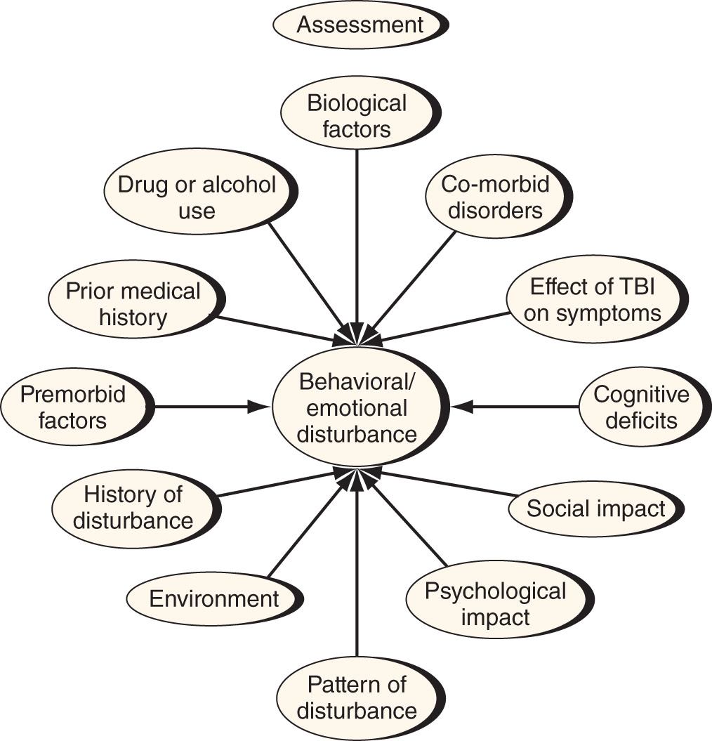
Figure E5 Assessment of the Behavioral and Emotional Changes after Traumatic Brain Injury (TBI)

From Stern TA et al: Massachusetts General Hospital comprehensive clinical psychiatry, ed 3, Philadelphia, 2025. Elsevier.
TABLE E6 Factors to Consider Regarding the Need for Computed Tomography in Head-Injured Patients
CT, Computed tomography; GCS, Glasgow Coma Scale; LOC, loss of consciousness.
From Jankovic J et al: Bradley and Daroffs neurology in clinical practice, ed 8, Philadelphia, 2022, Elsevier.
CT scan is the cornerstone of imaging modalities for head trauma; however, it is not always necessary. Patients over the age of 16 with minimal head injury (i.e., no history of loss of consciousness, amnesia, and confusion), not on blood thinners, and without associated seizure generally do not need a CT scan. Canadian CT head rules (Box E1) for patients with GCS 13 to 15 who do have history of loss of consciousness, amnesia, and/or confusion are a useful guide in determining utility of obtaining a CT scan. If any of these risk factors are present, a CT scan of the head should be performed.
CSF, Cerebrospinal fluid; CT, computed tomography; GCS, Glasgow Coma Scale.
BOX E1 Clinical Decision Rules for Neuroimaging in Adults With Mild Traumatic Brain Injury Canadian Computed Tomography Head Rule (CCHR)
From Walls RM et al: Rosens emergency medicine, concepts and clinical practice, ed 10, Philadelphia, 2023, Elsevier.
TABLE E7 Comparison of Head Imaging Modalities
| Computed Tomography Scans | Magnetic Resonance Imaging | Angiography | Skull Radiography | |
CT, Computed tomography.
From Marx JA et al: Rosens emergency medicine, concepts and clinical practice, ed 7, Philadelphia, 2010, Elsevier.
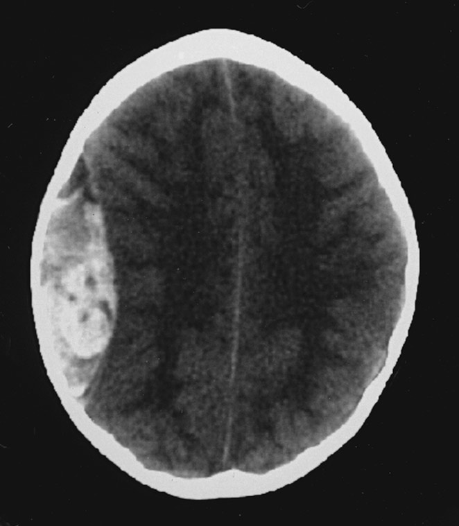
Usually, in addition to a plain CT of the head (Fig. E6), computed tomography angiography (CTA) head/neck or CT of the spine is necessary if arterial or C-spine injury is suspected, respectively. Other imaging modalities such as MRI can be helpful in certain situations but are typically adjuncts in the acute setting to CT-guided management.
Figure E6 Non-Contrast-Enhanced Computed Tomography Scan of Acute Epidural Hematoma at the Level of Right Midconvexity

There is an associated mass effect and moderate midline shift.
(From Walls RM et al: Rosens emergency medicine, concepts and clinical practice, ed 10, Philadelphia, 2023, Elsevier.)
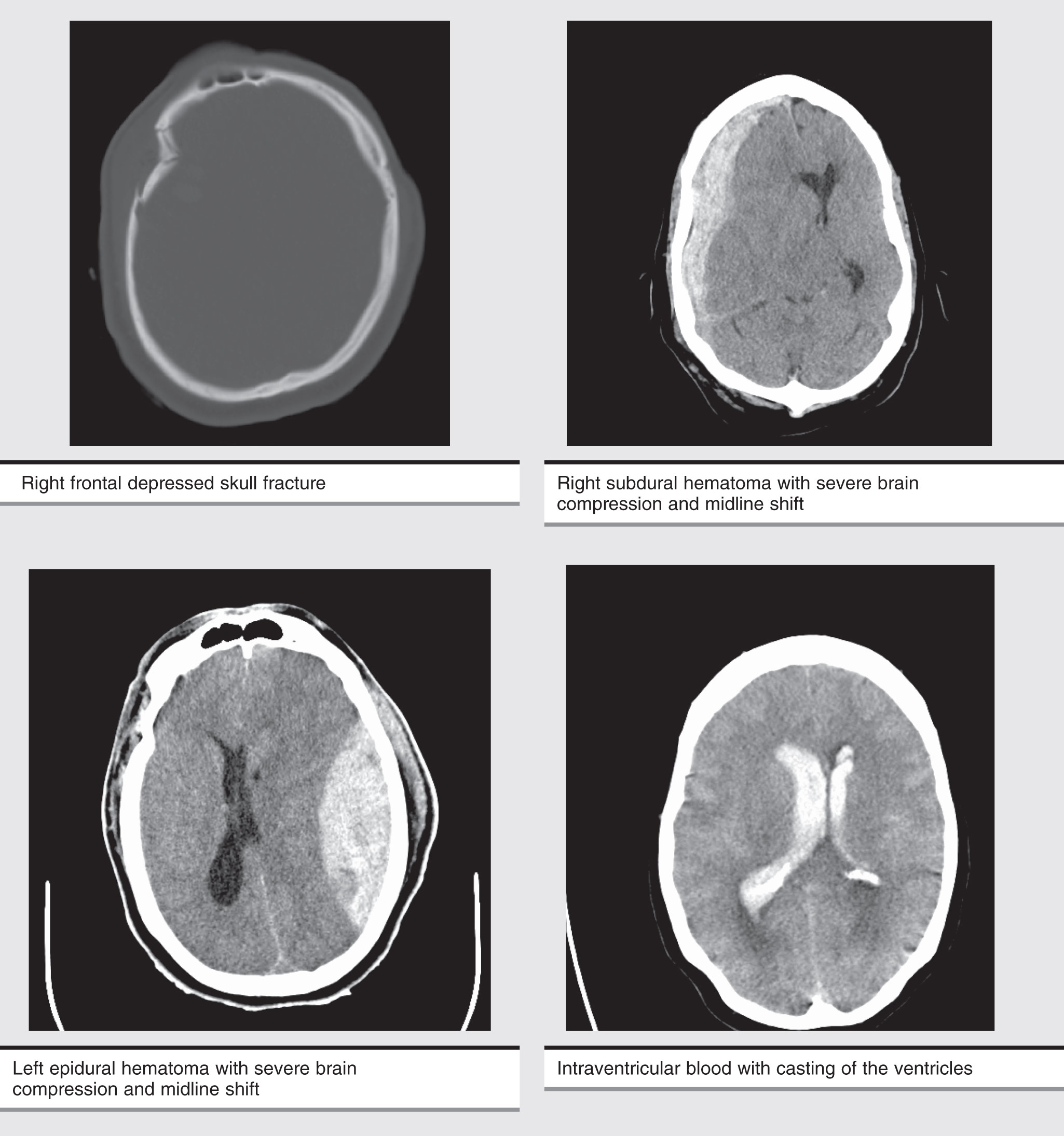
Pathologies that can be identified with imaging are noted in the following:
Prevention of secondary injury is the primary goal of prehospital and early in-hospital management. Most common mechanisms of secondary injury are either intracranial (increased intracranial pressure [ICP], hematoma) or systemic (hypoxia, hypovolemia, hypotension). Early categorization of head trauma patients according to the severity (based on GCS) and transport to facilities equipped with personnel and technology to deal with issues pertaining to head trauma has improved the overall management of head injury patients and prevention of secondary injury. Assessment and treatment recommendations for mild TBI are summarized in Table E8. Airway, breathing, and circulation along with bleeding control remain the most important parameters to be stabilized and both directly and indirectly affect GCS and overall outcome. Trauma guidelines suggest intubation should be performed in any patient with a GCS of 8 or less to prevent respiratory failure. Recent evidence suggests normo-ventilation for patients with severe TBI, with hyperventilation only being used as a temporary measure in patients with TBI and ICP crisis, until other methods of reducing ICP are employed.11 Hypotension should also be avoided in patients with TBI, as it has been shown to increase mortality.11 Transfer to and care in a Level 1 trauma center is associated with better outcomes. Monitoring and treatment recommendations for severe TBI are summarized in Table E9.
TABLE E8 Assessment and Treatment Recommendations for Mild Traumatic Brain Injury
| Function | Assessment | Treatment |
| Overall recovery | Standardized symptom checklist | Physical rest 1-2 days1 followed by subsymptomatic aerobic exercise2 |
| Headache | Determine the type | HA persisting more than 3-4 days may require abortive treatment tailored to phenotype (migraine, tension-type, occipital neuralgia, etc.) |
| Vertigo | Romberg test, dynamic standing, tandem gait | If Hallpike Dix is normal, or if Epley maneuver does not relieve symptoms, consider physical therapy for vestibular rehabilitation |
| Eye movements | Examine cranial nerves 3, 4, 6 for tracking, saccades, diplopia, nystagmus | Physical therapy evaluation for vestibular rehabilitation |
| Near vision | Near-point accommodation and binocular convergence | Ophthalmologic evaluation for vision therapy |
| Cognitive function | Symptoms, cognitive testing, neuropsychologist evaluation | Sleep hygiene, neuropsychology evaluation for cognitive rehabilitation3 |
HA, Headache.
1 Thomas DG et al: Benefits of strict rest after acute concussion: a randomized controlled trial, Pediatrics 135:213-223, 2015.
2 Kurowski BG et al: Aerobic exercise for adolescents with prolonged symptoms after mild traumatic brain injury: an exploratory randomized clinical trial, J Head Trauma Rehabil 32:79-89, 2017.
3 Cooper DB et al: Cognitive rehabilitation for military service members with mild traumatic brain injury: a randomized clinical trial, J Head Trauma Rehabil 32:E1-E15, 2017.
From Goldman L, Schafer AI: Goldman-Cecil medicine, ed 27, Philadelphia, 2024, Elsevier.
TABLE E9 Guidelines for the Management of Severe Traumatic Brain Injury
| Topic | Level 1 | Level 2 | Level 3 |
| Blood pressure and oxygenation | Insufficient data | Avoid systolic blood pressure <90 mm Hg. | Avoid hypoxia (PaO2<60 mm Hg or O2 saturation <90%). |
| Hyperosmolar therapy | Insufficient data | Mannitol is effective for control of raised ICP at doses of 0.25 g/kg to 1 g/kg body weight. | Restrict mannitol use prior to ICP monitoring in patients with signs of transtentorial herniation. |
| Prophylactic hypothermia | Insufficient data | Insufficient data | Pooled data indicate that prophylactic hypothermia is not significantly associated with decreased mortality as compared with normothermic controls. |
| Infection prophylaxis | Insufficient data | To reduce infection, routine ventricular catheter exchange or prophylactic antibiotic use for ventricular catheter placement is not recommended. | |
| Deep venous thrombosis prophylaxis | Insufficient data | Insufficient data |
|
| Indications for ICP monitoring | Insufficient data | ICP should be monitored in all salvageable patients with a GCS score of 3-8 after resuscitation and an abnormal CT scan. | ICP monitoring is indicated in patients with severe TBI with a normal CT scan if >40 yr of age with blood pressure <90 mm Hg. |
| ICP pressure-monitoring technology | N/A | N/A | N/A |
| ICP thresholds | Insufficient data | Treatment should be initiated with ICP >22 mm Hg | A combination of ICP values and clinical and brain CT findings should be used to determine the need for treatment |
| Cerebral perfusion thresholds | Insufficient data | Aggressive attempts to maintain CPP above 70 mm Hg with fluids and pressors should be avoided because of the risk of acute respiratory distress syndrome. | |
| Brain oxygen monitoring and thresholds | Insufficient data | Insufficient data | Jugular venous saturation (<50%) or brain tissue oxygen tension (<20 mm Hg) are treatment thresholds |
| Anesthetics, analgesics, sedatives | Insufficient data | Prophylactic administration of barbiturates to induce burst suppression electroencephalogram is not recommended. High-dose barbiturate administration is recommended to control elevated ICP refractory to maximum standard medical and surgical treatment. Hemodynamic stability is essential before and during barbiturate therapy. Propofol is recommended for the control of ICP but not for improvement in mortality or 6-mo outcome. | N/A |
| Nutrition | Insufficient data | Patients should be fed to attain full caloric replacement by day 7 postinjury. | N/A |
| Antiseizure prophylaxis | Insufficient data | Anticonvulsants are indicated to decrease the incidence of early PTS (within 7 days of injury). | N/A |
| Hyperventilation | Insufficient data | Prophylactic hyperventilation (PaCO2 of 25 mm Hg or less) is not recommended. | Hyperventilation is recommended as a temporizing measure for the reduction of ICP. Hyperventilation should be avoided during the first 24 h after injury, when cerebral blood flow is often critically reduced. If hyperventilation is used, jugular venous oxygen saturation (SjO2) or brain-tissue oxygen tension (PbtO2) measurements are recommended to monitor oxygen delivery. |
| Steroids | The use of high-dose methylprednisolone is associated with increased mortality and is contraindicated. | N/A | N/A |
CT, Computed tomography; CPP, cerebral perfusion pressure; ICP, intracranial pressure; GCS, Glasgow Coma Scale; PTS, posttraumatic seizures; TBI, traumatic brain injury.
From Jankovic J et al: Bradley and Daroffs neurology in clinical practice, ed 8, Philadelphia, 2022, Elsevier.
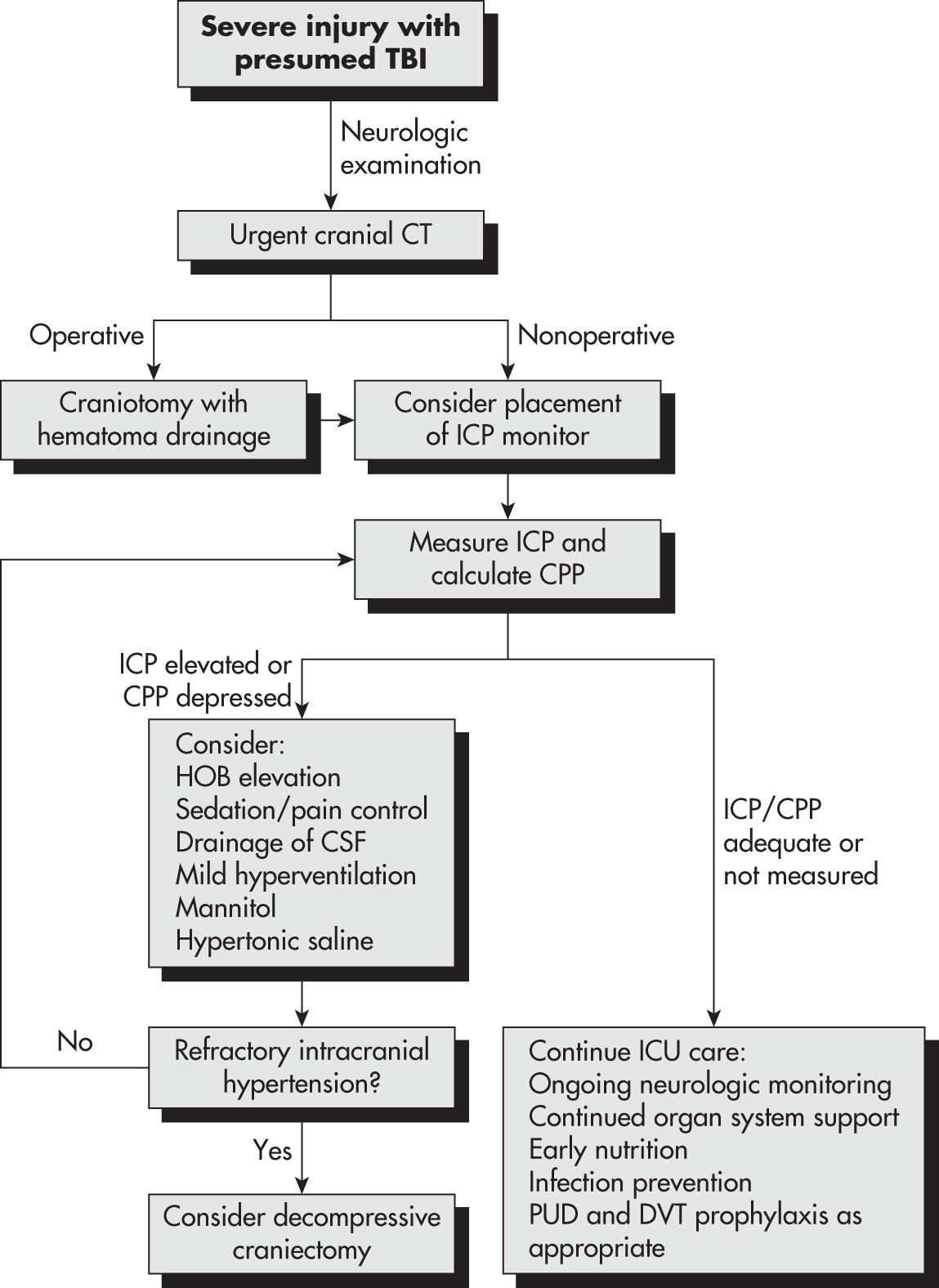
Figure E7 Algorithm for the Management of Traumatic Brain Injury (TBI)
Cpp, Cerebral Perfusion Pressure; CSF, Cerebrospinal Fluid; CT, Computed Tomography; DVT, Deep Venous Thrombosis; Hob, Head of Bed; ICP, Intracranial Pressure; ICU, Intensive Care Unit; Pud, Peptic Ulcer Disease.


(From Townsend CM et al: Sabiston textbook of surgery, ed 21, St Louis, 2022, Elsevier.)
Details of in-hospital management including critical care and surgical intervention is beyond the scope of this text. Some important points are summarized below:
TABLE E10 Goal-Directed Parameters for Head Injury
| Pulse oximetry ≥95% | ICP 20-25 mm Hg | Serum sodium 135-145 mEq/L |
| PaO2≥100 mm Hg | PbtO2≥15 mm Hg | INR ≤1.4 |
| PaCO2 35-45 mm Hg | CPP ≥60 mm Hg* | Platelets ≥75 × 103/mm3 |
| SBP ≥100 mm Hg | Temperature 36°-37.5°C | Hemoglobin ≥7 g/dl |
| pH 7.35-7.45 | Glucose 80-180 mg/dl |
CPP, Cerebral perfusion pressure; ICP, intracranial pressure; INR, international normalized ratio; PaCO2, partial pressure of carbon dioxide; PaO2, partial pressure of oxygen; PbtO2, brain tissue oxygen tension; SBP, systolic blood pressure.
* Depending on status of cerebral autoregulation.
From Vincent JL et al: Textbook of critical care, ed 8, Philadelphia, 2024, Elsevier.
Figure E8 Indications for acute seizure prophylaxis in severe head trauma.

(From Walls RM et al: Rosens emergency medicine, concepts and clinical practice, ed 7, Philadelphia, 2010, Elsevier.)
TABLE E11 Elevated ICP Management
|
CBF, Cerebral blood flow; CPP, cerebral perfusion pressure; EEG, electroencephalography; EVD, external ventricular drain; ICP, intracranial pressure; MAP, mean arterial pressure; PbtO2, Brain tissue oxygen.
Modified from Jankovic J et al: Bradley and Daroffs neurology in clinical practice, ed 8, Philadelphia, 2022, Elsevier.
TABLE E12 Personality and Behavioral Changes Associated With Traumatic Brain Injury
| Aggression | Need for immediate gratification | ||
| Apathy | Mood lability | ||
| Erratic and difficult to predict temper outbursts and mood swings | |||
| Lack of goal-directed activity | |||
| Lack of empathy | Abnormal jocularity | ||
| Distractible | Irritability and reduced tolerance for frustration | ||
| Difficulty learning from mistakes | Disinhibition | ||
| Impulsivity |
From Stern TA et al: Massachusetts General Hospital comprehensive clinical psychiatry, ed 3, Philadelphia, 2025. Elsevier.
| Impaired Attention | Impaired Memory | Language Deficits | Executive Dysfunction |
From Stern TA et al: Massachusetts General Hospital comprehensive clinical psychiatry, ed 3, Philadelphia, 2025. Elsevier.
Figure E9 Treatment of emotional and behavioral disturbances after traumatic brain injury.

(From Stern TA et al: Massachusetts General Hospital comprehensive clinical psychiatry, ed 3, Philadelphia, 2025. Elsevier.)
BOX E3 Medication Side Effects of Concern for Patients With Traumatic Brain Injury
From Stern TA et al: Massachusetts General Hospital comprehensive clinical psychiatry, ed 3, Philadelphia, 2025. Elsevier.
TABLE E14 Common Neurocognitive Sequelae of Moderate to Severe Traumatic Brain Injury
| Cognitive Domain | Clinical Manifestation of Impairment | ||
| Attention | Difficulty with sustained attention | ||
| Poor concentration | |||
| Psychomotor impersistence | |||
| Memory | Problems with acquiring and retaining new verbal or nonverbal information | ||
| Problems in retrieving verbal and nonverbal memories | |||
| Speed of information processing | Slowed sensorimotor skills and information processing | ||
| Executive functioning | Problems in convergent and divergent reasoning | ||
| Poor judgment | |||
| Difficulty planning | |||
| Problems in self-monitoring and self-correcting behavior | |||
| Awareness of symptoms | Difficulty recognizing deficits | ||
| Unrealistic expectations concerning the recovery of functions | |||
| Problems related to poor treatment compliance | |||
| Language and communication | Problems in word comprehension | ||
| Impaired reading, spelling, and writing ability | |||
| Tendency to become fragmented in free speech | |||
| Integrative functions | Problems in adequate or time-efficient execution of various perceptual-motor-spatial-sequential tasks |
From Jankovic J et al: Bradley and Daroffs neurology in clinical practice, ed 8, Philadelphia, 2022, Elsevier.
TBI is a major health care issue. Early recognition of high-risk patients, early imaging, and early evaluation at a Level 1 Trauma Center by a specialist are associated with improved outcomes. Risk factors for suicide following TBI are summarized in Table E15. The goal of health care providers in the field or in the community is to identify patients who need this attention.
TABLE E15 Risk Factors for Suicide Following Traumatic Brain Injury
| Depression | Cognitive and motor disturbances | ||
| Profound feelings of hopelessness, despair, worthlessness | Emotional lability | ||
| Loss of sense of integrity | Impulsivity | ||
| Prior history of suicide attempt | Inflexibility | ||
| Male | Hyperactivity | ||
| Mid-adolescence to mid-twenties | Poor problem-solving | ||
| Lower socioeconomic level | Inability to identify alternatives | ||
| Drug or alcohol use | |||
| Cluster B personality disorder | |||
| Social isolation |
From Stern TA et al: Massachusetts General Hospital comprehensive clinical psychiatry, ed 3, Philadelphia, 2025. Elsevier.