section name header

Basic Information

Author: Victoria Krzywicki, MDand Bharti Rathore, MD

Definition

Mycosis fungoides is an epidermotropic cutaneous T-cell lymphoma, a type of extranodal non-Hodgkin lymphoma, characterized by a proliferation of small to medium-size T lymphocytes with cerebriform nuclei with skin as their primary site.

Synonyms

  • Cutaneous T-cell lymphoma
  • MF

ICD-10CM CODES
C84.00Mycosis fungoides, unspecified site
C84.01Mycosis fungoides, lymph nodes of head, face, and neck
C84.02Mycosis fungoides, intrathoracic lymph nodes
C84.03Mycosis fungoides, intra-abdominal lymph nodes
C84.04Mycosis fungoides, lymph nodes of axilla and upper limb
C84.05Mycosis fungoides, lymph nodes of inguinal region and lower limb
C84.06Mycosis fungoides, intrapelvic lymph nodes
C84.07Mycosis fungoides, spleen
C84.08Mycosis fungoides, lymph nodes of multiple sites
C84.09Mycosis fungoides, extranodal and solid organ sites
Epidemiology & Demographics

  • In the U.S., the annual incidence of cutaneous lymphomas is 0.6 to 1.0 cases per 100,000, with mycosis fungoides (MF) constituting about 50% of the cases.
  • MF affects more males than females (2:1), and majority of patients are White (70%).
  • Median age of diagnosis is 55 to 60 yr, but it can be seen in young adults and children.
Physical Findings & Clinical Presentation

  • MF presents with pruritic skin lesions of varying sizes that may manifest as erythematous patches, plaques, or tumors (Fig. E1 ), affecting virtually any body surface.
  • Clinical course is typically indolent with slow progression over years and, in some cases, over decades.
  • Skin lesions in MF may evolve over time but initially start as scaly patches, developing into infiltrative plaques and eventually to tumors.
  • Patients may show a combination of patches, plaques, and tumors at the same time. If only tumors are present, without preceding or concurrent patches or plaques, a diagnosis of MF is highly unlikely, and another type of cutaneous T-cell lymphoma should be considered.
  • Scaling often occurs on patch and plaque lesions.
  • Rarely, lesions are atrophied and dyspigmented.
  • Some patients develop erythroderma later in the course of the disease.
  • Lesions can be intensely pruritic, which can significantly impact a patient’s quality of life.
  • The initial skin lesions have predilection for the buttocks and other sun-protected areas (“bathing suit” areas).
  • Sézary syndrome (SS) is considered in patients with erythroderma, circulating T cells, and with or without associated lymphadenopathy.
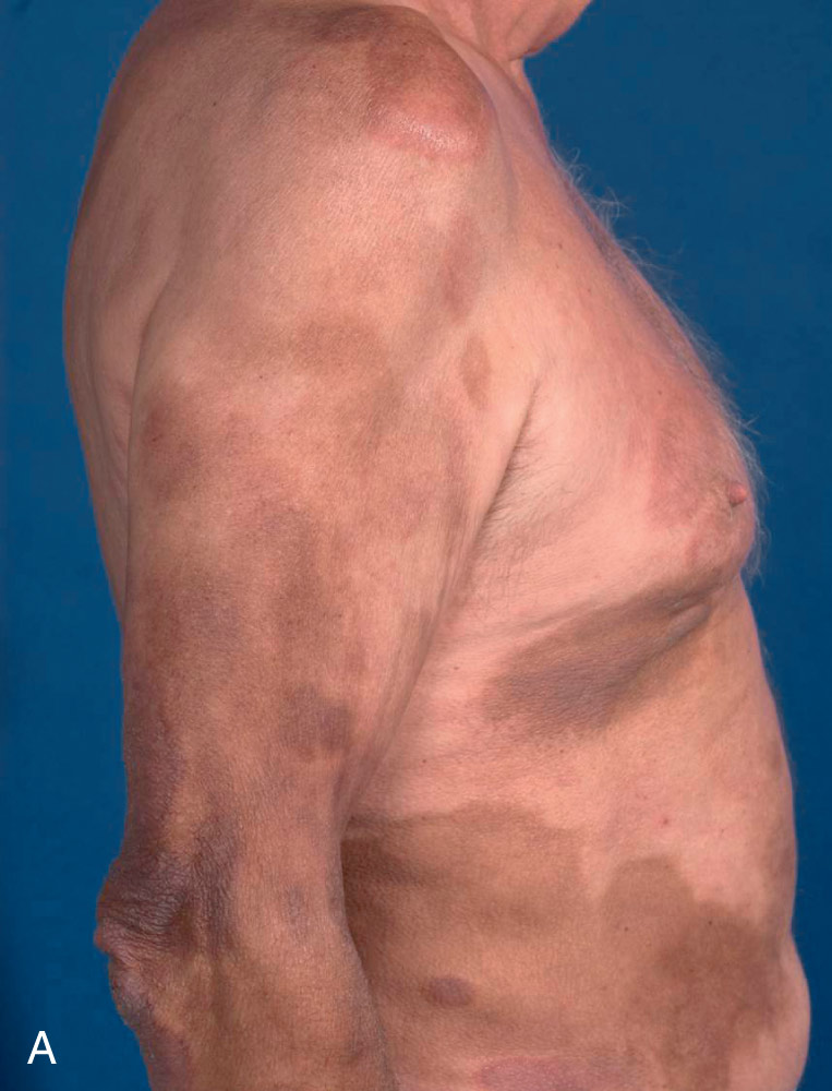
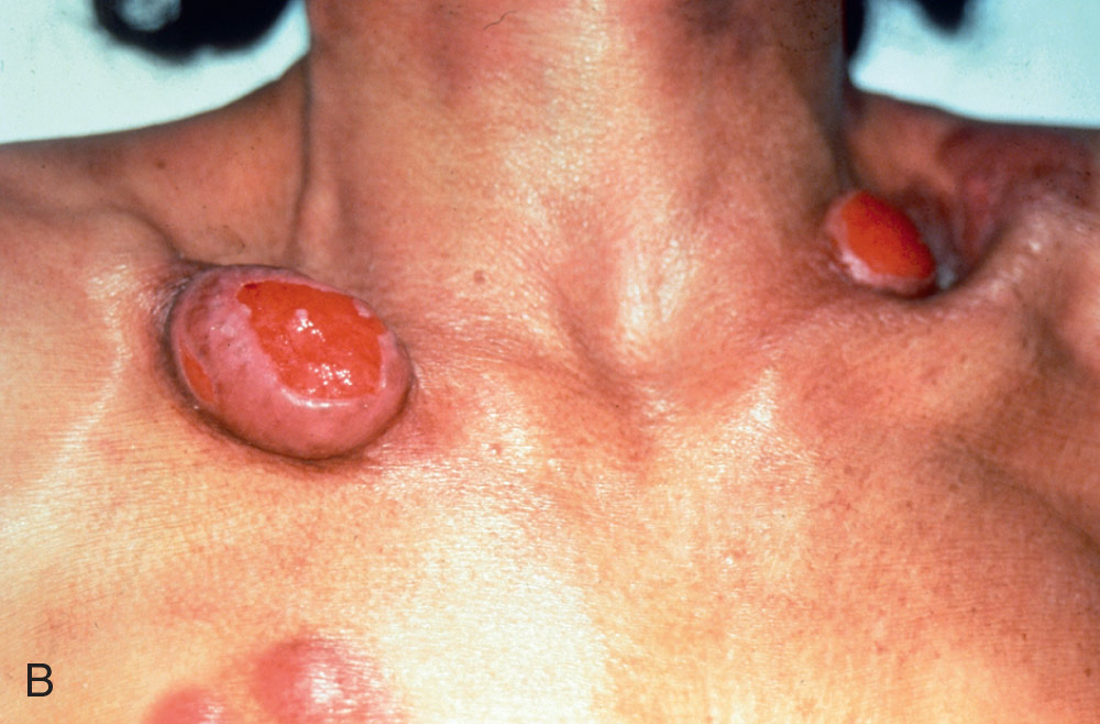
Figure E1 Mycosis fungoides.

A, Patch stage. B, Tumor stage.

From James WD et al: Cutaneous lymphoid hyperplasia, cutaneous T-cell lymphoma, other malignant lymphomas, and allied diseases. In: Andrews’ diseases of the skin clinical atlas, Philadelphia, 2018, Elsevier, pp. 501-516.

Staging

Staging (Table E1 ) is helpful in guiding therapy and has prognostic value.

TABLE E1 TNMB Classification and Staging for Patients With Mycosis Fungoides/Sézary Syndrome

T (Skin)
T1Limited patch or plaque (<10% of BSA)
T2Generalized patch or plaque (>10% of BSA)
T2a = patch only
T3One or more tumors (>1 cm)
T4Generalized erythroderma (>80% of BSA)
N (Nodes)
N0No clinically abnormal peripheral lymph nodes
N1Clinically abnormal peripheral lymph nodes
Dutch grade 1, NCI grade LN0-LN2
N1aClone negative
N1bClone positive
N2Clinically abnormal peripheral lymph nodes
Dutch grade 2, NCI grade LN3
N2aClone negative
N2bClone positive
N3Clinically abnormal peripheral lymph nodes
Dutch grade 3, NCI grade LN4, clone positive or negative
NxClinically abnormal peripheral lymph nodes
No histologic confirmation
M (Viscera)
M0No visceral organ involvement
M1Visceral organ involvement (pathology confirmation required)
B (Blood)
B0Atypical circulating cells not present (<5%)
B0aClone negative
B0bClone positive
B1Atypical circulating cells present (>5%)
B1aClone negative
B1bClone positive
B21000/µL Sézary cells, clone positive
StageTNMB
IA1000, 1
IB2000, 1
IIA1, 21, 200, 1
IIB30-200, 1
IIIA40-200
IIIB40-201
IVA11-40-202
IVA21-4300-2
IVB1-40-310-2

BSA, Body surface area; NCI, National Cancer Institute; TNMB, tumor, node, metastasis, blood.

From Niederhuber JE: Abeloff’s clinical oncology, ed 6, Philadelphia, 2020, Elsevier.

The lesions of mycosis fungoides are classified into four tumor (T) groups:

  • T1: Patches and plaques affecting less than 10% of body surface area
  • T2: Patches and plaques affecting more than 10% of body surface area
  • T3: Presence of tumors (i.e., raised, dome-shaped lesions >1 cm in diameter)
  • T4: Erythroderma affecting more than 80% of body surface area

Because patients with extensive MF lesions (T2 or T3 disease) can have substantial blood involvement in the absence of erythroderma, blood assessment is needed in these patients.1

  • Stage IA is defined as a patch or plaque skin disease involving <10% of the skin surface area and with absence of blood involvement or with low blood tumor burden (<5% of Sézary cells in the peripheral blood).
  • Stage IB is defined as a patch or plaque skin disease involving 10% of the skin surface area (Fig. E2 ) with absence of blood involvement or low blood tumor burden (<5% Sézary cells).
  • Stages IIA & IIB are defined by the presence of tumors with or without clinically abnormal peripheral lymph nodes with absence of blood involvement or low blood tumor burden. In approximately 5% of cases of MF, the presentation may be a diffuse, painful, pruritic erythroderma with Sézary cells (Fig. E3 ) in the peripheral blood (known as Sézary syndrome) (Fig. E4 ).
  • Stage III disease is defined by the presence of generalized erythroderma from the spread of cancer cells through the skin but not yet to the lymph nodes.
  • Stage IVA disease is defined by a lymph node biopsy showing large clusters of atypical cells, more than six cells, or total effacement by atypical cells.
  • Stage IVB disease is defined by the presence of visceral involvement.
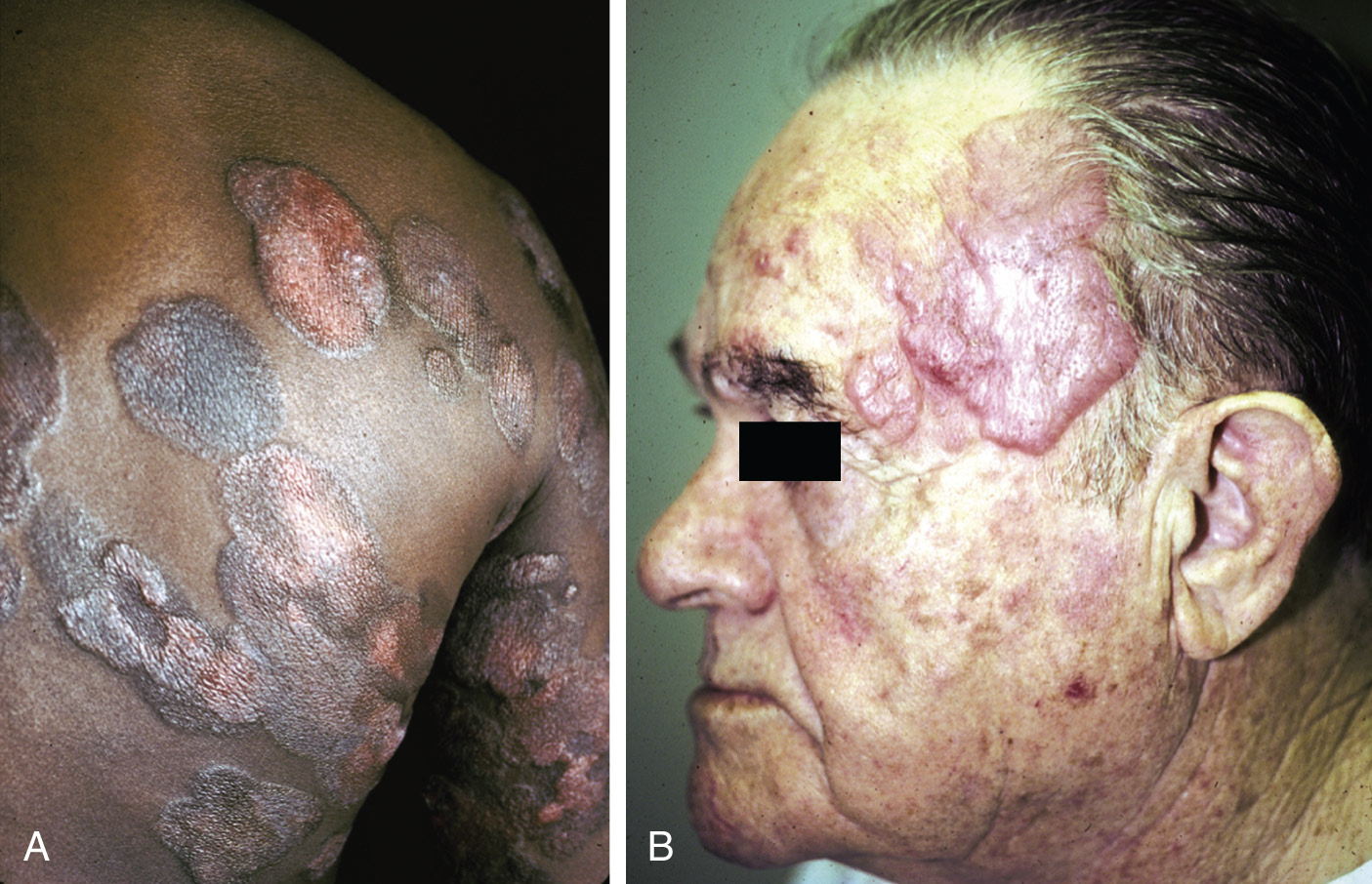
Figure E2 Mycosis fungoides.

A, Plaques of mycosis fungoides often have a polycyclic appearance. B, Tumors are more elevated above the skin surface. By the time tumors supervene, sun-exposed skin, such as the face, is often involved.

From Jaffe ES et al: Hematopathology, ed 1, Philadelphia, 2011, Saunders.
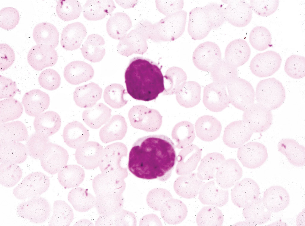
Figure E3 Sézary cells in a peripheral blood smear.

Recognition of these cells is no longer critical for the diagnosis since the advent of clonality studies and flow cytometry.

From Jaffe ES et al: Hematopathology, ed 1, Philadelphia, 2011, Saunders.
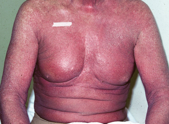
Figure E4 Sézary syndrome presents with erythroderma-diffuse red skin.

The term erythroderma is overused by clinicians; it should refer to confluent erythema, not just widespread erythematous lesions.

From Jaffe ES et al: Hematopathology, ed 1, Philadelphia, 2011, Saunders.

Etiology

Several etiologies have been postulated for MF. There is a supporting role of the skin microenvironment in the pathogenesis, as MF is thought to arise in the background of chronic inflammation. Extravasation and migration of malignant T cells are facilitated by chemokines, cytokines, and adhesion molecules. Th1 and Th2 cytokines are both involved in different stages of the course of MF.

Diagnosis

The diagnosis of early MF often needs integration of clinical, histologic, and molecular features since it can be confused with benign eczematous skin disease.1

Diagnosis is established by skin biopsy, and it may be difficult to differentiate from other skin lesions in the early phases of the disease (e.g., premycotic patch or early plaque lesions).

Differential Diagnosis

  • Contact dermatitis
  • Atopic dermatitis
  • Nummular dermatitis
  • Parapsoriasis
  • Superficial fungal infections
  • Drug eruptions
  • Psoriasis
  • Photodermatitis
  • Alopecia mucinosa
  • Lymphomatoid papulosis
Workup

Any patient who is suspected of having MF should have a staging workup. Prognosis in patients with mycosis fungoides depends on the type of skin lesions and the extent of disease. The workup should focus on:

  • Complete physical examination:
    1. 1.The type of skin lesion and the extent of skin involvement of the body (e.g., skin involvement is >10% or <10% of the skin surface)
    2. 2.Identification of palpable lymph nodes (especially those >1.5 cm in largest diameter)
    3. 3.Identification of organomegaly (e.g., lungs, liver)
  • Skin biopsy:
    1. 1.Biopsy of the most indurated area (multiple skin biopsies are often required)
    2. 2.Immunophenotyping
    3. 3.Evaluation for clonality
  • Blood testing:
    1. 1.CBC with differential, liver function tests, lactate dehydrogenase, chemistry
    2. 2.T-cell receptor gene rearrangement
    3. 3.Determination of Sézary cell count and/or flow cytometry
  • Radiologic tests: Depending on the stage of the disease, chest x-ray examination; ultrasound; and computed tomography scan of the chest, abdomen, and pelvis alone, with or without fluorodeoxyglucose PET scan
  • Lymph node biopsy:
    1. 1.Excisional biopsy of the largest lymph node should be done
    2. 2.If multiple nodes enlarged, order of preference is cervical, axillary, and inguinal areas
    3. 3.Histopathology, flow cytometry, T-cell receptor gene rearrangement

Treatment

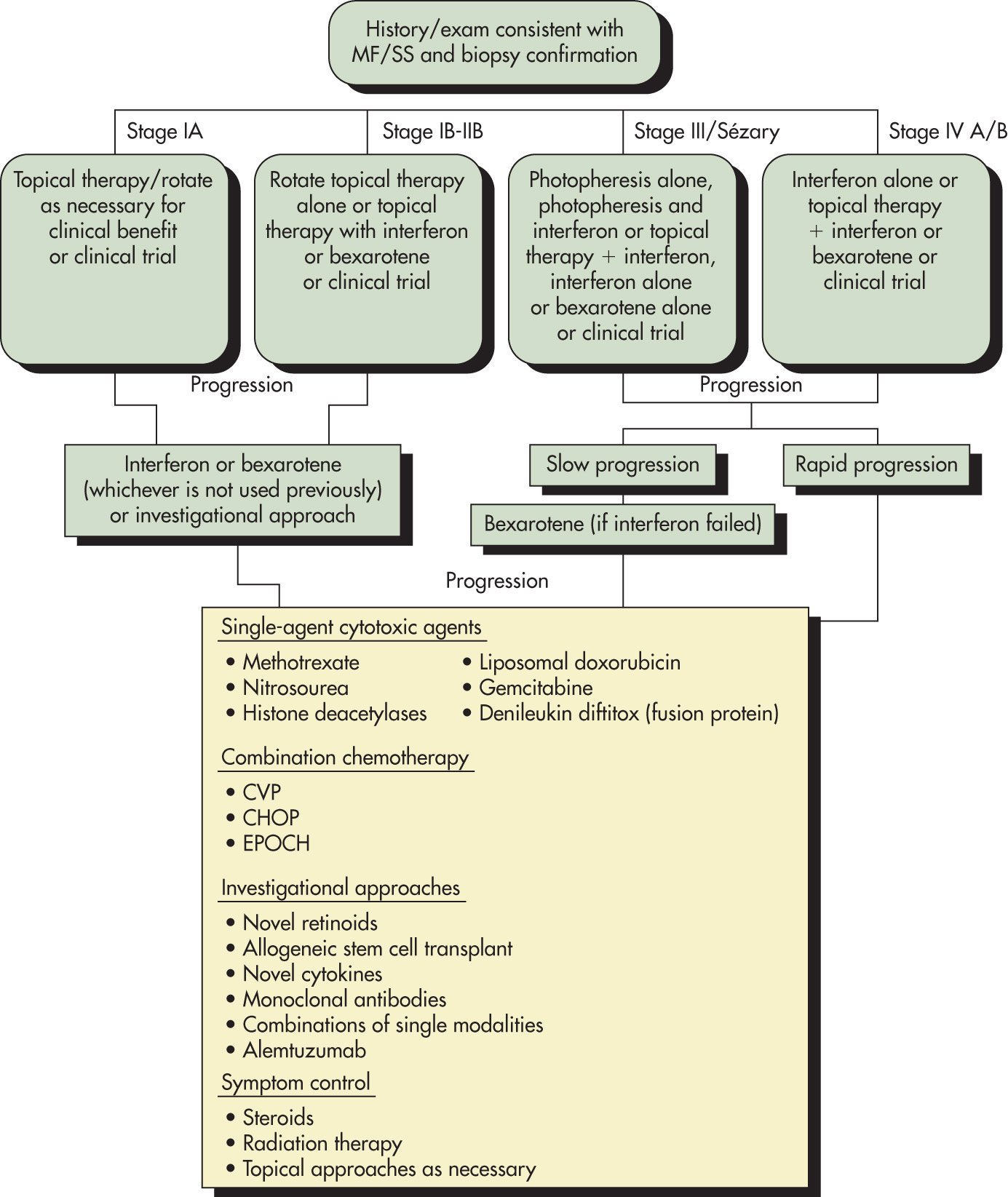
In general, MF is not curable, and the goal of therapy is to help control symptoms and prevent further progression of disease. Treatment is guided according to the stage of disease (Box E1 ) and involves a multidisciplinary team consisting of dermatopathologists, dermatologists, radiation oncologists, and medical oncologists. Therapeutic options are summarized in Table E2 , and a treatment algorithm is described in Fig. E5 .

BOX E1 Summary of Treatment Approaches for Patients With Mycosis Fungoides and Sézary Syndrome

Early-Stage Mycosis Fungoides

  • Topical
  • Corticosteroids
  • Phototherapy
  • Nitrogen mustard
  • Bexarotene
  • Radiation or TSEBT
Refractory Early-Stage Mycosis Fungoides

  • Combined therapy
  • PUVA or NB-UVB and INF-α
  • PUVA or NB-UVB and bexarotene (low dose)
Advanced Mycosis Fungoides/Sézary Syndrome

  • Biologic therapy
  • Interferons (INF-α, IFN-γ)
  • Retinoid (bexarotene)
  • Extracorporeal photopheresis
  • Alemtuzumab
  • HDAC inhibitors (romidepsin, vorinostat, panobinostat)
  • Bortezomib
  • Antifolates (methotrexate, pralatrexate)
  • Mogamulizumab (anti-CCR4)
  • Denileukin diftitox (Ontak)
  • Brentuximab vedotin
  • Combined therapy
  • INF-α and phototherapy
  • INF-α and retinoid or rexinoid
  • Retinoid and phototherapy
  • ECP and INF-α
  • ECP and retinoid or rexinoid
  • Systemic chemotherapy
  • Single agent
  • Pegylated doxorubicin
  • Purine or pyrimidine analogues (Pentostatin)
  • Gemcitabine
  • Temozolomide
  • Multiagent chemotherapy
  • CHOP and CHOP-like, CHOEP
  • Stem cell transplantation
  • Autologous
  • Allogeneic (including nonmyeloablative)
  • Investigational therapy
  • Lenalidomide

From Niederhuber JE: Abeloff’s clinical oncology, ed 6, Philadelphia, 2020, Elsevier.

TABLE E2 Therapeutic Options for Mycosis Fungoides

Topical Therapy
Ultraviolet A with psoralen
Ultraviolet B
External beam radiation therapy
Total-skin electron beam radiation
Topical chemotherapy
Topical retinoids
Systemic Therapy
Photopheresis
Interferon-α
Oral retinoids
Targeted therapies
Single-agent chemotherapy
Combination chemotherapy
Stem cell transplantation
Investigational agents

From Hoffman R et al: Hematology, basic principles and practice, ed 5, Philadelphia, 2009, Churchill Livingstone.

Figure E5 Algorithm for the care of patients with mycosis fungoides or Sézary syndrome.

!!flowchart!!

CHOP,Cyclophosphamide, doxorubicin (hydroxorubicin), Oncovin (vincristine), prednisone; CVP, central venous pressure; EPOCH, etoposide-prednisone-Oncovin-cyclophosphamide-hydroxydaunorubicin; MF, mycosis fungoides; SS, Sézary syndrome.

Modified from Hoffman R et al: Hematology, basic principles and practice, ed 5, Philadelphia, 2009, Churchill Livingstone.

Nonpharmacologic Therapy

  • For dry, cracking skin, emollients (e.g., lanolin and petrolatum) are applied bid.
  • Moisturizing lotion (e.g., ammonium lactate) applied bid.
  • Topical antibiotics (e.g., bacitracin) are used on ulcerative tumors.
Acute General Rx

Treatment is guided by accurate disease staging and presence of limited vs. advanced disease.2-4

Limited Disease

  • Stage IA disease: Treatment for limited patch or plaque phase disease includes skin-directed therapies:
    1. 1.Topical corticosteroids
    2. 2.Topical chemotherapy (nitrogen mustard or carmustine)
    3. 3.Topical imiquimod
    4. 4.Topical retinoid, phototherapy, and local radiation
    5. 5.There are no well-designed prospective randomized controlled trials, and most patients respond to one or another modality of therapy
  • Stage IB to IIA disease: Similar to stage IA. Skin-directed therapies alone or in combination are used with good results. In addition, other options include:
    1. 1.Total skin electron beam therapy
    2. 2.Phototherapy includes ultraviolet A light with oral methoxypsoralen (PUVA)
    3. 3.Combination of PUVA phototherapy with systemic interferon in stage IB to IIA patients has complete response rates of about 80%. Similarly, PUVA with systemic interferon has shown superior response rates and less time to response when compared to systemic interferon with retinoids.
  • Stage IIB disease: For generalized tumor and plaque disease
    1. 1.Total skin electron beam therapy has shown effectiveness in patients with IIB disease. The 5-yr disease-free survival is about 50%. Patients with <10% BSA involvement do better than patients with extensive involvement. Phototherapy in combination with interferon has shown a response rate of about 50%.
    2. 2.Patients not responding to local therapies require systemic therapy.
Advanced Disease

Topical therapies can be considered in stage III disease, but the response and duration of response are less. Hence, systemic therapies (biologic therapy or systemic chemotherapy) are considered in stage III and stage IV disease.

  • Biologic therapies used in these advanced diseases are:
    1. 1.Interferon alfa
    2. 2.Photopheresis
    3. 3.Systemic retinoids
    4. 4.Histone deacetylase inhibitors (vorinostat, romidepsin, belinostat, and panobinostat)
    5. 5.Monoclonal antibodies (alemtuzumab, mogamulizumab)
    6. 6.Anti-CD30 antibodies (brentuximab) can be used in CD30 expressing cases of MF

Unfortunately, most of these patients relapse and develop disease that is refractory to treatment.

  • Cytotoxic combination chemotherapy regimens have demonstrated activity, produce high responses, but are not durable and have significant toxicities. Hence, single-agent systemic chemotherapy is preferred over combination chemotherapy unless patients are refractory or immediate palliation is needed for extensive tumor burden.
  • Allogenic stem cell transplant has been shown to induce complete and durable remissions in a small number but carries a risk of transplant-related mortality. Hence, reduced intensity allogenic stem cell transplantation is currently being investigated.
  • JAK inhibitors have shown a modest benefit in treatment of relapsed/refractory disease; however, more studies are needed for further investigation.8
  • Immune checkpoint inhibitors (pembrolizumab, nivolumab) have shown encouraging results with durable outcomes in heavily pretreated MF/SS patients.5
Disposition

The 10-yr rates of overall survival are as follows:

  • Stage I disease: 65% to 90%
  • Stage II disease: 35% to 50%
  • Stage III disease: 25% to 35%
  • Stage IV disease: 15% to 20%

The Cutaneous Lymphoma International Consortium analyzed 10 variables in 1275 patients and found four (stage IV, age >60 yr, large-cell transformation, and increased lactate dehydrogenase) were independent prognostic markers for a worse survival in advanced MF/SS patients. Combining these four factors, a prognostic index model was constructed that identified three risk groups across stages with significantly different 5-yr survival rates: Low risk (68%), intermediate risk (44%), and high risk (28%).6

Referral

Patient referral to a dermatologist with expertise in this entity. Oncology consultation is indicated in patients with more advanced disease.

Pearls & Considerations

TABLE E3 Comparison of EORTC and WHO Classifications of Primary Cutaneous Lymphoma

EORTC ClassificationWHO Classification
Cutaneous T-Cell Lymphoma
Indolent clinical behaviorMycosis fungoides
Mycosis fungoides variantsMycosis fungoides variants
Follicular mycosis fungoidesFollicular mycosis fungoides
Pagetoid reticulosisPagetoid reticulosis
CTCL, large cell, CD30+Primary cutaneous CD30+ ALCL (CD30+ lymphoproliferative disease, including lymphomatoid papulosis)
Lymphomatoid papulosis
Aggressive clinical behavior
Sézary syndromeSézary syndrome
CTCL, large cell, CD30Peripheral T-cell lymphoma, unspecified (most); extranodal NK/T-cell lymphoma, nasal type
Provisional Entities
CTCL, pleomorphic, small/medium sized
Subcutaneous panniculitis-like T-cell lymphomaSubcutaneous panniculitis-like T-cell lymphoma
Cutaneous B-Cell Lymphoma
Indolent clinical behaviorExtranodal marginal zone B-cell lymphoma
Primary cutaneous immunocytoma (marginal zone B-cell lymphoma)
Follicle center cell lymphoma (any grade)
Intermediate clinical behavior
Primary cutaneous large B-cell lymphoma of the leg
Provisional Entities
Primary cutaneous plasmacytomaPlasmacytoma
Intravascular large B-cell lymphomaDiffuse large B-cell lymphoma (intravascular)

ALCL, Anaplastic large-cell lymphoma; CTCL, cutaneous T-cell lymphoma; EORTC, European Organization for Research and Treatment of Cancer; MF, mycosis fungoides; NK, natural killer; SS, Sézary syndrome; WHO, World Health Organization.

From Hoffman R et al: Hematology, basic principles and practice, ed 7, Philadelphia, 2018, Elsevier.

TABLE E4 ISCL/EORTC Revision to the Classification of MF and SS (Adaptation)

TNMB Stages
Skin
T1Limited patches, papules, and/or plaques covering <10% of the skin surface. May stratify into T1a (patch only) vs. T1b (plaque ± patch).
T2Patches, papules, or plaques covering 10% of skin surface. May stratify into T2a (patch only) vs. T2b (plaque ± patch).
T3One or more tumors (1-cm diameter)
T4Confluence of erythema covering 80% body surface area
Node
N0No clinically abnormal peripheral lymph nodes; biopsy not required
N1Abnormal peripheral lymph nodes; histopathology Dutch grade 1 or NCI LN0-2
N2Abnormal peripheral lymph nodes; histopathology Dutch grade 2 or NCI LN3
N3Abnormal peripheral lymph nodes; histopathology Dutch grades 3-4 or NCI LN4
NxAbnormal peripheral lymph nodes; no histologic confirmation
Visceral
M0No visceral organ involvement
M1Visceral involvement (must have pathology confirmation and involved organ should be specified)
Blood
B0Absence of significant blood involvement: 5% of peripheral blood lymphocytes are atypical (Sézary) cells
B1Low blood tumor burden: >5% of peripheral blood lymphocytes are atypical (Sézary) but does not meet the criteria for B2
B2High blood tumor burden: 1000/µL Sézary cells with positive clones
Clinical Staging
IAT1, N0, M0, B0,1
IBT2, N0, M0, B0,1
IIAT1,2, N1,2, M0, B0,1
IIBT3, N0-2, M0, B0,1
IIIAT4, N0-2, M0, B0
IIIBT4, N0-2, M0, B1
IVA1T1-4, N0-2, M0, B2
IVA2T1-4, N3, M0, B0-2
IVBT1-4, N0-3, M1, B0-2

CTCL, Cutaneous T-cell lymphoma; EORTC, European Organization of Research and Treatment of Cancer; ISCL, International Society for Cutaneous Lymphomas; MF, mycosis fungoides; NCI LN, National Cancer Institute lymph node grading system; SS, Sézary syndrome; TNMB, tumor-node-metastasis-blood.

From Hoffman R et al: Hematology, basic principles and practice, ed 7, Philadelphia, 2018, Elsevier.

Important quality-of-life considerations include pruritus, xerosis, and the prevention of skin infections.

Reference(s)

  1. Jawed SI : Primary cutaneous T-cell lymphoma (mycosis fungoides and Sézary syndrome): part I. Diagnosis: clinical and histopathologic features and new molecular and biologic markersJ Am Acad Dermatol. 70(2):e1-e16, 2014.
  2. Jawed SI : Primary cutaneous T-cell lymphoma (mycosis fungoides and Sezary syndrome): part II. Prognosis, management, and future directionsquiz 221-222J Am Acad Dermatol. 70(2):e1-e16, 2014.
  3. Foss FM, Girardi M : Mycosis fungoides and Sezary syndromeHematol Oncol Clin North Am. 31(2):297-315, 2017.
  4. Whittaker S : How I treat mycosis fungoides and Sézary syndromeBlood. 127(25):3142-3153, 2016.
  5. Pelcovits A : Advances in immunotherapy for the treatment of cutaneous T-cell lymphomaCancer Manag Res. 15:989-998, 2023.
  6. Scarisbrick JJ : Cutaneous Lymphoma International Consortium study of outcome in advanced stages of mycosis fungoides and Sézary syndrome: effect of specific prognostic markers on survival and development of a prognostic modelJ Clin Oncol. 33(32):3766-3773, 2015.
  7. Pimpinelli N : Defining early mycosis fungoidesJ Am Acad Dermatol. 53:1053, 2005.
  8. Vahabi SM : JAK inhibitors in cutaneous T-cell lymphoma: friend or foe? A systematic review of the published literatureCancers (Basel). 16(5):861, 2024.