Author: Victoria Krzywicki, MDand Bharti Rathore, MD

Mycosis fungoides is an epidermotropic cutaneous T-cell lymphoma, a type of extranodal non-Hodgkin lymphoma, characterized by a proliferation of small to medium-size T lymphocytes with cerebriform nuclei with skin as their primary site.
| ICD-10CM CODES | |||
| C84.00 | Mycosis fungoides, unspecified site | ||
| C84.01 | Mycosis fungoides, lymph nodes of head, face, and neck | ||
| C84.02 | Mycosis fungoides, intrathoracic lymph nodes | ||
| C84.03 | Mycosis fungoides, intra-abdominal lymph nodes | ||
| C84.04 | Mycosis fungoides, lymph nodes of axilla and upper limb | ||
| C84.05 | Mycosis fungoides, lymph nodes of inguinal region and lower limb | ||
| C84.06 | Mycosis fungoides, intrapelvic lymph nodes | ||
| C84.07 | Mycosis fungoides, spleen | ||
| C84.08 | Mycosis fungoides, lymph nodes of multiple sites | ||
| C84.09 | Mycosis fungoides, extranodal and solid organ sites | ||
- •In the U.S., the annual incidence of cutaneous lymphomas is 0.6 to 1.0 cases per 100,000, with mycosis fungoides (MF) constituting about 50% of the cases.
- •MF affects more males than females (2:1), and majority of patients are White (70%).
- •Median age of diagnosis is 55 to 60 yr, but it can be seen in young adults and children.
- •MF presents with pruritic skin lesions of varying sizes that may manifest as erythematous patches, plaques, or tumors (Fig. E1 ), affecting virtually any body surface.
- •Clinical course is typically indolent with slow progression over years and, in some cases, over decades.
- •Skin lesions in MF may evolve over time but initially start as scaly patches, developing into infiltrative plaques and eventually to tumors.
- •Patients may show a combination of patches, plaques, and tumors at the same time. If only tumors are present, without preceding or concurrent patches or plaques, a diagnosis of MF is highly unlikely, and another type of cutaneous T-cell lymphoma should be considered.
- •Scaling often occurs on patch and plaque lesions.
- •Rarely, lesions are atrophied and dyspigmented.
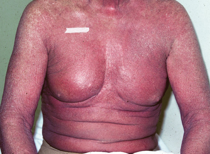
- •Some patients develop erythroderma later in the course of the disease.
- •Lesions can be intensely pruritic, which can significantly impact a patient’s quality of life.
- •The initial skin lesions have predilection for the buttocks and other sun-protected areas (“bathing suit” areas).
- •Sézary syndrome (SS) is considered in patients with erythroderma, circulating T cells, and with or without associated lymphadenopathy.
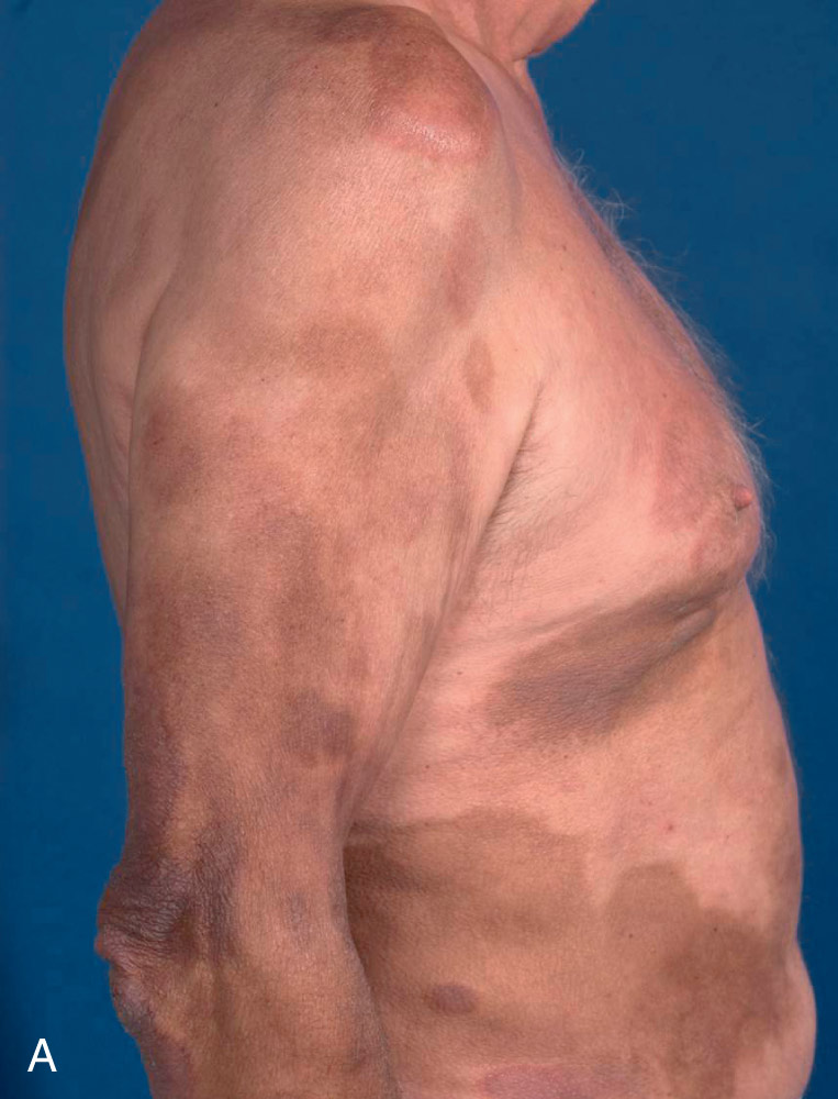
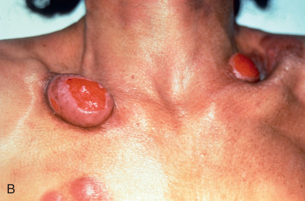
A, Patch stage. B, Tumor stage.
From James WD et al: Cutaneous lymphoid hyperplasia, cutaneous T-cell lymphoma, other malignant lymphomas, and allied diseases. In: Andrews’ diseases of the skin clinical atlas, Philadelphia, 2018, Elsevier, pp. 501-516.
Staging (Table E1 ) is helpful in guiding therapy and has prognostic value.
TABLE E1 TNMB Classification and Staging for Patients With Mycosis Fungoides/Sézary Syndrome
| T (Skin) | |||
| T1 | Limited patch or plaque (<10% of BSA) | ||
| T2 | Generalized patch or plaque (>10% of BSA) T2a = patch only | ||
| T3 | One or more tumors (>1 cm) | ||
| T4 | Generalized erythroderma (>80% of BSA) | ||
| N (Nodes) | |||
| N0 | No clinically abnormal peripheral lymph nodes | ||
| N1 | Clinically abnormal peripheral lymph nodes Dutch grade 1, NCI grade LN0-LN2 | ||
| N1a | Clone negative | ||
| N1b | Clone positive | ||
| N2 | Clinically abnormal peripheral lymph nodes Dutch grade 2, NCI grade LN3 | ||
| N2a | Clone negative | ||
| N2b | Clone positive | ||
| N3 | Clinically abnormal peripheral lymph nodes Dutch grade 3, NCI grade LN4, clone positive or negative | ||
| Nx | Clinically abnormal peripheral lymph nodes No histologic confirmation | ||
| M (Viscera) | |||
| M0 | No visceral organ involvement | ||
| M1 | Visceral organ involvement (pathology confirmation required) | ||
| B (Blood) | |||
| B0 | Atypical circulating cells not present (<5%) | ||
| B0a | Clone negative | ||
| B0b | Clone positive | ||
| B1 | Atypical circulating cells present (>5%) | ||
| B1a | Clone negative | ||
| B1b | Clone positive | ||
| B2 | 1000/µL Sézary cells, clone positive | ||
| Stage | T | N | M | B |
| IA | 1 | 0 | 0 | 0, 1 |
| IB | 2 | 0 | 0 | 0, 1 |
| IIA | 1, 2 | 1, 2 | 0 | 0, 1 |
| IIB | 3 | 0-2 | 0 | 0, 1 |
| IIIA | 4 | 0-2 | 0 | 0 |
| IIIB | 4 | 0-2 | 0 | 1 |
| IVA1 | 1-4 | 0-2 | 0 | 2 |
| IVA2 | 1-4 | 3 | 0 | 0-2 |
| IVB | 1-4 | 0-3 | 1 | 0-2 |
BSA, Body surface area; NCI, National Cancer Institute; TNMB, tumor, node, metastasis, blood.
From Niederhuber JE: Abeloff’s clinical oncology, ed 6, Philadelphia, 2020, Elsevier.
The lesions of mycosis fungoides are classified into four tumor (T) groups:
- •T1: Patches and plaques affecting less than 10% of body surface area
- •T2: Patches and plaques affecting more than 10% of body surface area
- •T3: Presence of tumors (i.e., raised, dome-shaped lesions >1 cm in diameter)
- •T4: Erythroderma affecting more than 80% of body surface area
Because patients with extensive MF lesions (T2 or T3 disease) can have substantial blood involvement in the absence of erythroderma, blood assessment is needed in these patients.1
- •Stage IA is defined as a patch or plaque skin disease involving <10% of the skin surface area and with absence of blood involvement or with low blood tumor burden (<5% of Sézary cells in the peripheral blood).
- •Stage IB is defined as a patch or plaque skin disease involving ≥10% of the skin surface area (Fig. E2 ) with absence of blood involvement or low blood tumor burden (<5% Sézary cells).
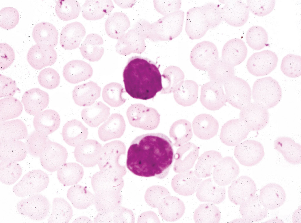
- •Stages IIA & IIB are defined by the presence of tumors with or without clinically abnormal peripheral lymph nodes with absence of blood involvement or low blood tumor burden. In approximately 5% of cases of MF, the presentation may be a diffuse, painful, pruritic erythroderma with Sézary cells (Fig. E3 ) in the peripheral blood (known as Sézary syndrome) (Fig. E4 ).
- •Stage III disease is defined by the presence of generalized erythroderma from the spread of cancer cells through the skin but not yet to the lymph nodes.
- •Stage IVA disease is defined by a lymph node biopsy showing large clusters of atypical cells, more than six cells, or total effacement by atypical cells.
- •Stage IVB disease is defined by the presence of visceral involvement.
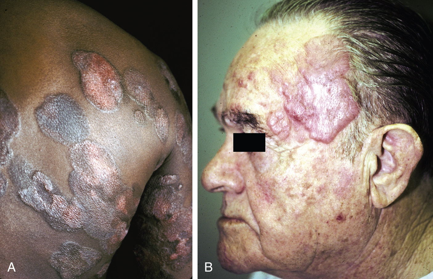
A, Plaques of mycosis fungoides often have a polycyclic appearance. B, Tumors are more elevated above the skin surface. By the time tumors supervene, sun-exposed skin, such as the face, is often involved.
From Jaffe ES et al: Hematopathology, ed 1, Philadelphia, 2011, Saunders.
Several etiologies have been postulated for MF. There is a supporting role of the skin microenvironment in the pathogenesis, as MF is thought to arise in the background of chronic inflammation. Extravasation and migration of malignant T cells are facilitated by chemokines, cytokines, and adhesion molecules. Th1 and Th2 cytokines are both involved in different stages of the course of MF.