Author: Joseph S. Kass, MD, JD, FAAN
Dementia is a syndrome characterized by progressive loss of previously acquired cognitive skills, including memory, language, insight, and judgment. Alzheimer disease (AD) is thought to account for the majority of all cases of dementia.
| ICD-10CM CODES | |||
| G30.0 | Alzheimer disease with early onset | ||
| G30.1 | Alzheimer disease with late onset | ||
| G30.8 | Other Alzheimer disease | ||
| G30.9 | Alzheimer disease, unspecified | ||
Risk doubles every 5 yr after the age of 65. The Chicago Health and Aging Population study found that the average annual incidence in people ages 65 and above was 2.3%, with Blacks having a significantly increased risk compared to Whites.1
Approximately 1 in 9 people (10.7%) ages ≥65 has Alzheimer dementia. Currently an estimated 6.5 million Americans have AD; 5% of the population between the ages of 65 and 74, 13.1% between 75 and 84, and 33.2% at ≥85 yr. Between 12% and 18% of Americans over age 60 are thought to have mild cognitive impairment (MCI).2
Females greater than males. In the U.S. 4 million women vs. 2.5 million men are affected (12% of women, 9% of men ≥65).2
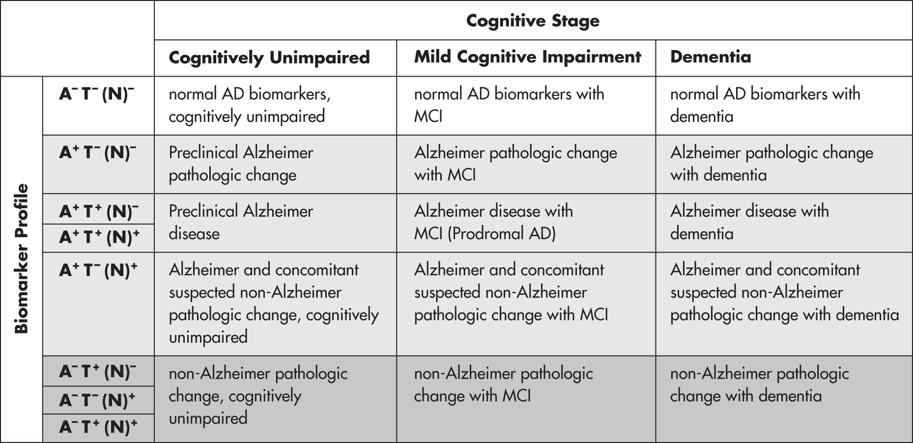
Diagnosis of AD has evolved with the development of biomarkers that indicate AD pathology in vivo such as brain amyloidosis and pathologic tau accumulation. The National Institute on Aging (NIA) and the Alzheimer Association (AA) recommended new diagnostic criteria and guidelines for AD in 2011, and these criteria were further revised in 2018 (Table 1). The NIA-AA criteria differed from prior DSM or NINDCS-ADRDA criteria in the following ways: (1) They recommend AD be considered a disease well before the onset of symptoms by incorporating biomarkers in diagnosis, and (2) they define three distinct stages of AD: (1) Preclinical AD, in which there is measurable biologic evidence of AD pathology but no symptoms; (2) MCI due to AD, in which the patient experiences mild memory loss but experiences no functional impairment at home or work but demonstrates biomarker evidence of AD; and (3) dementia due to AD, in which the patient experiences cognitive decline causing functional impairment and demonstrates biomarker evidence of AD. The 2018 NIA-AA criteria define AD not as three clinical syndromes but as a biologic process defined by biomarkers indicating the presence of beta amyloid (A+), pathologic tau (T+), and neurodegeneration or neuronal injury (N+). Using the ATN system and clinical status together allows an entire study population to be characterized (Fig. 1).
TABLE 1 New Diagnostic Criteria
| RESEARCH CRITERIA | |||
| Criteria for Probable Alzheimer Disease | DSM-5 2013 | NINCDS-ADRDA 2007 | NIA-AA 2018 |
| Insidious onset | X | X | X |
| Onset over months to years | X | X | |
| Progressive decline | X | X | X |
| Deficits are not explained by delirium or other medical or psychiatric conditions | X | X | X |
| Social/occupational impairment | X | X | |
| Presence of episodic memory deficit | X | X | |
| Cognitive deficits in at least two domains | X | X | |
| Neuropsychologic testing required for diagnosis? | Preferably | X | Only if routine history and mental status testing are inconclusive |
| Abnormal PET or MRI scan | Supportive feature* | Required if needed to show biomarker evidence of amyloidosis, tauopathy, neurodegeneration as part of the biomarker-based AT(N) diagnostic schema | |
| Genetic markers? | X | Supportive feature* | For research purposes |
| Required only if there is evidence of multiple causes and no clear evidence of progression and decline in memory and another cognitive domain | |||
| Abnormal cerebrospinal fluid marker required? | Supportive feature* | Required if needed to show biomarker evidence of amyloidosis, tauopathy, neurodegeneration as part of the biomarker-based AT(N) diagnostic schema | |
DSM-5, Diagnostic and Statistical Manual of Mental Disorders, fifth edition; MRI, magnetic resonance imaging; NIA-AA, National Institute on Aging-Alzheimers Association; NINCDS-ADRDA, National Institute of Neurological and Communicative Disorders and Stroke-Alzheimers Disease and Related Disorders Association; PET, positron emission tomography.
* At least one supportive feature is required for diagnosis of probable Alzheimer disease.
Modified from Fillit HM: Brocklehursts textbook of geriatric medicine and gerontology, ed 8, Philadelphia, 2017, Elsevier.
Figure 1 Descriptive nomenclature: Syndromal cognitive staging combined with biomarkers.

AD, Alzheimer disease; MCI, mild cognitive impairment. NOTE: Formatting denotes three general biomarker "categories" based on biomarker profiles: Those with normal AD biomarkers (no color), those with non-AD pathologic change (dark gray), and those who are in the Alzheimer continuum (light gray).
(From Clifford RJ Jr et al: NIA-AA research framework: toward a biological definition of Alzheimers disease, Alzheimer Dement 14:535-562, 2018.)
Biomarkers have not been commonly used in the clinic but have invariably been used in AD clinical trials. However, with the advent of monoclonal antibodies targeting beta amyloid, biomarker-based diagnosis-especially in patients with early-stage disease who may be eligible for new, disease modifying treatment-is becoming an important part of the diagnostic process. Biomarkers transform the diagnosis of AD into one that can be established definitively while the patient is still alive. In clinical practice, the diagnosis has been commonly made based on clinical history, a thorough physical and neurologic examination, and use of reliable and valid diagnostic criteria (i.e., DSM or NINDCS-ADRDA) such as the following:
Red flags for an AD diagnosis are summarized in Box 1.
BOX 1 Red Flags for an Alzheimer Disease Diagnosis
|
Modified from Kaufman DM et al: Kaufmans clinical neurology for psychiatrists, ed 9, Philadelphia, 2023, Elsevier.
TABLE 2 Cognitive Disorders in Older Adults
| Diagnosis (% of Dementias Attributable) | History | Physical Examination Findings | Imaging Findings | Comment |
| ||||
DM, Diabetes mellitus; EEG, electroencephalogram; HTN, hypertension; MCI, mild cognitive impairment; MRI, magnetic resonance imaging; PET, positron emission tomography; REM, rapid eye movement; TIA, transient ischemic attack.
From Warshaw G et al: Hams primary care geriatrics, ed 7, Philadelphia, 2022, Elsevier.
TABLE 3 Features Distinguishing Alzheimer Disease and Frontotemporal Dementia
| Feature | Alzheimer Disease | Frontotemporal Dementia |
| Age at onset (yr) | >65 | 53 (mean) |
| Memory impairments | Early, pronounced | Subtle, at least initially, with preserved visuospatial ability |
| Behavior abnormalities | None until middle or late stage | Early and prominent perseverative and compulsive behavior; hyperorality; impaired executive ability |
| Language impairment | Except for anomia, none until late stage | Paraphasias, anomia, decreased fluency |
| CT/MRI appearance | General atrophy, but especially parietal and temporal lobes | Frontal and temporal lobe atrophy |
| Histologic marker | Aβ accumulation | Tau accumulation |
CT, Computed tomography; MRI, magnetic resonance imaging.
From Kaufman DM et al: Kaufmans clinical neurology for psychiatrists, ed 9, Philadelphia, 2023, Elsevier.
TABLE 4 Clinical Features of Delirium, Depression, and Alzheimer Disease
| Delirium | Depression | Alzheimers Disease | |
| Onset of initial symptoms | Abrupt | Relatively discrete | Insidious |
| Difficulty with attention and disturbed consciousness | Dysphoric mood or lack of pleasure | Memory deficits-verbal and/or spatial | |
| Course | Fluctuating-over days to weeks | Persistent-usually lasting months if untreated | Gradually progressive, over years |
| Family history | Not contributory | May be positive for depression | May be positive for AD |
| Memory | Poor registration | Patchy/inconsistent | Recent > remote |
| Memory complaints | Absent | Present | Variable-usually absent |
| Language deficits | Dysgraphia | Increased speech latency | Confrontation naming difficulties |
| Affect | Labile | Depressed/irritable | Variable-may be neutral |
From Stern TA: Massachusetts General Hospital handbook of general hospital psychiatry, ed 8, Philadelphia, 2025, Elsevier.
TABLE 5 Symptoms and Preserved Abilities by Cognitive Domain Across Various Stages of Alzheimer Dementia
| Mild | Moderate | Severe | |
| Memory | |||
| Symptoms | |||
| Preserved abilities | |||
| Executive Function | |||
| Symptoms | |||
| Preserved abilities | |||
| Language and Communication | |||
| Symptoms | |||
| Preserved abilities | |||
| Sensory/Perceptual | |||
| Symptoms | |||
| Preserved abilities | |||
ADLs, Activities of daily living.
From Warshaw G et al: Hams primary care geriatrics, ed 7, Philadelphia, 2022, Elsevier.
Brief mental status testing can be done easily and quickly in the office. Formal neuropsychologic testing offers more nuanced data about a patients current cognitive and emotional function but is not required for straightforward cases. Formal neuropsychologic testing is indicated when patients present with atypical symptoms, have significant psychiatric comorbidities, and when patients or families report of cognitive dysfunction differs from findings on a bedside cognitive assessment. Also, neuropsychologic testing may be beneficial if there are concerns that in the future, the patients testamentary capacity will be challenged.
Commonly used cognitive tests to detect dementia include the Folstein Mini-Mental State Examination (MMSE), the Mini-Cog test, and the Montreal Cognitive Assessment. A newer self-administered gerocognitive examination (SAGE) that patients complete by themselves, usually in 15 minutes, is now available and consists of a validated 11-item instrument that compares favorably with MMSE and has the advantage of self-administration at home.3 A meta-analysis examining the performance of commonly used screening tests for dementia identified 11 commonly used tests, with the MMSE having the most data. The combined sensitivity and specificity for detecting dementia were 0.81 and 0.89, respectively, for the MMSE and 0.91 and 0.86, respectively, for the Mini-Cog. Subgroup analysis revealed that only the Montreal Cognitive Assessment had comparable performance to the MMSE for detecting MCI with 0.89 sensitivity and 0.75 specificity.
The Mini-Cog (https://mini-cog.com/) is a 3-min instrument consisting of a 3-item recall test for memory and a simply scored clock drawing test. The Montreal Cognitive Assessment (MoCA, www.mocatest.org/) is a 30-point test that takes approximately 10 min to administer and includes tests of visuospatial function, attention, verbal recall, language, abstraction, and orientation. A score of 25 points or less (26 points if the patient has <12 yr of education) indicates cognitive impairment. The test is available in >35 languages, and multiple forms in English allow for repeated assessments over time. A summary of commonly used tests may be found in Table E6.
TABLE E6 Commonly Used Neuropsychologic Tests
| Domains to Be Assessed | Tests Used With Age-Corrected and/or Education Norms for Adults Older Than 65 Yr | ||
| Premorbid ability | |||
| Verbal memory | |||
| Visual memory | |||
| Simple attention | |||
| Language | |||
| Executive function | |||
| Visuospatial | |||
| Motor | |||
| Mood |
WAIS-IV, Wechsler Adult Intelligence Scale, fourth edition; WMS-IV, Wechsler Memory Scale, fourth edition.
From Fillit HM: Brocklehursts textbook of geriatric medicine and gerontology, ed 8, Philadelphia, 2017, Elsevier.
Mental status testing should include tests that assess the following cognitive functions:
Patients with AD typically have trouble with verbal recall in addition to experiencing visuospatial or language deficits. Attention is usually preserved until the later stages of AD, so consider alternative diagnoses in patients who perform poorly on tests of attention early in their disease. A summary of the pattern of cognitive deficits associated with different dementias and depression may be found in Table 7.
TABLE 7 Patterns of Cognitive Impairment by Domain and Dementia
| Episodic Memory | Attention | Language | Executive | Visuospatial | Behavioral Symptoms | |
| ||||||
FTLD, Frontotemporal lobar degeneration; I, impaired; P, preserved; PPA, primary progressive aphasia; V, variable.
From Fillit HM: Brocklehursts textbook of geriatric medicine and gerontology, ed 8, Philadelphia, 2017, Elsevier.
In addition to the common amnestic presentation, AD rarely presents as one of three rare nonamnestic syndromes that affect memory later in the course of the disease. These three rare presentations should also raise suspicion of another dementia type. A primary language variant presents as either logopenic expressive aphasia or progressive nonfluent aphasia and may be either a form of AD or of frontotemporal lobar degeneration. A primary visuospatial variant called posterior cortical atrophy presents with disturbances in complex visual processing and may be a form of either AD or dementia with Lewy bodies. An executive/behavioral variant presents with impaired executive function and/or behavior derangement and may represent either a frontal variant of AD or behavioral variant frontotemporal dementia.
TABLE 8 Laboratory Evaluation of Patients With Dementia
| Type of Study | Examples | ||
| Basic studies, excluding reversible with specific indication from history for causes of dementia or examination |
| ||
| Adjuvant Studies to Aid Diagnosis | |||
| Other tests as indicated by history or physical or neurologic examination | |||
From Fillit HM: Brocklehursts textbook of geriatric medicine and gerontology, ed 8, Philadelphia, 2017, Elsevier.
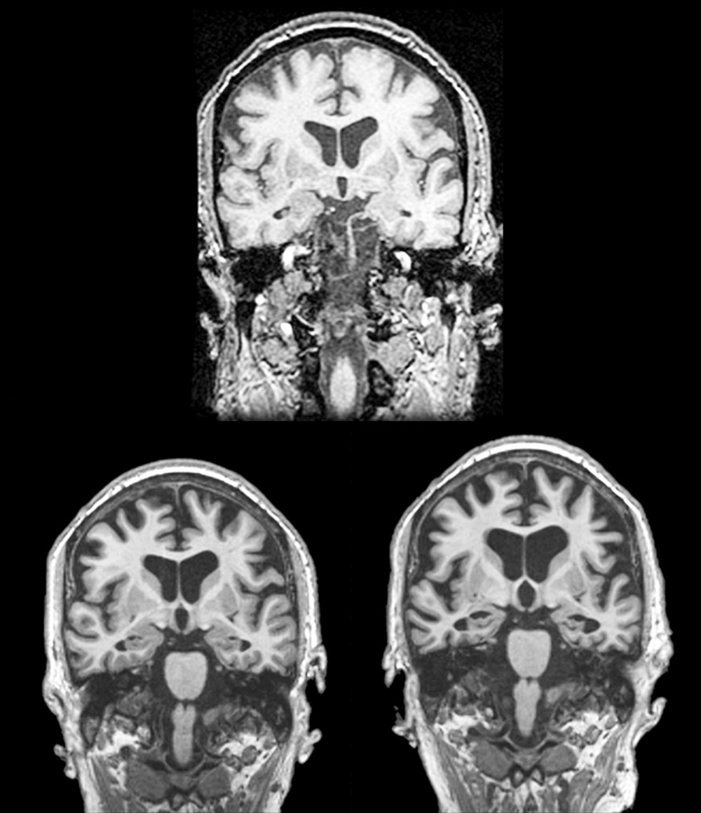
Figure E3 Longitudinal Coronal T1 Magnetic Resonance Imaging in a Patient that Progressed from Normal Cognition to Amnestic Mild Cognitive Impairment (Amci) to Dementia Due to Alzheimer Disease (AD)
Note Progressive Hippocampal and Cortical Atrophy. Top Image: Normal Cognition Age 75. Bottom Left Image: Amci Age 81. Bottom Right Image: Dementia Due to AD Age 86.

(From Jankovic J et al: Bradley and Daroffs neurology in clinical practice, ed 8, Philadelphia, 2022, Elsevier.)
TABLE 9 Person-Centered Care Approach Applied to Care of People Living With Dementia
| Key Component | Early Stage | Middle Stage | Late Stage |
| Develop a personalized, goal-oriented care plan, based on a thorough medical, functional, and social assessment | |||
| Periodically review the persons goals and care plan to assess ongoing effectiveness and to address evolving goals | |||
| Engage an interprofessional team that adapts its composition in response to the needs of the person living with dementia | |||
| A specified team leader to facilitate information transfer, care coordination, and continuity | |||
BPSD, Behavioral and psychologic symptoms of dementia; CHe-I, acetylcholinesterase inhibitors; OT, occupational therapy; PT, physical therapy; SLP, speech language pathology.
From Warshaw G et al: Hams primary care geriatrics, ed 7, Philadelphia, 2022, Elsevier.
TABLE 12 Instruments Used to Monitor Clinical Response of Alzheimer Disease (AD) to Pharmacologic Therapy
From Fillit HM: Brocklehursts textbook of geriatric medicine and gerontology, ed 8, Philadelphia, 2017, Elsevier.
TABLE 11 Acetylcholinesterase Inhibitor Dosing
| Drug | Initial Dose | Recommended Dose | Minimum Therapeutic Dose | Formulations |
| Donepezil | 5 mg daily | 10 mg daily | 5 mg daily | 5, 10, 23 mg |
| Galantamine IR | 4 mg bid | 12 mg bid | 8 mg bid | 4, 8, 12 mg |
| Galantamine ER | 8 mg daily | 24 mg daily | 16 mg daily | 8, 12, 24 mg |
| Rivastigmine | 1.5 mg bid | 6 mg bid | 3 mg bid | 1.5, 3, 4.5, 6 mg |
| Rivastigmine patch | 4.6 mg daily | 9.5 mg daily | 9.5 mg daily | 4.6, 9.5, 13.3 mg |
bid, Twice daily.
Acetylcholinesterase inhibitor dosing and suggested titration intervals. From US Department of Veterans Affairs. Pharmacy Benefits Management Services. 2018 [cited October 15, 2019]. Available at: http://www.pbm.va.gov/. Note that medication doses can be increased every 4 wk as patient tolerates.
From Warshaw G et al: Hams primary care geriatrics, ed 7, Philadelphia, 2022, Elsevier.
TABLE 10 Symptomatic Treatment of Memory Disturbance
| Initial Dose | Target Dose | |
| Donepezil | 5 mg qd for 4-6 wk | 10 mg qd |
| Rivastigmine | 1.5 mg bid with food, increase by 1.5 mg bid weekly | 3-6 mg bid |
| Galantamine | 4 mg bid with food, increase by 4 mg bid every 4 wk | 8-12 mg bid |
| Memantine | 5 mg qd, increase by 5 mg weekly | 10 mg bid |
bid, Twice daily; qd, once daily.
TABLE 13 Treatment of Behavioral and Neuropsychiatric Symptoms
| Initial Dose | Maximum Dose | |
| Atypical Antipsychotics | ||
| Olanzapine | 2.5 mg qd to bid, may increase by 2.5 mg as needed | 7.5 mg bid |
| Quetiapine | 25 mg bid, may increase by 25 mg every 2 days | 250 mg tid |
| Antidepressants | ||
| Sertraline | 25-50 mg qd, may increase by 25 mg every wk | 200 mg qd |
| Escitalopram | 10 mg qd, may increase after 1 wk to 20 mg qd | 10 mg qd |
bid, Twice daily; qd, once a day.
The physician should make a thorough search for the treatable causes of dementia. Current American Academy of Neurology practice parameters recommend:
In addition, refer patients with MCI or early AD to centers with experience with the new anti-amyloid monoclonal antibodies.
TABLE 14 Strategies for Prevention of Dementia
| Recommendation | Quality of Evidence | ||
| Engage in physical activity | Moderate | ||
| In adults with mild cognitive impairment, engage in physical activity to slow cognitive decline | Low | ||
| Tobacco cessation | Low | ||
| Do not exceed maximum daily recommended amount of alcohol intake | Moderatea | ||
| Follow a healthy diet based on WHO recommendationsb | Moderatec | ||
| Follow a Mediterranean diet | Moderate | ||
| Maintain a healthy weight | Low | ||
| Participate in cognitively stimulating activities or cognitive training | Low | ||
| Treatment of hypertension | High | ||
| Treatment of diabetes mellitus | Moderate | ||
| Treatment of dyslipidemia | Low |
Strategies for Prevention of Dementia, as based on World Health Organization (WHO) 2019 Guidelines for Risk Reduction of Cognitive Impairment and Dementia.
a 4 units of alcohol per wk for men, 7 units of alcohol per wk for women.
b Components include 5 daily servings of nonstarchy vegetables, <10% dietary intake of free sugars, <30% dietary intake of fats (preferentially unsaturated fats), <5 g daily of salt.
c Strength of evidence is variable based on individual dietary components.
From Warshaw G et al: Hams primary care geriatrics, ed 7, Philadelphia, 2022, Elsevier.
TABLE 15 Genes Implicated in the Development of Alzheimer Disease
| Gene | Comment | ||
| APP | >30 known mutations associated with EOAD; located on chromosome 21; associated with elevated risk of AD in Down syndrome | ||
| PSEN1 | >150 known mutations associated with EOAD | ||
| PSEN2 | <20 known mutations associated with EOAD | ||
| APOE |
AD, Alzheimer disease; EOAD, early-onset Alzheimer disease; LOAD, late-onset Alzheimer disease.
From Warshaw G et al: Hams primary care geriatrics, ed 7, Philadelphia, 2022, Elsevier.
For additional information for patients, families, and clinicians, contact the following organizations:
Available at Ebooks.health.elsevier.com.